The effective removal of Cr(VI) ions by carbon dot–silica hybrids driven by visible light

Yun Liua, Yu-jie Maab, Chun-yan Liu*a, Zhi-ying Zhanga, Wen-dong Yanga, Shi-dong Niea and Xue-hua Zhoub
aKey Laboratory of Photochemical Conversion and Optoelectonic Materials, Technical Institute of Physics and Chemistry, Chinese Academy of Sciences, Beijing 100190, People's Republic China. E-mail: cyliu@mail.ipc.ac.cn
bEnvironment Research Institute, Shandong University, Jinan, 250100, P. R. China

Received 25th May 2016 , Accepted 7th July 2016

First published on 7th July 2016


Abstract

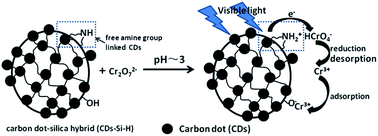
Methods to economically and efficiently remove highly toxic hexavalent chromium (Cr(VI)) from discharged industrial effluents have attracted critical attention due to the carcinogenic, mutagenic and teratogenic effects of Cr(VI). In the present work, we have explored the potential of carbon dots (CDs) in the elimination of Cr(VI) contamination. A series of carbon dot–silica hybrid photocatalysts (CD–Si–H) were firstly prepared by the co-hydrolysis and condensation of amine silane functionalized carbon dots and tetraethyl orthosilicate (TEOS). Due to the action of the carbon dots in the silica matrix, CD–Si–H demonstrated significant visible light absorption and good blue fluorescence with a longer fluorescent lifetime. With the increase of CDs, however, CD–Si–H suffered from a gradual reduction in the mesoporous structure, surface area and pore volume. In spite of this, the incorporation of a high concentration of CDs in the hybrid led to better visible light-harvesting, a stronger ability to capture Cr(VI) ions and a more effective visible-light-driven photo-reduction of Cr(VI). The presence of Cr(VI) resulted in a decrease of the fluorescent lifetime of the CDs embedded in CD–Si–H, suggesting that the electron transfer from the excited CDs to Cr(VI) is responsible for the effective photoreduction of Cr(VI). This work provides new methods to easily synthesize carbon dot–silica hybrids and to eliminate Cr(VI) contaminants under visible light.


Introduction

In the past decades, pollution caused by various toxic heavy metal ions has become a worldwide issue due to their great menace to our health and environment.1 As one of the most toxic heavy metal contaminants, chromium (Cr) is widely used and commonly found in the wastewater of various industrial processes including leather tanning, electroplating and metal finishing as well as processes associated with paint, textile and ceramic industries.2–4 Cr generally exists in hexavalent (Cr(VI)) and trivalent (Cr(III)) forms. Compared with Cr(III), Cr(VI) is regarded as one of the top-priority toxic pollutants by the U.S. Environmental Protection Agency (EPA) and the World Health Organization (WHO) due to its carcinogenic, mutagenic and teratogenic effects on the biological food chain and its very wide mobility in environments.5,6 Thus, discovering methods to economically, effectively and efficiently remove toxic Cr(VI) from discharged industrial effluents has become a very urgent issue.

Until now, various techniques, for instance adsorption, reduction, electrodialysis, reverse osmosis, ion-exchange, chemical precipitation as well as foam flotation etc., have been developed to remove Cr(VI).1,4,7–10 However, the practical applications of these techniques generally suffer from either large amounts of expensive reductants, high operational costs, the generation of highly toxic sludge etc.11 Considering the simple, clean, operable and economical features of photocatalytic processes, much attention has been paid to the conversion of Cr(VI) to the less toxic Cr(III) through photochemical processes with light, especially solar light.12 In addition, various photocatalysts, including wide or narrow band gap semiconductors and their heterojunctions, have been developed. However, for practical applications, they have some serious shortcomings. For instance, wide band gap photocatalysts, such as TiO2,11–13 CeO2,14 NaTaO3,12 ZnO15 etc., can only utilize ultraviolet light, which accounts for 5% of the total energy of the sun. Many narrow band gap semiconductors, such as SnS2,16 CdS,17 Cu2O,18 BiVO4 19 and Ag@Ag(Br, I)20 demonstrated poor photocatalytic efficiency and lower stability. Composite photocatalysts, including CdS/CdO,21 CdS/TiO2,22 SnS2/TiO2,23–25 SnS2/TiO2,26 Ag@Fe3O4@SiO2@TiO2,27 AgS quantum dots/TiO2,28 CuO/ZnO29 and so on, could realize the photoreduction of Cr(VI) in visible light. However, the net reduction of Cr(VI) is a multi-proton process and is more favourable in acidic or neutral media.12 Thus, the composite photocatalysts of metal chalcogenides or copper oxides reported could possibly sustain chemical etching and photo-corrosion, which would probably result in the bleaching of heavy metal ions and could consequently cause potential hazards to our environment. Plasmonic metal composite photocatalysts demonstrated better visible light action in some cases, but the expensive cost of noble metals limited their industrial applications. Thus, it is necessary to pursue new visible light-responsive photocatalysts that are low-cost, light and chemically stable and highly efficient.

Recently, carbon and carbon related quantum dots have attracted more and more attention not only for their excellent and tunable photoluminescence (PL), resistance to photobleaching and easy functionalization, but also for their chemical inertness, non-toxicity and elemental abundance.30–33 Associated applications in bioimaging,33,34 biolabeling,35,36 phototherapy,37 selective detection of chemical and biological species,38–42 photoelectric conversion,43 photochemical energy transition44 etc. have been frequently reported. However, techniques to effectively remove heavy metal ions using carbon dots, not only to detect these ions, have been less frequently reported. In the present work, we used amine silane-functionalized carbon dots (Si–CDs) and tetraethyl orthosilicate (TEOS) as co-precursors to synthesize CD–silica hybrids (CD–Si–H) and investigated their potential in the removal of Cr(VI) in visible light. The experimental results concluded that for the incorporation of CDs, CD–Si–H showed a good ability in the harvesting of visible light and the effective photocatalytic reduction of Cr(VI) in visible light. Moreover, for the a large number of amine groups in CD–Si–H, their adsorption capacity of Cr(VI) could reach approximately 74 mg g−1, significantly higher than that reported for many photocatalysts, some oxide adsorbents and modified silica.12–26,28,45–47 All these results suggested that through careful selection of the functional agents on the CDs and the reaction conditions, CD hybrids could not only be a good adsorbent for Cr(VI) and Cr(III), but also could be a promising photocatalyst to realize the effective photo-reduction of harmful Cr(VI) in visible light.

Experimental

Materials

The analytic grade reagents citric acid monohydrate (C6H8O7·H2O), cetyltrimethyl ammonium bromide (CTAB), TEOS, sodium hydroxide (NaOH), anhydrous ethanol, acetic acid and potassium dichromate (K2Cr2O7) were obtained from Beijing Chemical Reagent Co. N-(β-2-aminoethyl)-γ-aminopropyltrimethoxysilane (AEAPTMS) was purchased from Xiangqian Chemical Factory (Nanjing, China). Except for the citric acid, all other reagents were used as received without further purification. Deionized water was used in the experiments. Before use, citric acid monohydrate was dried under vacuum at 60 °C for 3 h, and then at 120 °C for 3 h to remove the crystallized water.

Synthesis of CD–Si–H

The silane-functionalized CDs (Si–CDs) were prepared by the pyrolysis of citric acid in the presence of AEAPTMS at 234 °C for 6 min.48 For the surface-passivation with silane, Si–CDs demonstrated a bright blue PL with a quantum yield of ∼34% (excited at 360 nm, quinine sulfate in 0.5 M sulfuric acid as a reference).49 Dynamic Light Scattering (DLS) measurements showed that the particle size of Si–CDs mainly centered around 1.8 nm.

The carbon dot–silica hybrid (CD–Si–H) was synthesized according to the following procedure. Under vigorous magnetic stirring at 80 °C, 0.343 g CTAB was dissolved in 149 ml of a NaOH aqueous solution (∼1.5 × 10⁻² mol L−1). Then, 1.95 ml of the mixture of TEOS and Si–CDs was quickly injected into the above solution. After stirring for 3 h at 80 °C, the precipitate was separated and washed with deionized water until the supernatant was close to neutrality. The obtained slurry was re-dispersed in a 100 ml mixture solution of acetic acid and ethanol (volume ratio: 95[thin space (1/6-em)]:[thin space (1/6-em)]5) and then stirred at room temperature for 24 h to remove the residual CTAB. The final product was washed with deionized water several times and then dried in air at room temperature. To prepare CD–Si–H with different concentrations of CDs, the volume ratio of TEOS to silane-functionalized CDs was varied at 12[thin space (1/6-em)]:[thin space (1/6-em)]1, 5.5[thin space (1/6-em)]:[thin space (1/6-em)]1, 3.3[thin space (1/6-em)]:[thin space (1/6-em)]1 and 1.6[thin space (1/6-em)]:[thin space (1/6-em)]1 (the total volume of Si-precursors (TEOS and Si–CDs) remained constant (1.95 ml)) and the resulting hybrids were named CD–Si–H1, CD–Si–H2, CD–Si–H3 and CD–Si–H4, respectively. Silica without carbon dots (m-Si) was prepared in the absence of the silane-functionalized CDs by the same procedures.

Characterization

Transmission electron microscopy (TEM) images were recorded with a JEOL JEM-2100 microscope operating at an accelerating voltage of 200 kV. The infrared (IR) spectra of the products were measured using a Hitachi Fourier transform IR (FT-IR) spectroscope with a resolution of 4 cm−1 and a scan time of 49. A Cary 5000 spectrophotometer with a 110 mm integrating sphere was used to record the ultraviolet-visible-near IR (UV-Vis-NIR) absorption spectra of CD–Si–H. Fluorescence spectra were recorded with a F-4500 fluorescence spectrophotometer. Time-resolved PL spectroscopy was performed with a time-correlated single photon counting (TCSPC) module (Edinburgh Instruments F900) and a temporal resolution of 4 ps. A pulse laser (∼375 nm) with an average power of 1 mW, operating at 40 MHz with a duration of 70 ps was used to excite CD–Si–H. The samples were prepared by ultrasonically dispersing CD–Si–H powders in an aqueous solution containing different concentrations of Cr(VI) (pH ∼ 3). To fit the fluorescence decay of the silane-functionalized CDs or CD–Si–H, two exponential functions were used to reach acceptable residuals (χ2 ≤ 1.2). The intensity-weighted average fluorescence lifetime (τav) was calculated with the following equation:50
τav = ∑Biτi2/∑Biτi
where Bi is the fractional weight of the various decay time components τi of the multi-exponential fitting. N2 adsorption–desorption measurements were performed on a Micrometrics ASAP 2020 instrument and the pore size distribution was calculated from the desorption branch of the sorption isotherms with the Barrett–Joyner–Halenda (BJH) method. Before measurements, the samples were dried under vacuum at 100 °C for 6 h. The size of the silane-functionalized CDs was measured with DLS on a Dynapro NanoStar (Wyatt, USA). To estimate the mass concentration change of total Cr during the photo-reaction, an inductively coupled plasma (ICP) optical emission spectrometer (Varian 710-ES) was used. X-ray photoelectron spectroscopy (XPS) analysis was carried out on an ESCAlab250xi electron spectrometer from Thermo Scientific with Al-Kα radiation and a spot size of 650 μm.

Catalytic experiments

Cr(VI) was used as a probe to evaluate the photocatalytic behavior of CD–Si–H in visible light. The typical experimental procedure is described as follows. The photocatalyst was added into an aqueous solution of K2Cr2O7 (8 × 10⁻⁴ mol L−1). After ultrasonic agitation for about 20 min, the pH of the dispersion was adjusted to 3 with a 0.1 mol L−1 HCl aqueous solution and the concentration of the photocatalyst was maintained at 1.0 g L−1. After stirring and purging with nitrogen gas for 30 min, the photocatalytic reaction was started under the illumination of a 300 W high pressure Hg lamp with a Pyrex glass tube and an appropriate filter to cut off light shorter than 400 nm. During the reaction, stirring and a stream of nitrogen gas was maintained. At given intervals, 5 ml aliquots were collected and then centrifuged to remove the photocatalyst. The supernatants were analyzed by recording their absorption spectra with a Shimadzu UV-1601 UV-Vis spectrophotometer. The typical absorption of Cr(VI) at about 350 nm was followed to survey the degradation rates of Cr(VI).

CD–Si–H4 was used as a model to estimate the photocatalytic activity in different cycles. After each cycle, all the photocatalyst (including the centrifuged and separated sample during the spectral analysis) was collected and dispersed in 5 ml of a 4 mol L−1 ammonia aqueous solution for 10 min, followed by centrifugation. This process was repeated twice and then the solid sample was washed with deionized water until the supernatant was close to neutral. The obtained photocatalyst was dried at 60 °C and the photocatalytic activity was tracked by the procedure introduced above.

Results and discussion

As Fig. 1 shows, CD–Si–H1, CD–Si–H2 and CD–Si–H3 consisted of mesoporous spheres with sizes of approximately 120, 128 and 155 nm; comparatively, the duplex and triplex grains tended to increase. In contrast, the particles of CD–Si–H4 (Fig. 1(d)) obviously adhered to each other and the porous structure disappeared.
image file: c6ra13593e-f1.tif
Fig. 1 TEM images of the CD–Si–H hybrids: (a) CD–Si–H1; (b) CD–Si–H2; (c) CD–Si–H3 and (d) CD–Si–H4 (the inserts in the figures are the corresponding magnified images).

A dominant band at 357 nm and a shoulder peak at about 450–500 nm emerged in the UV-Vis absorption spectrum of the Si–CDs (insert of Fig. 2). For the hybrid of the CDs in the SiO2 matrix, the dominant and shoulder peaks of CD–Si–H were red shifted to 373 nm and 475–700 nm, respectively (Fig. 2). With the dosage increase of CDs in the silica matrix, the absorption intensity became stronger and stronger. As Fig. 3 shows, the fluorescence emissions of Si–CDs and CD–Si–H1 were excitation wavelength-dependent. Comparing with Si–CDs, the optimal emission and excitation wavelengths of CD–Si–H1 red shifted about 10 nm, in accordance with the variation in the UV-Vis absorption, possibly due to the increase in the molecular rigidity of the CDs in the solid matrix. This could be confirmed by the change in the fluorescence lifetime (τ) from (9.76, 1.71) ns (original Si–CDs) to (10.87, 2.09) ns (CD–Si–H1). The optimal emission and excitation wavelengths of CD–Si–H continued to red shift with the increase in the concentration of the CDs in the silica matrix (Fig. S1), possibly resulting from the inner-filter effect of the fluorescent species with a high concentration.51


image file: c6ra13593e-f2.tif
Fig. 2 UV-Vis-NIR absorption spectra of the CD–Si–H powders. The insert is the UV-Vis absorption spectrum of Si–CDs in an ethanol solution.

image file: c6ra13593e-f3.tif
Fig. 3 Emission and excitation spectra at different excitation wavelengths: (a) Si–CDs ethanol solution and (b) CD–Si–H1 aqueous solution.

The N2 adsorption–desorption isotherms of CD–Si–H1, CD–Si–H2 and CD–Si–H3 were typical IV isotherms with a H3 hysteresis loop at relatively high pressure (Fig. 4), indicating the presence of mesoporous structures,52 agreeing well with the results observed in Fig. 1. The corresponding surface area and pore volumes are listed in Table 1. With the increase of Si–CDs introduced, the surface area and pore volume of CD–Si–H decreased gradually. Especially for the sample CD–Si–H4, the surface area was only 20.95 m2 g−1, ten times smaller than other CD–Si–H samples. Clearly, most of the pores disappeared in this case. As described in the experimental section, CD–Si–H was prepared in a basic medium and the formation of mesoporous structures should be related to the interaction between >Si–O (ionized product of Si–OH) and the micelles of CTA+ (ionized CTAB in aqueous solution).53 Excess organosilane would obviously reduce the amount of Si–OH produced during the hydrolysis and co-condensation of Si–CDs and TEOS, which sequentially affected the static interaction of >Si–O with CTA+ and the formation of pores, and at last led to a less porous structure of CD–Si–H4.53


image file: c6ra13593e-f4.tif
Fig. 4 (a) Nitrogen adsorption–desorption isotherms and (b) the corresponding pore size distribution obtained from the desorption branches of the different CD–Si–H samples.
Table 1 Structural parameters of the CD–Si–H samples
Samples CD–Si–H1 CD–Si–H2 CD–Si–H3 CD–Si–H4
a The surface area was calculated with the Brunauer–Emmett–Teller (BET) method.b Pore volume was calculated by the N2 amount adsorbed at P/P0 of 0.98.
Surface areaa/m2 g−1 644.1 398.1 366.9 20.95
Pore volumeb/cm3 g−1 1.03 0.459 0.405 0.074


As the FT-IR spectra of CD–Si–H demonstrated, with the dosage increase of Si–CDs introduced, the bands at 1414, 1560 and 1638 cm−1 related to the NH deformation and the C–N stretching vibrations became more obvious (Fig. S2), implying an increase in the presence of nitrogen-containing groups. The XPS results demonstrated that CD–Si–H mainly consisted of silicon, oxygen, nitrogen and carbon and that the atomic concentration of nitrogen was enhanced from 2.37% to 6.30% as the samples changed from 1 to 4 (Table S1 and Fig. S3a), further confirming the increase in the nitrogen-containing functional groups, which is in agreement with the results of the FT-IR. The high resolution spectrum of N 1s could be separated into two bands at 398.4 and 400.2 eV (Fig. S3b), indicating the presence of amine and amide groups.54

In the present work, the toxic Cr(VI) ion was used as a probe to evaluate the ability of CDs–H–Si to remove heavy metal ion pollution in visible light. As shown by curve a of Fig. 5, in the absence of any photocatalyst, the Cr(VI) aqueous solution showed no significant variation, even after illumination by visible light for 180 min. The introduction of m-SiO2 resulted in a small decrease in Cr(VI), whereas CD–Si–H caused the concentration of Cr(VI) to decrease significantly, especially in the case of CD–Si–H4. Moreover, the photocatalytic activity of CD–Si–H increased in the order CD–Si–H1 < CD–Si–H2 < CD–Si–H3 < CD–Si–H4, which is in good agreement with the dosage of CDs introduced. Simultaneously, the absorption of visible light (Fig. 2) became stronger, proving that the visible light harvesting ability of the photocatalyst played a crucial role in the efficient photoreduction of Cr(VI).


image file: c6ra13593e-f5.tif
Fig. 5 The relative concentration change of Cr(VI) with irradiation time of visible light in the presence of different photocatalysts: (a) no photocatalyst; (b, c, d, e and f) m-Si, CD–Si–H1, CD–Si–H2, CD–Si–H3 and CD–Si–H4.

Besides the response to visible light, the adsorption performance of the reactants is another important factor in the good photocatalytic activity of a photocatalyst. Therefore, we investigated the adsorption ability of different CD–Si–H samples toward Cr(VI) ions (Fig. S4 and Table 2). Clearly, CD–Si–H4 had the largest adsorption capacity for Cr(VI) ions, up to ∼73.7 mg g−1 (Table 2), significantly higher than those of many other photocatalysts reported, which were about 10 mg g−1[thin space (1/6-em)]12–26,28,45 As is well known, the adsorption property of materials is not only related to their surface area, but is also significantly impacted by the available adsorptive sites. The surface area of CD–Si–H4 was only 20.95 m2 g−1 (Table 1), dozens of times smaller than the other samples, but showed the largest adsorption capacity toward Cr(VI) ions (Table 2). As we know, AEAPTMS contains two amine groups, one is a primary amine, which was used to modify the carbon dots,48 and the other is a secondary amine, which can be protonated and become positively charged in acidic aqueous media.55 In aqueous solution, Cr(VI) generally exists as various oxyanions such as HCrO4, CrO42− and Cr2O72−. HCrO4 is the dominant Cr(VI) species in the pH range of 1–6.8.55,56 In our work, the photoreduction of Cr(VI) was carried out in a pH 3 aqueous solution. Thus, the protonated and positively charged secondary amine groups in CD–Si–H could act as specific sites to adsorb HCrO4 through electrostatic attraction. The maximum concentration of CDs is in CD–Si–H4, which contained the highest concentration of protonated amine groups. Clearly, despite having the smallest surface area, CD–Si–H4 showed the best ability for the capture of Cr(VI) in acidic media.

Table 2 Cr(VI) adsorption capacity of the CD–Si–H powders
Sample m-SiO2 CD–Si–H1 CD–Si–H2 CD–Si–H3 CD–Si–H4
Adsorbed Cr(VI) (mg g−1) 8.7 21.4 30.9 45.3 73.7


To explore the photoreduction mechanism of Cr(VI), we investigated the influence of the Cr(VI) concentration on the PL of CD–Si–H. Considering the dispersion of different CD–Si–H samples, CD–Si–H1 was used as a model. Fig. 6(A) exhibited that Cr(VI) evidently quenched the PL of CD–Si–H1 and the quenching degree increased along with the concentration of Cr(VI) added. The PL of CD–Si–H1 was focused in the range of 400–600 nm, whereas Cr(VI) showed little absorption in this wavelength range (Fig. 6(B)). Thus, energy transfer is not thought to be the key reason for the evident PL decrease observed here. Table 3 revealed that with an increase in the concentration of Cr(VI) from 0 to 32 × 10−6 mol L−1, the average fluorescence lifetime of CD–Si–H1 (τav) decreased from 9.89 ns to 5.98 ns, confirming the effective electron transfer from the excited carbon dots to Cr(VI) and consequently its evident reduction. In addition, the linear relationship between the relative τav(τav0/τav) and the concentration of Cr(VI) (Fig. S5) illustrated that the dynamic (collisional) quenching mechanism was mainly responsible for the decrease in fluorescence. The quenching constant was closely related to the diffusion velocity of Cr(VI). Thus, enriching Cr(VI) near CDs is a valid way to accelerate the electron transfer from excited CDs to Cr(VI) and then their photoreduction.


image file: c6ra13593e-f6.tif
Fig. 6 (A) Fluorescence change of CD–Si–H1 with varying concentration of Cr(VI) (pH ∼ 3); (B) curve a: the fluorescent spectrum of CD–Si–H1 (excited at 370 nm); curve b: the UV-Vis absorption of Cr(VI).
Table 3 Fluorescence lifetime of CD–Si–H1 in the presence of Cr(VI)a
Cr(VI) concentration × 10−6 mol L−1 τ1 (ns) (Rel%) τ2 (ns) (Rel%) τav (ns) χ2
a τ1, τ2 and τav correspond to the fluorescence lifetimes of the CDs hybridized in CD–Si–H1 and the average fluorescent lifetime, respectively. The fluorescence decay curves are fitted with the double exponential model and two fluorescence lifetimes were obtained.
0 10.9 (88.87) 2.09 (11.13) 9.89 1.11
8 10.1 (82.79) 2.20 (17.21) 8.70 1.10
16 9.33 (78.37) 2.13 (21.63) 7.77 1.15
24 8.33 (77.19) 1.84 (22.81) 6.85 1.20
32 7.65 (71.80) 1.73 (28.20) 5.98 1.12


As described in Fig. 5, CD–Si–H4 illustrated the highest efficiency for the photoreduction of Cr(VI) driven by visible light. After the reaction, the colour of the photocatalysts changed from yellow to bluish-grey (Fig. S6), confirming the formation and adsorption of Cr(III) ions on CD–Si–H4. The concentration of total Cr (Cr(VI) and Cr(III)) before and after the photocatalytic treatment was measured by ICP. The original total dose of Cr was about 89 mg g−1 and decreased to 15 mg g−1 due to adsorption onto CD–Si–H4. After the photocatalytic reaction, the residual Cr dropped to 4 mg g−1, indicating the effective photocatalytic reduction of Cr(VI) and adsorption of Cr(III) onto CD–Si–H4. In other words, through photocatalytic reduction and adsorption processes, as much as 95% of the total Cr species was removed from the reaction solution.

Understandably, the excess accumulation of Cr(III) in the environment is still harmful. Thus, besides Cr(VI), the WHO set a limit of 50 mg L−1 as the maximum allowable emission standard for total Cr.57 Therefore, Cr(III) from the reduced or photoreduced Cr(VI) is generally removed by precipitation, adsorption and separation.58,59 In the present work, CD–Si–H4 could not only convert Cr(VI) to Cr(III) under the illumination of visible light, but could also eliminate Cr(III) efficiently from the reaction system through the adsorption process. Without a doubt, it is more favourable.

The reusability of CD–Si–H4 was evaluated in Fig. S7. Clearly, the photoreduction efficiency of Cr(VI) decreased with the increase in recycle number. Similar results have also been reported by some other groups11,24,25 and they are generally recognized as the masking effect of Cr(OH)3 on the photoactive sites of the photocatalysts. We suggest that the detachment of the CDs from CD–Si–H4 during the regeneration process is the critical factor in the decrease in the photoreduction efficiency of Cr(VI) observed here. When 4 mol L−1 of ammonia solution was used to regenerate the photocatalysts used, a strong fluorescence was observed in the supernatant, indicating the presence of CDs in the supernatant. The adsorption capacity of the photocatalyst gradually reduced with the recycle number. Meanwhile, the color of the photocatalyst became lighter after regeneration with an alkaline solution. As is well known, silica can dissolve in strong alkaline solution through the breaking of Si–O–Si bonds. Correspondingly, N-(β-2-aminoethyl)-γ-aminopropyl rendered the Si–O–Si bond neighbouring CDs easily attacked by hydroxyl ions, which possibly resulted in the leakage of CDs from the hybrids. Thus, to improve the durability of CD-based photocatalysts, a new CD-based composite structure or system should be designed in the future.

Fig. 7 illustrates how to effectively remove Cr(VI) and Cr(III) by CD–Si–H under visible light illumination. As mentioned above, AEAPTMS was used to functionalize the CDs and then was used with TEOS as co-precursors to synthesize CD–Si–H photocatalysts. AEAPTMS contains two amine groups in an alkyl substitute, one of which is linked with the carboxyl groups of the CDs through amidation48 and the other (secondary amine) is free. The XPS results concluded the presence of two types of nitrogen-containing groups (Fig. S3b). Thus, there are plenty of free amine groups persisting in CD–Si–H, which could be protonated and positively charged in acidic media. For the electrostatic attraction, Cr(VI) was adsorbed on the sites close to the CDs hybridized in the silica matrix. Under illumination of visible light, Cr(VI) was reduced to Cr(III) by an electron transferred from the conduction band of the excited CDs. With the help of electrostatic repulsion, this produced positively charged Cr(III) left from the protonated amine sites and then adsorbed on CD–Si–H via the interaction with Si–O groups derived from ionized silanols groups on the surface of the SiO2 micro-domain.53 Through these processes, the effective removal of total Cr could be realized.


image file: c6ra13593e-f7.tif
Fig. 7 Schematic illustration of the removal of Cr(VI) and Cr(III) by CD–Si–H in acidic media (pH ∼ 3).

Conclusions

In summary, CDs functionalized with trimethoxysilane containing two amine groups in the alkyl substituent were successfully used to synthesize carbon dot–silica–hybrids (CD–Si–H). Due to the rich free amine groups and the covalent incorporated CDs with a high dosage, CD–Si–H demonstrated significant visible light harvesting, a large Cr(VI) adsorption capacity (73.7 mg g−1), a strong photocatalytic activity for the reduction of Cr(VI) into Cr(III) in visible light and the concurrent effective adsorption and elimination of Cr(III). In the future, CD-based photocatalysts with desirable properties could be developed through careful selection of the molecular structure of the passivate agents, by the employment of CDs with the absorption of visible light or even NIR light and by increasing the rate of the interface electron transfer from the CDs to Cr(VI). In order to more effectively remove Cr pollution, this is vital.

Acknowledgements

The authors are grateful for the support of the National Natural Science Foundation of China (21103209 and 21273256).

Notes and references

  1. K. Ikehata, Y. Jin, N. Malkey and J. Lin, Heavy Metals in Water: Presence, Removal and Safety, ed. S. K. Sharma, the Royal Society of Chemistry, 2015, ch. 7, p. 141 Search PubMed.
  2. W. Liu, J. Zhang, C. Zhang and L. Ren, Chem. Eng. J., 2012, 189–190, 295 CrossRef CAS.
  3. Y. G. Abou El-Reash, M. Otto, I. M. Kenawy and A. M. Ouf, Int. J. Biol. Macromol., 2011, 49, 513 CrossRef CAS PubMed.
  4. Z. Song, W. Li, W. Liu, Y. Yang, N. Wang, H. Wang and H. Gao, RSC Adv., 2015, 5, 13028 RSC.
  5. S. Basha, Z. Murthy and B. Jha, Chem. Eng. J., 2008, 137, 480 CrossRef CAS.
  6. J. Fuller, D. I. Stewart and I. T. Burke, Ind. Eng. Chem. Res., 2013, 52, 4704 CrossRef.
  7. X.-Q. Zhang, Y. Guo and W.-C. Li, RSC Adv., 2015, 5, 25896 RSC.
  8. Y. Xing, X. Chen and D. Wang, Environ. Sci. Technol., 2007, 41, 1439 CrossRef CAS PubMed.
  9. F. Gode and E. Pehlivan, J. Hazard. Mater., 2005, 119, 175 CrossRef CAS PubMed.
  10. V. A. Okello, N. Du, B. Deng and O. A. Sadik, J. Environ. Monit., 2011, 13, 1236 RSC.
  11. (a) H. Eskandarloo, A. Badiei, M. A. Behnajady and G. M. Ziarani, RSC Adv., 2014, 4, 28587 RSC; (b) Y. Yang, G. Wang, G. Gu, Q. Li, S. Kang, Y. Zhang, D. H. L. Ngc and H. Zhao, RSC Adv., 2015, 5, 11349 RSC.
  12. Q. Cheng, C. Wang, K. Doudrick and C. K. Chan, Appl. Catal., B, 2015, s176–177, 740 CrossRef.
  13. Z. Yang, B. Wang, J. Zhang, H. Cui, Y. Pan, H. An and J. Zhai, Phys. Chem. Chem. Phys., 2015, 17, 18670 RSC.
  14. J. Wu, J. Wang, Y. Du, H. Li, Y. Yang and X. Jia, Appl. Catal., B, 2015, 174–175, 435 CrossRef CAS.
  15. J. Doménech and J. Muñoz, Electrochim. Acta, 1987, 32, 1383 CrossRef.
  16. Y. C. Zhang, J. Li, M. Zhang and D. D. Dionysiou, Environ. Sci. Technol., 2011, 45, 9324 CrossRef CAS PubMed.
  17. X. Liu, L. Pan, T. Lv, G. Zhu, Z. Sun and C. Sun, Chem. Commun., 2011, 47, 11984 RSC.
  18. S.-K. Li, X. Guo, Y. Wang, F.-Z. Huang, Y.-H. Shen, X.-M. Wang and A.-J. Xie, Dalton Trans., 2011, 40, 6745 RSC.
  19. B. Xie, H. Zhang, P. Cai, R. Qiu and Y. Xiong, Chemosphere, 2006, 63, 956 CrossRef CAS PubMed.
  20. P. Wang, B. Huang, Q. Zhang, X. Zhang, X. Qin, Y. Dai, J. Zhan, J. Yu, H. Liu and Z. Lou, Chem.–Eur. J., 2010, 16, 10042 CrossRef CAS PubMed.
  21. W. Yang, Y. Liu, Y. Hu, M. Zhou and H. Qian, J. Mater. Chem., 2012, 22, 13895 RSC.
  22. H. Park, W. Choi and M. R. Hoffmann, J. Mater. Chem., 2008, 18, 2379 RSC.
  23. J. Wang, X. Li, X. Li, J. Zhu and H. Li, Nanoscale, 2013, 5, 1876 RSC.
  24. J. Li, T. Wang and X. Du, Sep. Purif. Technol., 2012, 101, 11 CrossRef CAS.
  25. Y. C. Zhang, J. Li and H. Y. Xu, Appl. Catal., B, 2012, 123–124, 18 CrossRef CAS.
  26. Y. C. Zhang, L. Yao, G. Zhang, D. D. Dionysiou, J. Li and X. Du, Appl. Catal., B, 2014, 144, 730 CrossRef CAS.
  27. J. Su, Y. Zhang, S. Xu, S. Wang, H. Ding, S. Pan, G. Wang, G. Li and H. Zhao, Nanoscale, 2014, 6, 5181 RSC.
  28. H. Wang, X. Yuan, Y. Wu, X. Chen, L. Leng and G. Zeng, RSC Adv., 2015, 5, 32531 RSC.
  29. J. Yu, S. Zhuang, X. Xu, W. Zhu, B. Feng and J. Hu, J. Mater. Chem. A, 2015, 3, 1199 CAS.
  30. X. Li, M. Rui, J. Song, Z. Shen and H. Zeng, Adv. Funct. Mater., 2015, 25, 4929 CrossRef CAS.
  31. H. T. Li, Z. H. Kang, Y. Liu and S.-T. Lee, J. Mater. Chem., 2012, 22, 24230 RSC.
  32. C. Ding, A. Zhu and Y. Tian, Acc. Chem. Res., 2014, 47, 20 CrossRef CAS PubMed.
  33. C. Zhou, R. Booker, R. Li, X. Zhou, T. K. Sham and X. Sun, et al., J. Am. Chem. Soc., 2007, 129, 744 CrossRef PubMed.
  34. E. J. Goh, K. S. Kim, Y. R. Kim, H. S. Jung, S. Beack, W. H. Kong, G. Scarcelli, S. H. Yun and S. K. Hahn, Biomacromolecules, 2012, 13, 2554 CrossRef CAS PubMed.
  35. Q. Li, T. Y. Ohulchanskyy, R. Liu, K. Koynov, D. Wu, A. Best, R. Kumar, A. Bonoiu and P. N. Prasad, J. Phys. Chem. C, 2010, 114, 12062 CAS.
  36. C. H. Lee, R. Rajendran, M.-S. Jeong, H. Y. Ko, J. Y. Joo, S. Cho, Y. W. Chang and S. Kim, Chem. Commun., 2013, 49, 6543 RSC.
  37. J. C. Ge, M. H. Lan, B. J. Zhou, W. M. Liu, L. Guo, H. Wang, Q. Y. Jia, G. L. Niu, X. Huang, H. Y. Zhou, X. M. Meng, P. F. Wang, C. S. Lee, W. J. Zhang and X. D. Han, Nat. Commun., 2014, 5, 4596 CAS.
  38. M. Zheng, Z. Xie, D. Qu, D. Li, P. Du, X. Jing and Z. Sun, ACS Appl. Mater. Interfaces, 2013, 5, 13242 CAS.
  39. Y.-L. Zhang, L. Wang, H.-C. Zhang, Y. Liu, H.-Y. Wang, Z.-H. Kang and S.-T. Lee, RSC Adv., 2013, 3, 3733 RSC.
  40. J. Hou, J. Yan, Q. Zhao, Y. Li, H. Ding and L. Ding, Nanoscale, 2013, 5, 9558 RSC.
  41. X. Li, S. Zhu, B. Xu, K. Ma, J. Zhang, B. Yang and W. Tian, Nanoscale, 2013, 5, 7776 RSC.
  42. D. Wu, X. Huang, X. Deng, K. Wang and Q. Liu, Anal. Methods, 2013, 5, 3023 RSC.
  43. (a) V. Gupta, N. Chaudhary, R. Srivastava, G. D. Sharma, R. Bhardwaj and S. Chand, J. Am. Chem. Soc., 2011, 133, 9960 CrossRef CAS PubMed; (b) X. Yan, X. Cui, B. Li and L. Li, Nano Lett., 2010, 10, 1869 CrossRef CAS PubMed.
  44. (a) J. Liu, Y. Liu, N. Liu, Y. Han, X. Zhang, H. Huang, Y. Lifshitz, S.-T. Lee, J. Zhong and Z. Kang, Science, 2015, 347, 970 CrossRef CAS PubMed; (b) L. Cao, S. Sahu, P. Anilkumar, C. E. Bunker, J. Xu, K. A. S. Fernando, P. Wang, E. A. Guliants, K. N. Tackett and Y.-P. Sun, J. Am. Chem. Soc., 2011, 133, 4754 CrossRef CAS PubMed.
  45. A. Walcarius and L. Mercier, J. Mater. Chem., 2010, 20, 4478 RSC.
  46. (a) X.-Q. Zhang, Y. Guo and W.-C. Li, RSC Adv., 2015, 5, 25896 RSC; (b) W. Cai, J. Yu and M. Jaroniec, J. Mater. Chem., 2010, 20, 4587 RSC; (c) Y. Cantu, A. Remes, A. Reyna, D. Martinez, J. Villarreal, H. Ramos, S. Trevino, C. Tamez, A. Martinez, T. Eubanks and J. G. Parsons, Chem. Eng. J., 2014, 254, 374 CrossRef CAS PubMed.
  47. (a) L. Zhu, C. Zhang, Y. Liu, D. Wang and J. Chen, J. Mater. Chem., 2010, 20, 1553 RSC; (b) V. Kumari, M. Sasidharan and A. Bhaumik, Dalton Trans., 2015, 44, 1924–1932 RSC; (c) M. K. Dinker and P. S. Kulkarni, New J. Chem., 2015, 39, 3687 RSC.
  48. F. Wang, Z. Xie, H. Zhang, C. Liu and Y. Zhang, Adv. Funct. Mater., 2011, 21, 1027 CrossRef CAS.
  49. J. N. Demas and G. A. Crosby, J. Phys. Chem., 1971, 75, 991 CrossRef.
  50. G. Z. Chen, X. Z. Huang, Z. S. Zheng, J. G. Xu and Z. B. Wang, Fluorescence Spectroscopy, Science Press, Beijing, 3rd edn, 1990 Search PubMed.
  51. Y. Liu, C. Liu and Z. Zhang, J. Colloid Interface Sci., 2011, 356, 416 CrossRef CAS PubMed.
  52. K. S. D. Sing, D. H. Everett, R. A. W. Haul, L. Moscou, R. A. Pierotti, J. RouQuerol and T. Siemiewska, Pure Appl. Chem., 1985, 57, 603 CrossRef CAS.
  53. D. K. Padhi and K. Parida, J. Mater. Chem. A, 2014, 2, 10300 CAS.
  54. (a) M.-C. Hsiao, S.-H. Liao, M.-Y. Yen, P.-I. Liu, N.-W. Pu, C.-A. Wang and C.-C. M. Ma, ACS Appl. Mater. Interfaces, 2010, 2, 3092 CrossRef CAS PubMed; (b) F. Beguin, M. Inagaki, R. Benoit and R. Erre, Internal symposium on carbon, 1990, vol. 2, pp. 806–809 Search PubMed.
  55. B. Chen, X. Zhao, Y. Liu, B. Xu and X. Pan, RSC Adv., 2015, 5, 1398 RSC.
  56. Y. J. Jiang, X. Y. Yu, T. Luo, Y. Jia, J. H. Liu and X. J. Huang, J. Chem. Eng. Data, 2013, 58, 3142 CrossRef CAS.
  57. F. Zhang, N. Du, H. Li, X. Liang and W. Hou, RSC Adv., 2014, 4, 46823 RSC.
  58. S. Deng and R. Bai, Water Res., 2004, 38, 2424 CrossRef CAS PubMed.
  59. V. Sarin and K. K. Pant, Bioresour. Technol., 2006, 97, 15 CrossRef CAS PubMed.

Footnote

Electronic supplementary information (ESI) available: Fluorescent spectra, FT-IR spectra, adsorption property, fluorescent quench by Cr(VI) and reusability of CDs–Si–H. See DOI: 10.1039/c6ra13593e

This journal is © The Royal Society of Chemistry 2016
Click here to see how this site uses Cookies. View our privacy policy here.