
Antifouling behavior of chitosan adorned zinc oxide nanorods
Tamilselvan Abiramana,
Ganapathy Kavithab,
Ramasamy Rengasamyb and
Sengottuvelan Balasubramanian*a
aDepartment of Inorganic Chemistry, University of Madras, Chennai-600025, India. E-mail: bala2010@yahoo.com
bCAS in Botany, University of Madras, Chennai-600025, India
First published on 15th July 2016
Abstract
Chitosan adorned zinc oxide nanorods (CAZO NRs) were synthesized by a chemical conversion method. The synthesis was carried out by the conversion of chitosan biopolymer capped zinc(II) ions using a sodium hydroxide solution under atmospheric conditions at 70 °C. The resulting CAZO NRs were investigated by UV-Visible spectroscopy, Fourier transform infra-red spectroscopy, Raman spectroscopy, X-ray diffraction, X-ray photoelectron spectroscopy, photoluminescence spectroscopy, field emission scanning electron microscopy and high resolution transmission electron microscopy. The CAZO NRs are mixed with commercial paints (polyurethane clear, polyurethane white and acrylic emulsion) and applied on three different surfaces (wood, mild steel and cement slab panels). The hydrophilicity and uniform surface smoothness of CAZO nanopaint coated panels were analyzed by water contact angle measurement. These panels were investigated for their antifouling behavior against three different green and marine algae viz. Arthrospira, Chlorella and Amphora. The CAZO NRs exhibit 75–90% antifouling efficiency against the growth of algae.
Introduction
Marine biofouling is defined as the growth of unwanted micro (bacteria, algae, spores) and macro (barnacles, mussels) organisms on the surface of natural and artificial structures immersed in seawater.1–3 It is well known that marine biofouling has an enormous harmful impact such as increased fuel consumption, decreased vessel speed, elevated dry-dock cleaning expenses, loss of hull strength, biocorrosion of naval vessels and seawater pipelines.4–6 The government and industry spend nearly US-$ 6.5 billion annually to control marine biofouling. Marine antifouling coating is the most effective way for avoiding marine organism attachment till date. Generally, according to the toxicity of coating materials, the antifouling strategies can be divided into two main categories viz. nontoxic coating and chemically active coating.7There are two major environmental pollutants released by the fouling of hulls; (i) an increase in gas emissions (CO2, CO and SO2) into the atmosphere and (ii) the dissemination of potentially rare fouling species. Antifouling paints are mixtures of toxic materials/biocides (lead, mercury and arsenic) used to control fouling organisms. However, organotin and tributyltin (TBT) are the most effective antifoulants. Studies pertaining to toxic biocides indicated that a high TBT concentration was detected in many marine organisms such as invertebrates, vertebrates and marine plants. The use of toxic organotin-based antifoulants and their application on ships has been prohibited a few years ago.8 The alternative solution for antifouling agents employs polyvinyl formal membranes, PEG-b-cationic polycarbonate, poly(ethyleneglycol) nanofibrous mesh, poly(N-hydroxyethylacrylamide)/salicylate hydrogels, poly(N-vinyl pyrrolidone), and zwitterionic silane that could minimize the adhesion of fouling organisms, through topography, novel surface chemistry and pulsed electrical fields.9–11 But the cost associated with these materials and their maintenance is quite high. Therefore, it is essential to develop eco-friendly antifouling compounds.
In the recent times, the physical, chemical and mechanical properties of nanomaterials have considerably improved the potential applications ranging from environment and energy to healthcare compared with those of bulk materials. International ecological restrictions have directed research toward developing environment-friendly nanomaterials for biofield applications. Environmental exposure of nanomaterials is inevitable because of their widespread use in many technological and biological fields. Several materials such as V2O5 NPs, Ag NPs modified surfaces, hybridpolymer/Au NPs, zwitterionic SiO2 NPs and dye-doped Si NPs have been employed as antifouling agents.12–14
Among the nanoparticles, ZnO is an attractive and promising material due to its wide band gap (3.37 eV at room temperature), large exciton binding energy (60 meV), n-type conductivity, abundance in nature and ecofriendly property.15 These characteristics make this material attractive for many applications such as solar cells, optical coatings, photo catalysts, electrical devices, antibacterial coatings and biofouling-resistant membranes. With one-fourth the cost, ZnO nanoparticles is clearly more economical than TiO2 and Al2O3 nanoparticles.16
Chitosan is one of the most abundant biopolymers on earth and it contains cationic polysaccharide of (1,4)-linked 2-amino-2-deoxy-β-D-glucose and 2-acetamido-2-β-D-glucose units. The primary amine and hydroxyl groups of chitosan exhibit very strong affinity (like chelating agent) towards metal ion to reduce the particle size and prevent agglomerisation.17 The zinc oxide NPs have been synthesized by various methods such as chemical conversion, sonochemical, micro emulsion, pulsed laser irradiation, polyol process and reverse micelles method. Especially, the chemical conversion method is very easy to handle and less time consuming for the synthesis of metal oxide NPs.18–23
Herein, we report the preparation of chitosan adorned zinc oxide NRs in aqueous medium with green capping agent (chitosan) under atmospheric condition at 70 °C. The chitosan adorned zinc oxide NRs were mixed with ordinary paints and applied on three different surfaces (wood, MS and concrete panels). The water contact angle measurement was carried out to ascertain the hydrophilicity and uniform surface smoothness of CAZO nanopaint coated panels. These panels were tested for their antifouling behavior against three different fouling algae (Arthrospira, Chlorella and Amphora) in both fresh and marine environments.
Experimental section
Materials
Zinc sulfate pentahydrate (Merck), sodium hydroxide (Merck) and chitosan (Sigma-Aldrich) were used without any purification. Polyurethane clear (MRF corp, vapocure paints), polyurethane white (MRF corp, vapocure paints), acrylic emulsion paints (premium coatings and chemicals pvt Ltd) and sanding sealer (Esdee paints) were obtained from local suppliers. Wood, mild steel and cement slab panels were also obtained from local suppliers. All other chemicals were used without any further purification.Synthesis of zinc oxide nanorods with chitosan
Zinc sulfate pentahydrate (0.0017 mol, 500 mg), was dissolved in 50 mL of double distilled water and kept at 70 °C. 10 mL of 1% chitosan solution was added with stirring for 10 min to get a homogeneous solution. Sodium hydroxide (0.0025 mol) solution was added drop-wise until the pH of the solution was raised to 10. The appearance of white solid indicates the formation of zinc oxide NRs. The reaction was continued for further 1 h. The colloidal solution was centrifuged at 10![[thin space (1/6-em)]](https://www.rsc.org/images/entities/char_2009.gif) 000 rpm for 30 min, the precipitate was collected, washed several times with double distilled water. The sample was dried in a vacuum desiccator and used for further studies.
000 rpm for 30 min, the precipitate was collected, washed several times with double distilled water. The sample was dried in a vacuum desiccator and used for further studies.
Synthesis of zinc oxide without chitosan
Zinc sulfate pentahydrate (0.0017 mol, 500 mg), was dissolved in 50 mL of double distilled water and kept at 70 °C. Sodium hydroxide (0.0025 mol) solution was added drop by drop, till the pH of the solution was raised to 10. The appearance of white precipitate indicates the formation of zinc oxide. The reaction was continued for 1 h. The solution was centrifuged at 10000 rpm for 30 min, the precipitate was collected, washed several times with double distilled water. The sample was dried in a vacuum desiccator and used for further studies.
Chitosan adorned zinc oxide nanopaint preparation
:2 ratio of the above mixture and water were taken in another beaker and then it was stirred well for further 30 min.Panels preparation
:1 ratio of zirconic acid and poly acrylic acid mixture was made up to 700 mL and the pH of the solution was maintained at 3–3.5 by the addition of water) and dried at room temperature. Then, the CAZO nanopaint was uniformly applied on the surface of the metal steel panels. The panels were dried at room temperature.:1 ratio of zirconic acid and poly acrylic acid was made up to 700 mL and the pH of the solution was maintained at 3–3.5 by the addition of water) and dried at room temperature. Then, the CAZO nanopaint was evenly applied on the surface of the concrete panels. The panels were dried at room temperature.Tanks preparation
Algae growth study
The growth of the algae on the surface area of the each panels was studied by the measurement of chlorophyll-a concentration (tank-I) and cell counts (tank-II & III) for a period of 30 days with an interval of 10 days.| Chl a (mg L−1) = 11.24 × A661.6 − 2.404 × A644.8 |
Characterization
The chitosan, zinc oxide and chitosan adorned zinc oxide nanorods were characterized by following methods:The optical absorbance of the three samples was recorded on a Perkin-Elmer lambda 650 spectrophotometer. The spectra were recorded in the wavelength range of 200 to 800 nm. FT-IR spectra of the samples were recorded using Perkin-Elmer 8300 FT-IR spectrometer and the spectra were recorded in the frequency range of 400 to 4000 cm−1 using KBr. Raman analysis was carried out using Laser Raman Microscope model 11I, Nanophoton, Japan, with Ne–Ar laser source of 532 nm wavelength, grating 600 mm−1. The crystalline nature of the samples were determined by powder X-ray diffraction using Bruker D8 Advance X-ray diffractometer with monochromatic Cu-Kα radiation (λ = 1.5418 Å). The samples were recorded over the diffraction angle (2θ) range between 5° and 70°. The analytical characterization of the material preceded with the XPS surface speciation analysis of the pristine CAZO NRs. The elemental XPS analysis was carried out on a XM1000 Omicron Nanotechnology XPS system with Al-α K radiation (1486.7 eV energy). The instrument was operated at 300 W. The CAZO NRs were made as pellet and used for XPS analysis. The XPS spectra were calibrated to C 1s core peak. The fluorescence spectra of the samples were recorded using Jobin Yvon Fluoromax-4P spectrofluorimeter and also Perkin-Elmer MPF-44B fluorescence spectrophotometer interfaced with computer through Rishcom multimeter.
The surface morphology of the samples was examined with a HITACHI-S3890 scanning electron microscope. The powder samples were directly used for the analysis. The particles size of the samples was determined on a T-30 HRTEM with an acceleration voltage of 250 kV. The samples were prepared on a copper grid after sonication. The water contact angles of CAZO nanopaint coated and uncoated panels were measured at room temperature using the Holmarcopto Mechatronics instrument. The water (Milli-Q) droplet volume was 10 μL and the contact angle was measured 5 s after the drop was deposited on the CAZO nanopaint coated and uncoated panels. The water contact angle measurement was carried out at five different places of each panel. The visual changes of CAZO nanopaint coated and uncoated panels were observed using a Cannon power shot A3400 camera. The images were obtained both before and after the study.
Results and discussion
The chitosan adorned zinc oxide nanorods were synthesized according to Scheme 1.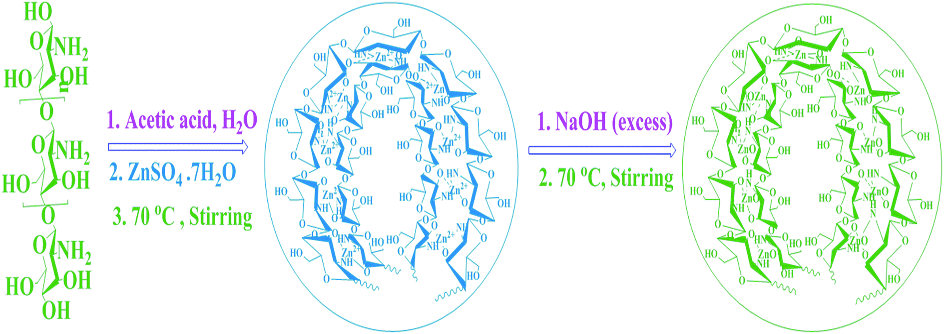 | ||
| Scheme 1 Synthesis of CAZO NRs. | ||
Mechanism for the formation of CAZO NRs
The following mechanism has been suggested for the formation of chitosan adorned zinc oxide nanorods: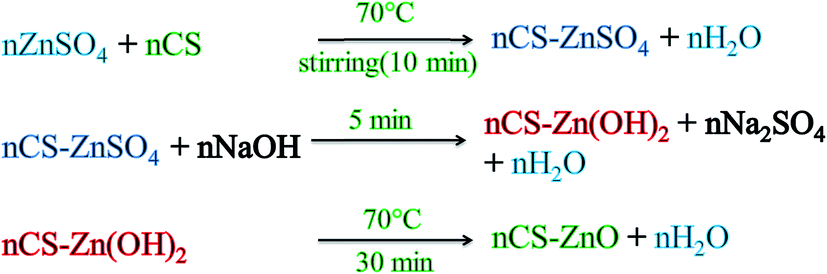
Various factors such as pH, temperature, capping agent, modulator reagent and reaction time play an important role in the synthesis of controllable size and morphology of nanostructures. The zinc(II) ions are chelated by chitosan biopolymer and subsequently they react with sodium hydroxide resulting in the formation of colloidal zinc hydroxide solution which was subsequently converted to zinc oxide nanorods. The synthesis of zinc oxide NRs in aqueous solution at low temperature and without capping agent was not easy since they immediately aggregate and some impurities like Zn(OH)2 are also present. Therefore in the present case, the mixture of precursor and the capping agent was stirred vigorously at 70 °C for 10 min. The capping agent prevents the aggregation and control the particles size. Avali et al. have used acetic acid as a modulator reagent to grow nanorods. Modulator reagent prevents the coordination interaction between the metal ions and the organic linkers, which generate a competitive situation that regulates the rate of framework extension and crystal growth. In our reaction few drops of acetic acid were added to increase the solubility of chitosan which is derived from chitin and has acetate group. In the presence of modulator reagent of acetic acid and excess of sodium hydroxide solution only the zinc hydroxide was completely converted from zinc hydroxide to zinc oxide nanorods without any impurity like zinc hydroxide or starting material.
The optical properties of pure chitosan, ZnO and CAZO NRs are investigated by UV-Visible spectroscopy. The (Fig. 1(a)) shows the absorbance spectrum of pure chitosan which shows the maximum absorbance at 286 nm and it corresponds to characteristic absorbance of chitosan. The peak at 365 nm confirms the formation of pure zinc oxide (Fig. 1(b)). The UV-Vis absorption spectrum of CAZO NRs (Fig. 1(c)) shows a strong absorption band at 362 nm, which corresponds to SPR of zinc oxide NRs28 and there is a blue shift relative to pure zinc oxide (365 nm) and the bulk compound (380 nm). The blue shift denotes the decrease in size of particle and increase in band gap energy. The band gap energy (Ebg) of ZnO can be calculated from the following equation:
| Ebg = 1240/λ (eV) |
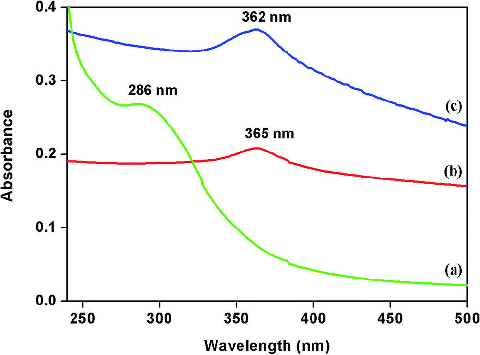 | ||
| Fig. 1 UV-Visible spectra of (a) pure chitosan, (b) ZnO and (c) CAZO NRs. | ||
The Fig. 2 shows FT-IR spectra of pure ZnO, chitosan and chitosan adorned ZnO NRs. The (Fig. 2(a)) shows FT-IR spectrum of pure ZnO. The spectrum clearly shows characteristic absorption bands at 447 and 501 cm−1 which correspond to two transverse optical stretching modes of ZnO and confirm the formation of ZnO. Fig. 2(b) shows the characteristic frequencies of pure chitosan. The band at 3367 cm−1 is assigned to both –O–H and –N–H stretching vibration. The frequencies at 2884 and 2143 cm−1 correspond to aliphatic –C–H symmetric and asymmetric stretching vibrations respectively. The band at 1651 cm−1 corresponds to –NH2 bending vibration. The bands at 1421, 1387 and 1317 cm−1 correspond to –C–H bending vibration. The bands near 1069 cm−1 are attributed to C–O and C–N stretching vibrations respectively.30 The intensity of these characteristic bands significantly increase in the spectrum of CS-ZnO NRs, indicating that C–O and C–N groups are involved in ZnO coordination (Fig. 2(c)). The peak at 3400 cm−1 corresponding to the stretching vibration of –OH and –NH2 became broader and of higher intensity indicating some interaction between these groups and ZnO NRs. Compared with pure chitosan, new bands appearing at 503 and 436 cm−1 can be attributed to the stretching vibration of N–Zn–O and O–Zn–O.31 The shape of FT-IR spectrum is generally influenced by factors such as particle size and morphology of NPs. Verge's et al. have reported, the theoretically established relationship between the shape of the FT-IR spectrum and the morphology of ZnO NRs. The strong absorption band is located at 494 cm−1 for spherical particles, but two bands at 406 (strong) and 580 cm−1 (weak) are observed for slab-type particles. In the present case, the ZnO exhibits rod shape, which is confirmed by HRTEM analysis.
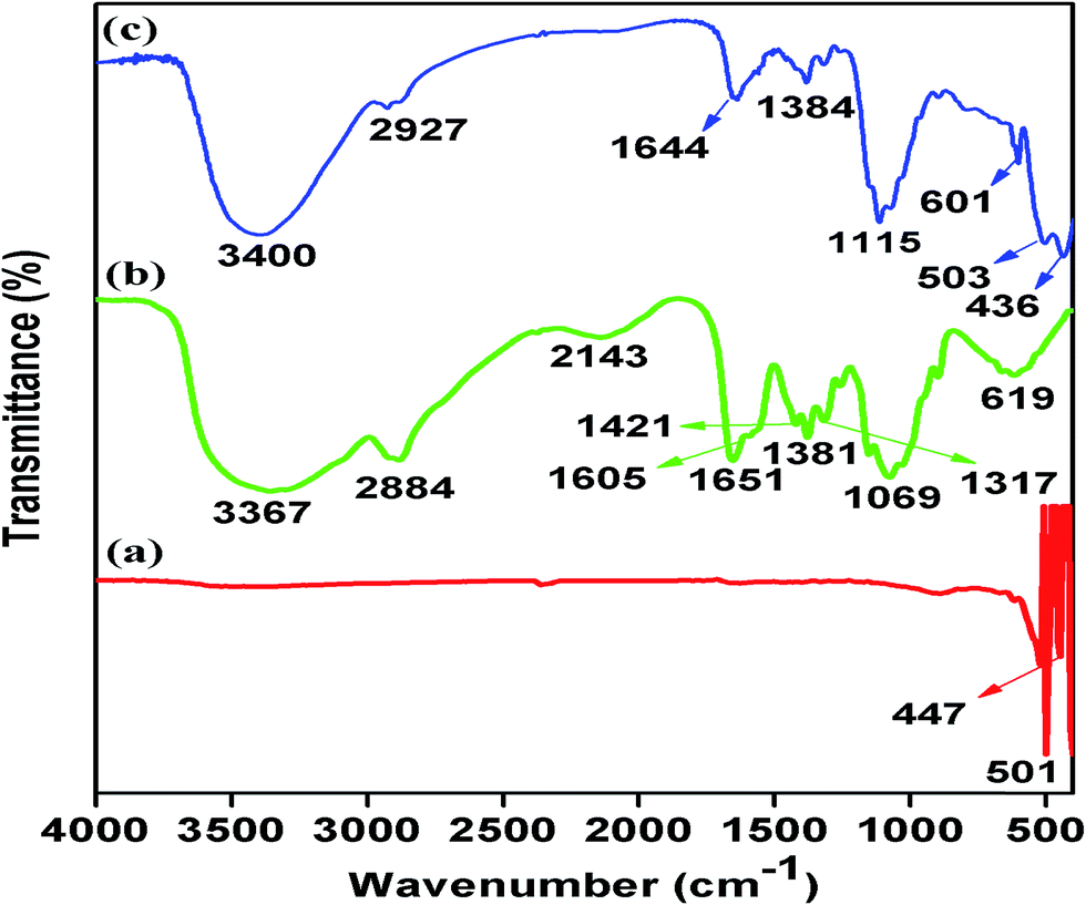 | ||
| Fig. 2 FT-IR spectra of (a) pure ZnO, (b) chitosan and (c) CAZO NRs. | ||
Raman spectrum is very useful and sensitive for determining the crystal perfection and structural defects. It is employed here to confirm the structure of ZnO nanostructures. Wurtzite-type ZnO belongs to the space group C46v with two formula units in the primitive cell. According to the group theory, single-crystal ZnO has eight sets of optical phonon modes at Γ point of the Brillouin zone, such as A1 + E1 + 2E2 modes (Raman active), 2B1 modes (Raman inactive), and A1 + E1 modes (IR active). Both A1 and E1 modes are polar and split into transverse (TO) and longitudinal optical (LO) phonons. Non-polar phonon modes with symmetry E2 have two frequencies such as E2 (H) and E2 (L). The E2 (H) is associated with oxygen atoms and E2 (L) is associated with Zn sub-lattice.32
Fig. 3 shows the Raman spectra of pure chitosan, zinc oxide and CAZO NRs in the range of 80–750 cm−1. The (Fig. 3(a)) shows Raman spectrum of pure chitosan which clearly indicate that the chitosan does not exhibit Raman bands in the case of visible region excitation. The pure ZnO exhibits major bands at 97.2, 326.6, 432.6 and 570 cm−1 which confirm the formation of ZnO (Fig. 3(b)). The main band observed at 432.6 cm−1 for ZnO NRs could be assigned to the nonpolar optical phonon E2 (high) mode in ZnO (Fig. 3(c)). The existence of the E2 (high) mode indicates the hexagonal wurtzite structure, which is in agreement with the XRD analysis. However, the non-polar phonon modes with symmetry E2 have two frequencies, E2 (high) is associated with oxygen atoms and E2 (low) is associated with Zn sublattice. The shift of wave numbers and change of band shapes were due to the chitosan strongly adorned with ZnO. The recorded Raman spectra clearly demonstrate that the formation CAZO NRs is possible only in the presence of chitosan and they have a hexagonal wurtzite structure with good crystal quality, which is in good agreement with the XRD analysis.
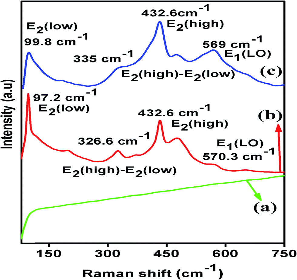 | ||
| Fig. 3 Raman spectra of (a) chitosan, (b) zinc oxide and (c) CAZO NRs. | ||
The purity, crystalline nature and average particle size of chitosan, zinc oxide and CAZO NRs were analyzed by PXRD with 2θ values in the range of 5° and 70°. The (Fig. 4(a)) shows XRD pattern of pure chitosan which indicates 2θ values at 10.04° and 19.9° which are characteristic peaks of pure chitosan. Fig. 4(b) shows the XRD pattern of CAZO NRs. The main characteristic peaks for zinc oxide NRs observed at 31.69°, 34.35°, 36.30°, 47.55°, 56.58°, 62.80° and 68.04° correspond to (100), (002), (101), (102), (110), (103) and (112) planes respectively. The additional two peaks appear at 10.2° and 21.3° which indicate the formation of ZnO adorned by chitosan. These peaks confirm the formation of wurtzite crystalline nature of CAZO NRs without any impurity which is in good agreement with JCPDS file # 003-1005.33 The average crystallite particle size of CAZO NRs calculated by using Scherrer formula to the most intense peak (002) is 16 nm. The main characteristic peaks for pure zinc oxide was observed at 31.69°, 34.35°, 36.30°, 47.55°, 56.58°, 62.80° and 68.04° which correspond to (100), (002), (101), (102), (110), (103) and (112) planes. The peaks confirm the formation of pure ZnO without any impurity which is in good agreement with JCPDS file # 089-0510 (Fig. 4(c)). Even though, the above results suggest that we can synthesize pure ZnO without capping agent, but it does not form in definite structure and nanosized shape. In the presence of capping agent only we can get definite structure and nanosized shape which is confirmed by FESEM and HRTEM studies. The nanomaterials exhibit totally different physical and chemical properties when compared to aggregate or bulk materials which are confirmed by photoluminescence study of pure ZnO and CAZO NRs.
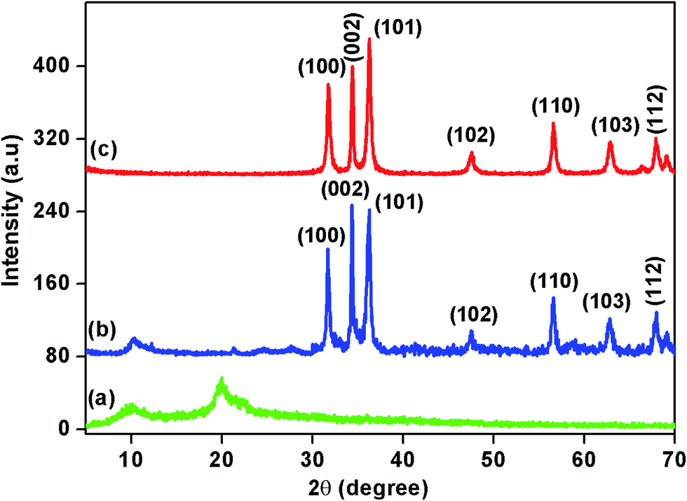 | ||
| Fig. 4 PXRD pattern of (a) chitosan, (b) zinc oxide and (c) CAZO NRs. | ||
The elemental analysis of CAZO NRs was carried out by high resolution XPS. The survey spectrum clearly indicates the presence of only Zn, C, O and N in CAZO NRs (Fig. 5(a)). The Zn2+ core level peak fitting spectrum (Fig. 5(b)) shows the major peaks at 1021.4 and 1044.7 eV which correspond to the core level of Zn 2P3/2 and 2P1/2.34 The C 1s peak fits at 284.5, 285.5, 287.2, 290.5, 293.5 and 295.6 eV (Fig. 5(c)) correspond to (C–C/C–H), (C–OH/C–N), (C–O–C), (C![[double bond, length as m-dash]](https://www.rsc.org/images/entities/char_e001.gif) O), (OC–O) and (π–π*) respectively. Fig. 5(d) shows the O 1s peak fitting spectrum and it clearly indicates the binding energies at 531.8, 530.6, 524.4, 522.5 and 520.7 eV which correspond to (Zn–O), (C–OH), (C–O–C), (OC–O) and (CO) respectively. The N 1s peak fit at 401.5 eV for (C–N) and 399.5 eV for (N–ZnO) have also been observed (Fig. 5(e)).35 The spectra (5a–e) indicate that the zinc oxide NRs are strongly adorned by chitosan.
O), (OC–O) and (π–π*) respectively. Fig. 5(d) shows the O 1s peak fitting spectrum and it clearly indicates the binding energies at 531.8, 530.6, 524.4, 522.5 and 520.7 eV which correspond to (Zn–O), (C–OH), (C–O–C), (OC–O) and (CO) respectively. The N 1s peak fit at 401.5 eV for (C–N) and 399.5 eV for (N–ZnO) have also been observed (Fig. 5(e)).35 The spectra (5a–e) indicate that the zinc oxide NRs are strongly adorned by chitosan.
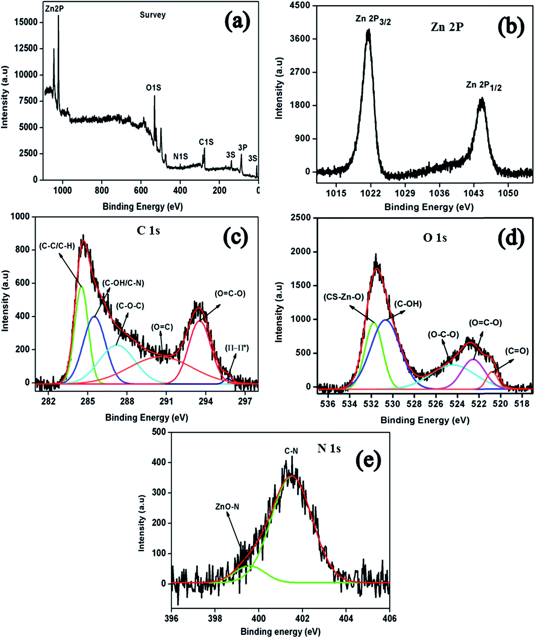 | ||
| Fig. 5 XPS analysis of CAZO NRs: (a) survey spectrum of all elements, (b) peak fitting of Zn 2P3/2, (c) peak fitting of C 1s, (d) peak fitting of O 1s, (e) peak fitting of N 1s. | ||
The photoluminescence (PL) spectrum of pure chitosan is shown in (Fig. 6(a)) which indicates a strong emission peak at 423 nm with excitation at 295 nm. The (Fig. 4(b)) shows the PL spectrum of pure ZnO with excitation at 375 nm which exhibit emissions at 453 nm with a shoulder at 436 nm which are attributed to the radiative annihilation of excitons of aggregated ZnO. The PL results show the typical two emissions of narrow blue (470 nm) and broad green bands (562 nm) of CAZO NRs (Fig. 6(c)) with excitation at 372 nm.36 The broad emission band observed in the visible region in the present case is due to the superposition of blue and green emissions. ZnO commonly exhibits luminescence in the visible spectral range due to different intrinsic or extrinsic defects. According to the synthetic process as well as the assignments of defects in the literature, the defects formed in our sample are likely due to an oxygen vacancy and interstitial oxygen, corresponding to blue and green bands in PL, respectively.
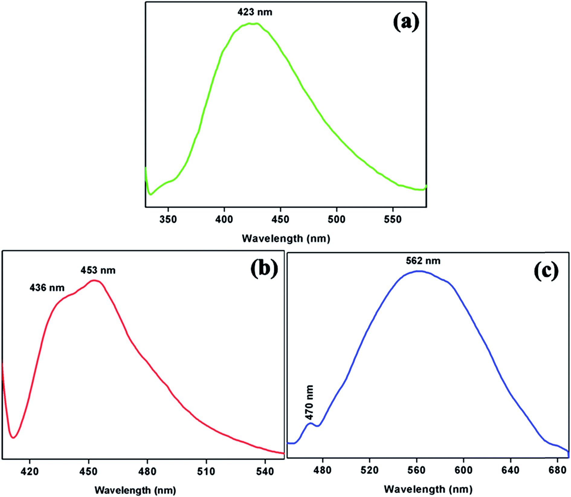 | ||
| Fig. 6 Photoluminescence spectra of (a) chitosan, (b) zinc oxide and (c) CAZO NRs. | ||
The morphology of pure chitosan, synthesized zinc oxide and zinc oxide NRs loaded chitosan was investigated by FESEM analysis. The (Fig. 7(a)) shows high magnification image of pure chitosan which indicates that the chitosan has sheet like morphology. Fig. 7(b) indicates high magnification image of synthesized zinc oxide which clearly shows that the zinc oxide has aggregated featureless morphology. The (Fig. 7(c)) shows the high magnification image of zinc oxide NRs loaded chitosan biopolymer, which indicates that the zinc oxide is formed in rod shape inside the chitosan matrix. Based on the above results, it is concluded that the formation of rod shaped zinc oxide is possible only in the presence of chitosan.
 | ||
| Fig. 7 FESEM images of (a) chitosan, (b) zinc oxide and (c) zinc oxide NRs loaded chitosan. | ||
The elemental composition of CAZO NRs was analyzed by EDAX. Fig. 8 shows that the CAZO NRs contain only Zn, C, O and N and other impurities are not present.
 | ||
| Fig. 8 EDAX spectrum of zinc oxide NRs loaded chitosan. | ||
The shape and size of pure chitosan, zinc oxide and CAZO NRs were analyzed by HRTEM. The (Fig. 9(a)) shows HRTEM image of pure chitosan which clearly indicates that the chitosan has sheet shape. The pure zinc oxide exhibits only aggregated shape which is confirmed in (Fig. 9(b)). Fig. 9(c) shows the high magnification HRTEM image of CAZO NRs and it indicates the rod shape dark parts surrounded by white features, which correspond to zinc oxide NRs uniformly decorated by chitosan biopolymer. The mean particle size was predicted to be around 15 nm width and 110 nm length using image J software. The image clearly demonstrates that the zinc oxide NRs are well dispersed without any agglomeration. The selected area electron diffraction (SAED) pattern was taken from a part of the NRs which confirmed the formation of wurtzite structure of chitosan adorned zinc oxide NRs (Fig. 9(d)).37 The above results suggest that the nanorod shape zinc oxide is formed only in the presence of chitosan and without that the particles are aggregated.
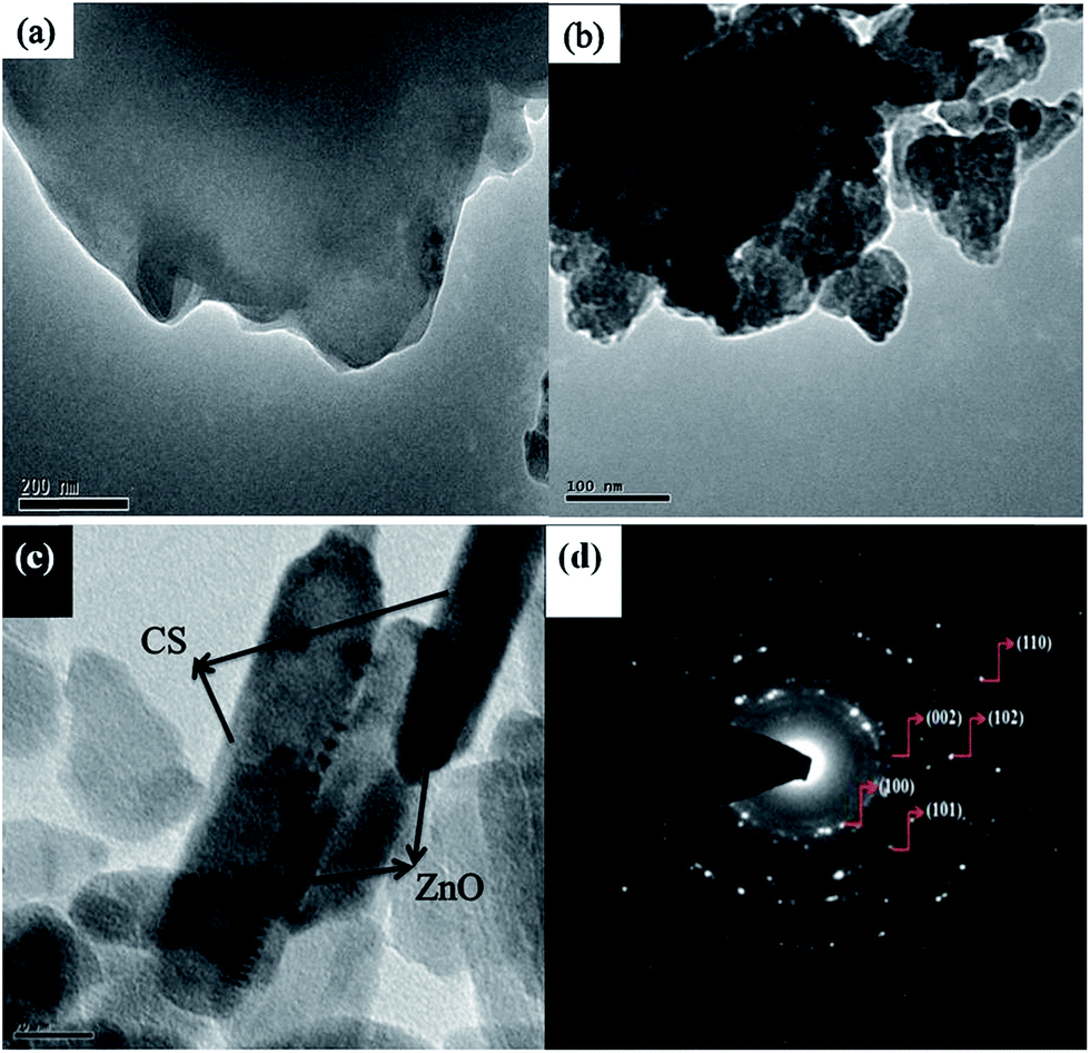 | ||
| Fig. 9 HRTEM images of (a) pure chitosan, (b) zinc oxide, (c) CAZO NRs and (d) SAED pattern of CAZO NRs. | ||
The fouling decreases by increasing the hydrophilicity and smoothness of the coated materials. The higher hydrophilicity and smoothness were confirmed by the contact angle measurements. The contact angle depends on surface hydrophilicity (or) hydrophobicity, roughness and porosity of the surface of substrates. The general rule states that lower the water contact angle the stronger is the interaction. Fig. 10(a)–(e) show the water contact angle of CAZO nanopaint coated and uncoated (exception wood) W, MS and CS panels. The water contact angle was measured at five different places of CAZO nanopaint coated wood panel surface (69 ± 0.1°, 69 ± 0.3°, 69 ± 0.5°, 69 ± 0.3° and 69 ± 0.2°), which shows that coated panel is hydrophilic in nature. The values are very close to each other indicating that the coated panels have uniform smooth surface. The water contact angle values of coated MS, coated CS, uncoated MS and uncoated CS panels are given in Table 1. The values obtained at different points on coated CS and coated MS panels indicate that they are hydrophilic and has smooth surface. However, the uncoated MS and uncoated CS panels are hydrophobic in nature.
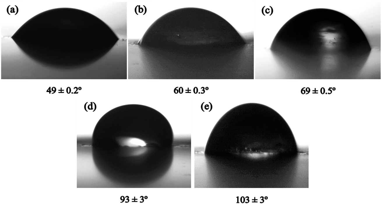 | ||
| Fig. 10 Water contact angle images of CAZO nanopaint coated panels (a–c) and uncoated panels (d & e). | ||
| Panels | Water contact angle (θ) |
|---|---|
| Coated MS | 49 ± 0.3, 49 ± 0.1, 49 ± 0.2, 49 ± 0.4, 49 ± 0.3 |
| Coated CS | 60 ± 0.2, 60 ± 0.1, 60 ± 0.3, 60 ± 0.2, 60 ± 0.4 |
| Uncoated MS | 90.2 ± 2, 88 ± 1, 93 ± 3, 87 ± 2, 91 ± 4 |
| Uncoated CS | 99 ± 3, 100.2 ± 1, 103 ± 3, 101.2 ± 2, 98.2 ± 4 |
Based on the above results, it is inferred that the CAZO nanopaint coated panels are more hydrophilic and have smooth surfaces when compared to that of the uncoated panels. Since chitosan is hydrophilic, hydrophilicity is increased in the case of CAZO nanopaint coated panels. Considering that surface hydrophilicity and smoothness of panels were sequentially increased by the CAZO nanopaint coating, the improved hydrophilicity and smoothness resulted in the reduction of algal growth on the surfaces. The water contact angle for uncoated wood panel has not been measured as it has lot of pores on the surface and hence the water drops are completely absorbed.
Antifouling behavior of CAZO nanopaint coated and uncoated panels against algae
In general, a number of factors are involved in the algal growth like temperature, pH, roughness, medium etc. The roughness is one of the main factors for the algal growth. If the surface of the substrates is rough then the algal growth is high when compared to substrates with smooth surface.38Fig. 11(a) and (b) shows, the variation between uncoated and CAZO nanopaint panel surfaces (W, MS and CS) before algal treatment. The surface of the uncoated panels is very rough, but in the case of CAZO nanopaint coated panel, surfaces are very smooth and homogeneous.
 | ||
| Fig. 11 (a) Uncoated and (b) CAZO nanopaint coated panels before algal treatment. | ||
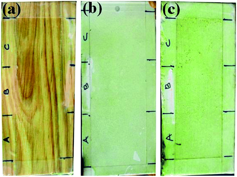
Fig. 12 shows the comparison of visual images of algal growth on the surface of uncoated and CAZO nanopaint coated panels after algal treatment. The surface of uncoated (W, CS and MS) panels were fully affected by both fresh and marine algae (Fig. 12(a), (c) and (e)). But CAZO nanopaint coated panels are not fully affected by the algae (Fig. 12(b), (d) and (f)) since the CAZO NRs exhibit good antifouling properties against both fresh and marine algae.
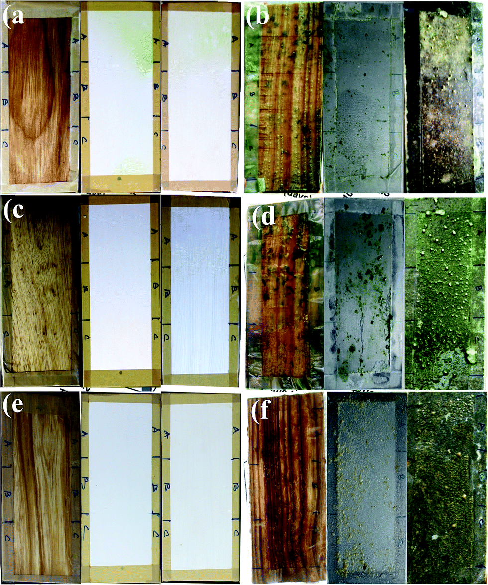 | ||
| Fig. 12 (a, c & e) CDZO nanopaint coated and (b, d & f) uncoated panels after algal treatment. | ||
It has been reported that the algal growth occurs in 4 stages: (1) the algal growth commences on new surfaces in the lag phase; here the algal growth increases slowly (lag phase). (2) Later the algal growth increases appreciably due to multiplication (log phase). (3) The algal growth is maintained in the stationary phase. (4) Finally, the algal growth decrease due to the nutrient deficiency (death phase).39
Fig. 13 shows the comparative study of three algal (Arthrospira, Chlorella and Amphora) growth on three different surfaces (CAZO nanopaint coated and uncoated W, MS and CS panels) via chlorophyll-a concentrations for Arthrospira algae, cell counts for Chlorella and Amphora algae. The experiments were carried out in the time period of 0 to 30 days in 10 days time interval. The chlorophyll-a concentration and cell counts were increased very gradually for the uncoated panels in 0 to 10 days (Fig. 13(b), (d) and (e)). In the case of CAZO nanopaint coated panels the chlorophyll-a concentration and cell counts were very less (Fig. 13(a), (c) and (f)). After 10 days, these values increase very steeply for uncoated panels when compared to that of CAZO nanopaint coated panels. The cell multiplication of algae is very high and the aeration spread algae to the entire tank, hence some of the algae are deposited on the surface of coated panels. In 30 days, these values steadily increase for the uncoated panels, but in the case of CAZO nanopaint coated panels they decrease when compared to 20 days values since CAZO NRs destroy the deposited algae on the surface of the panels.
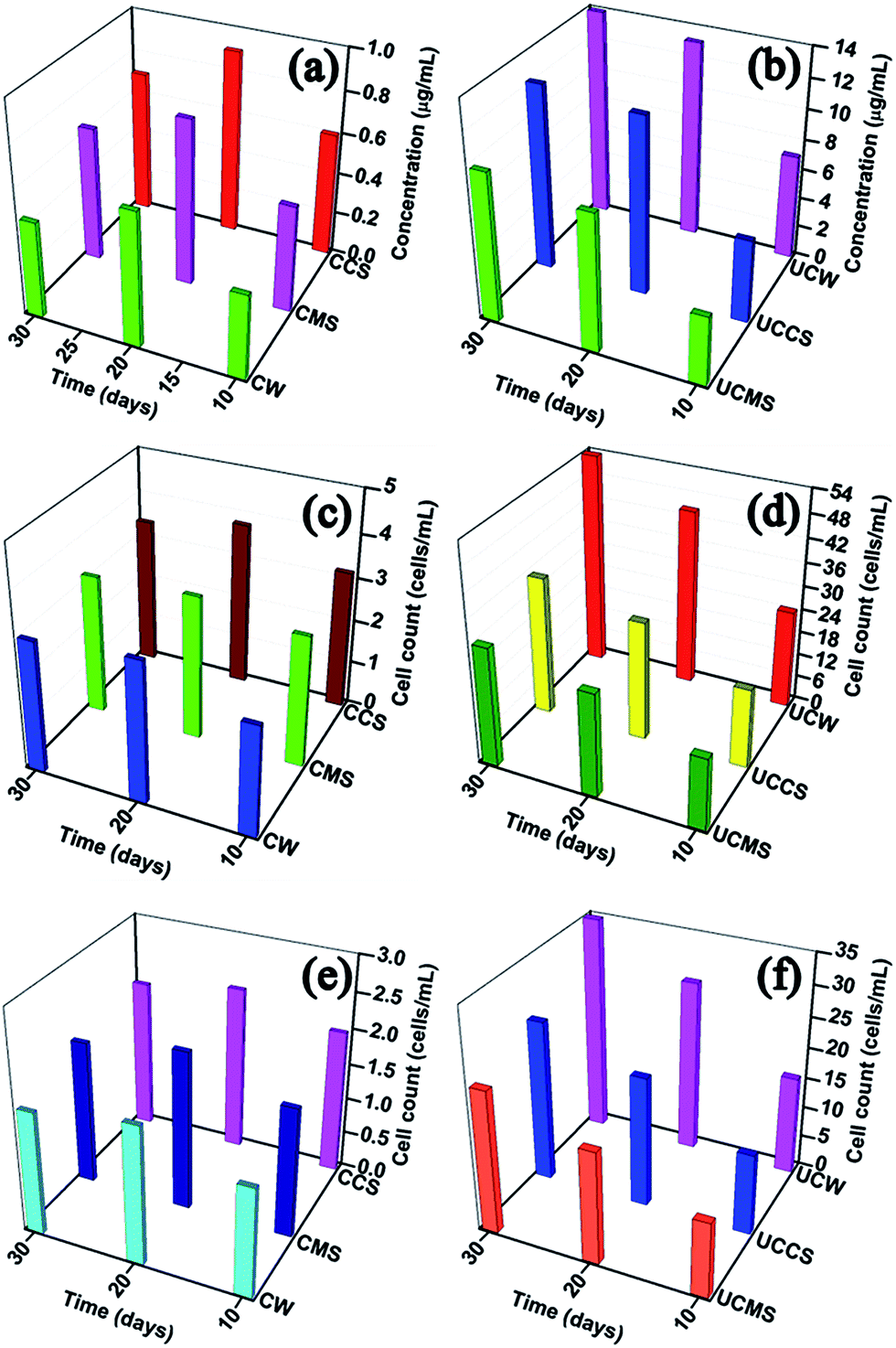 | ||
| Fig. 13 Comparison of algal growth between CAZO nanopaint coated and uncoated panels. | ||
However, to confirm the antifouling behavior of CAZO NRs, the panels coated with commercial paint without CAZO NRs were tested for the antifouling behavior. The Fig. 14 shows the images of commercial paint coated (without CAZO NRs) panels after algal treatment, which shows that the panels are completely covered by algae. This observation once again confirms the role of CAZO NRs regarding the growth of algae.
 | ||
| Fig. 14 Commercial paint (without CAZO NRs) coated (a) wood, (b) mild steel and (c) cement slab panels-after algal growth (tank-2). | ||
Algal growth study in bulk tank
The main drawback of commercial antifouling paints is their easy leach out from the surface of the substrate which is the major issue in the marine environment. The other untargeted algae were also affected by the antifouling paints which are leached out. In the present case, the CAZO nanopaint leach out from the surface of the panels is negligible. If the CAZO nanopaint is leached out, then it will reduce the algal growth in all the tanks. However, the algal growth is maintained in the entire tank till the end of the study period (Fig. 15). The base coat results in stronger binding between panel surface and CAZO nanopaint as well as it prevents the leaching of CAZO nanopaint from the surface of the panels. It has been found that the CAZO NRs exhibit better inhibition against the fouling organisms. | ||
| Fig. 15 The measurement of algal growth for bulk tanks. | ||
Suggested mechanism for antifouling behavior of CAZO NRs against the algae
ZnO shows a broad spectrum of antibacterial activity through a number of mechanisms such as (i) generation of reactive oxygen species (ROS) mostly hydroxyl radicals and singlet oxygen and deposition of the nanoparticles on the surface of bacteria, (ii) accumulation of nanoparticles either in the cytoplasm or in the periplasmic region causing disruption of cellular function and (iii) disruption and disorganization of membranes. Wang et al. indicate that the binding of ZnO NPs to the bacterial surface by electrostatic forces results in the death of bacteria. The surface abrasiveness of ZnO NPs is suggested to be the cause for high antibacterial performance of ZnO NPs by initiating disorganization of the cell membrane.The release of toxic Zn2+ from the dissolution of NPs is another possibility. The dispersion of NPs can be increased in bio-compatible solvents by capping ZnO-NPs with polymeric agents, such as chitosan, polyethyleneimine (PEI) and polyvinyl alcohol (PVA) in order to enhance the bactericidal activity. The death of bacteria can be attributed to the chemical damage to membrane biomolecules, robbing of lipid molecules through adsorption of NPs or membrane gelation/fluidization after NPs attachment.40,41
Conclusions
The CAZO NRs were synthesized by a simple chemical conversion method using green capping agent (chitosan). The CAZO NRs were characterized by UV-Visible, FT-IR, XPS, PL, FESEM and HRTEM analysis. The CAZO NRs were mixed with commercial paints and coated on to the surface of three different panels (wood, mild steel and cement slab). The surface smoothness of CAZO nanopaint coated panels was ascertained by water contact angle measurement. The resulting CAZO nanopaint coatings exhibit significant antifouling effect (75–90%) against the micro algal adhesion in freshwater and seawater environments. The efficacy of CAZO nanopaint to manage the biofouling by means of direct coating on the surface of the panels has been demonstrated in the present investigation. This also paves the way for the development of antifouling coating which is environmentally friendly.Acknowledgements
We thank the University of Madras, Chennai, India, for financial support (UGC, NON-NET) for this investigation. We acknowledge the National Centre for Nano Science and Nanotechnology, University of Madras for FESEM, HRTEM and XPS analyses. We thank Dr Aruna Dhathathreyan, Bio-Physics Laboratory, CLRI for water contact angle measurements.References
- J. A. Callow and M. E. Callow, Nat. Commun., 2011, 2, 244–254 CrossRef PubMed.
- W. Zhao, Q. Ye, H. Hu, X. Wang and F. Zhou, RSC Adv., 2015, 5, 100347–100353 RSC.
- M. Lejars, A. Margaillan and C. Bressy, Chem. Rev., 2012, 112, 4347–4390 CrossRef CAS PubMed.
- W. J. Yang, T. Cai, K. Neoh and E. Kang, Langmuir, 2011, 27, 7065–7076 CrossRef CAS PubMed.
- M. S. Selim, S. A. El-Safty, M. A. El-Sockary, A. I. Hashem, O. M. A. Elenien, A. M. Saeed and N. A. Fatthallah, RSC Adv., 2015, 5, 63175–63185 RSC.
- K. R. Goode, K. Asteriadou, P. T. Robbins and P. J. Fryer, Compr. Rev. Food Sci. Food Saf., 2013, 12, 121–144 CrossRef.
- X. Zhu, D. Jańczewski, S. S. C. Lee, S. L. Teo and G. J. Vancso, ACS Appl. Mater. Interfaces, 2013, 5, 5961–5968 CAS.
- M. S. Majik, S. Tilvi, S. Mascarenhas, V. Kumar, A. Chatterjee and M. Banerjee, RSC Adv., 2014, 4, 28259–28264 RSC.
- J. Jiang, L. Zhu, H. Zhang, B. Zhu and Y. Xu, ACS Appl. Mater. Interfaces, 2013, 5, 12895–12904 CAS.
- B. P. Tripathi, N. C. Dubey, R. Subair, S. Choudhury and M. Stamm, RSC Adv., 2016, 6, 4448–4457 RSC.
- S. Yeh, C. Chen, W. Chen and C. Huang, Langmuir, 2014, 30, 11386–11393 CrossRef CAS PubMed.
- F. Natalio, R. Andre, A. F. Hartog, B. Stoll, K. P. Jochum, R. Wever and W. Tremel, Nat. Nanotechnol., 2012, 7, 530–535 CrossRef CAS PubMed.
- L. Zhu, L. Zhu, Y. Zhao, B. Zhu and Y. Xu, J. Mater. Chem. A, 2014, 2, 15566–15574 CAS.
- V. Moghimifar, A. Esmaili Livari, A. Raisi and A. Aroujalian, RSC Adv., 2015, 5, 55964–55976 RSC.
- R. Sharma, F. Alam, A. K. Sharma, V. Dutta and S. K. Dhawan, J. Mater. Chem. A, 2015, 3, 22227–22238 CAS.
- V. B. Schwartz, F. Thétiot, S. Ritz, S. Pütz, L. Choritz, A. Lappas, R. Förch, K. Landfester and U. Jonas, Adv. Funct. Mater., 2012, 22, 2376–2386 CrossRef CAS.
- P. Petkova, A. Francesko, M. M. Fernandes, E. Mendoza, I. Perelshtein, A. Gedanken and T. Tzanov, ACS Appl. Mater. Interfaces, 2014, 6, 1164–1172 CAS.
- B. Ludi and M. Niederberger, Dalton Trans., 2013, 42, 12554–12568 RSC.
- S. Mehra, E. M. Chan and A. Salleo, J. Mater. Chem. C, 2015, 3, 7172–7179 RSC.
- D. Morselli, A. Scarpellini, A. Athanassioua and D. Fragouli, RSC Adv., 2016, 6, 11412–11418 RSC.
- B. Liu and H. C. Zeng, J. Am. Chem. Soc., 2003, 125, 4430–4431 CrossRef CAS PubMed.
- R. Zhang, X. Yang, D. Zhang, J. Qin, C. Lu, H. Ding, X. Yan, H. Tang, M. Wang and Q. Zhang, Cryst. Res. Technol., 2011, 46, 1189–1194 CrossRef CAS.
- L. Guo, Y. L. Ji and H. Xu, J. Am. Chem. Soc., 2002, 124, 14864–14865 CrossRef CAS PubMed.
- C. Zarrouk, Geitler, Ph.D. thesis, University of Paris, Paris, France, 1966.
- H. C. Bold, Bull. Torrey Bot. Club, 1949, 76, 101–108 CrossRef.
- R. R. L. Guillard, Culture of phytoplankton for feeding marine invertebrates, Plenum Publishers, New York, 1975, pp. 29–60 Search PubMed.
- H. K. Lichtenthaler, Methods Enzymol., 1987, 148, 350–382 CAS.
- S. Chatterjee, A. Shit and A. K. Nandi, J. Mater. Chem. A, 2013, 1, 12302–12309 CAS.
- X. Zhang, J. Qin, Y. Xue, P. Yu, B. Zhang, L. Wang and R. Liu, Sci. Rep., 2014, 4, 4596–4604 Search PubMed.
- R. Kumar, A. M. Isloor, A. F. Ismail, S. A. Rashid and T. Matsuura, RSC Adv., 2013, 3, 7855–7861 RSC.
- M. A. Verges, A. Mifsud and C. J. Serna, J. Chem. Soc., Faraday Trans., 1990, 9, 959–963 RSC.
- N. R. Panda, B. S. Acharya, P. Nayak and B. P. Bag, Ultrason. Sonochem., 2014, 21, 582–589 CrossRef CAS PubMed.
- B. Ludi, M. J. Suess, I. A. Werner and M. S. Niederberger, Nanoscale, 2012, 4, 1982–1995 RSC.
- I. Perelshtein, E. Ruderman, N. Perkas, T. Tzanov, J. Beddow, E. Joyce, J. T. Mason, M. Blanes, K. Mollá, A. Patlolla, A. I. Frenkel and A. Gedanken, J. Mater. Chem. B, 2013, 1, 1968–1976 RSC.
- H. Maachou, M. J. Genet, D. Aliouche, C. C. Dupont-Gillainb and P. G. Rouxhet, Surf. Interface Anal., 2013, 45, 1088–1097 CrossRef CAS.
- C. Cheng, A. Amini, C. Zhu, Z. Xu, H. Song and N. Wang, Sci. Rep., 2014, 4, 4181–4186 Search PubMed.
- N. Tripathy, R. Ahmad, H. A. Ko, G. Khang and Y. Hahn, Nanoscale, 2015, 7, 4088–4096 RSC.
- W. Admiraal, Mar. Biol., 1977, 39, 1–9 CrossRef.
- Y. S. Mohite and P. S. J. Wakte, J. Algal Biomass Utln., 2011, 2, 53–68 Search PubMed.
- X. Wang, F. Yang, W. Yang and X. Yang, Chem. Commun., 2007, 4419–4421 RSC.
- X. Xu, D. Chen, Z. Yi, M. Jiang, L. Wang, Z. Zhou, X. Fan, Y. Wang and D. Hui, Langmuir, 2013, 29, 5573–5580 CrossRef CAS PubMed.
| This journal is © The Royal Society of Chemistry 2016 |