
Synergic therapy of melanoma using GNRs-MUA-PEI/siIDO2-FA through targeted gene silencing and plasmonic photothermia
Yujuan Zhang*ab,
Na Songab,
Jiamin Fuab,
Yanling Liuab,
Xuelin Zhanab,
Shanshan Pengab,
Zhi Yangab,
Xianfang Zhuc,
Yiguo Chenab,
Zhigang Wangab,
Yanrong Yuab,
Qiaofa Shia,
Yingyuan Fua,
Keng Yuanab,
Nanjin Zhouab,
Thomas E. Ichimd and
Weiping Min*abe
aInstitute of Immunotherapy and College of Basic Medicine, Nanchang University, Jiangxi Academy of Medical Sciences, Nanchang, China. E-mail: yujuanzhang@ncu.edu.cn
bJiangxi Provincial Key Laboratory of Immunotherapy, Nanchang, China
cChina-Australia Joint Laboratory for Functional Nanomaterials, School of Physics and Mechanical and Electrical Engineering, Xiamen University, Xiamen, China
dBatu Biologics Inc, San Diego, California, USA
eDepartment of Surgery, Pathology and Oncology, University of Western Ontario, London, Canada. E-mail: mweiping@uwo.ca
First published on 11th August 2016
Abstract
Indoleamine dioxygenase 2 (IDO2) is a newly discovered enzyme that contributes to tumor immune escape. Our previous studies demonstrated that small interfering RNA (siRNA)-mediated IDO2 knock-down induces tumor cell apoptosis and inhibits tumor growth. Modified gold nanorods (GNRs) are excellent siRNA nano-carriers as well as photothermal therapy agents, with potential to augment siRNA efficacy and target specificity. In this study, we developed a novel nanostructure of GNRs-MUA-PEI/siIDO2-FA for targeted therapy of melanoma. Firstly, we demonstrated that these new nanostructures possess excellent biocompatibility, as well as efficiently and specifically deliver RNA to melanoma cells. Secondly, IDO2 knockdown significantly enhanced the photothermal therapeutic efficacy of GNRs as evidenced by inducing increased tumor cell apoptosis in vitro as well as inhibiting the growth of tumors in vivo under low laser power density. Finally, we firstly demonstrated that GNRs-MUA-PEI/siIDO2-FA exerts synergistic effects on photothermal therapy and gene silencing IDO2 in anti-tumor therapy of inhibiting tumor growth, which holds great promise for anti-cancer therapeutics as an alternative strategy for the treatment of melanoma.
1. Introduction
Metastatic melanoma is one of the most aggressive and lethal malignancies.1 In the last decades, its global incidence has increased faster than for any other solid tumor with 160![[thin space (1/6-em)]](https://www.rsc.org/images/entities/char_2009.gif) 000 new cases per year and 48000 deaths.2,3 The conventional chemotherapy treatment of metastatic melanoma is using dacarbazine, but the response rate is low at only 15–20%.4 Immunotherapy has also been applied for metastatic melanoma therapy, which employs cytokines that stimulate the patient's immune system to fight cancer, such as interleukin-2 (IL-2), but high-dose IL-2 also has a low response rate of only 16%.5
000 new cases per year and 48000 deaths.2,3 The conventional chemotherapy treatment of metastatic melanoma is using dacarbazine, but the response rate is low at only 15–20%.4 Immunotherapy has also been applied for metastatic melanoma therapy, which employs cytokines that stimulate the patient's immune system to fight cancer, such as interleukin-2 (IL-2), but high-dose IL-2 also has a low response rate of only 16%.5
Cancer immunotherapy is based on the recognition of tumor-associated antigens (peptides) on cancer cells by the patient's T cells. The US Food and Drug Administration (FDA)-approved immunotherapies such as IL-2, interferon (IFN)-α, anti-CTLA-4 (cytotoxic T lymphocyte antigen-4) and anti-PD-L1 (programmed death-ligand) all appear to activate and/or expand tumor-specific T cells of antigen specificity. This process requires the delivery of appropriate signals that initiate immune responses from being ineffective to being capable of rejecting established tumors.6 One of the limiting factors of successful cancer immunotherapy is direct and indirect suppression of the immune response by the tumor, or by tumor-reprogrammed immune elements.7–9 One of the major means of tumor mediated immune suppression is inhibition of T cell effector functions and generation of Treg by tryptophan catabolism due to the overexpression of tryptophan-degrading enzymes in human tumors and tumor associated macrophages, such as indoleamine-2,3-dioxygenase (IDOs) including IDO1 and IDO2. These enzymes are intracellular heme-containing enzymes that initiate the first and rate-limiting step of tryptophan degradation along the kynurenine pathway.10,11 We and others have demonstrated that RNAi-mediated suppression of IDOs enhances anti-tumor immunity in a variety of preclinical systems.12–14 In addition to IDOs playing a role in immune suppression, our recent studies revealed that IDO2 may play a role in promotion of tumor cell viability in that knocking down expression of this enzyme resulted in enhanced apoptosis, chemosensitivity, and reduced tumor growth in the B16-BL6 model of metastatic melanoma.15,16
Ever since RNA interference (RNAi) was discovered, this technology has emerged as a promising treatment strategy for cancer, which could effectively knockdown or silence target genes.17 However, small interfering RNA (siRNA) is unstable in the blood stream, and are unable to penetrate cell membranes, in part due to their large size and negative charge.18 Successful application of siRNA for cancer therapy is highly dependent on the development of delivery vehicles that are nontoxic and enable the selective and efficient transport of siRNA to a specific tissue.
Utilization of nanoparticles for targeting neoplastic tissue has been successfully performed since 1995.19 Physical features of nanoparticles such as size and shape play a critical role in overall tissue permeability, as well as selectivity of delivery to malignant versus non-malignant tissue. Additionally, the characteristics of the nanoparticles are fundamental factors in determining pharmacokinetics and biodistribution.20 A limited number of nanoparticle systems have been developed that were successful in crossing the threshold to clinical entry.21 Various shapes of nanoparticles have been developed in order to provide a higher level of control over pharmacokinetic and delivery parameters.22 For example, cylindrical shaped nanoparticles possess enhanced ability to remain in systemic circulation as compared to spherical shapes. This is believed to be due to differences that the shapes cause on rigidity, surface area, and endothelial interactions.23 In particular, cylindrical shapes are believed to be more advantageous for preclinical development by allowing passive accumulation in tumors and enhanced circulation time.20
Hyperthermia, the elevation of tumor temperature, is an established method used in cancer therapy.24,25 In cancer treatment, thermal therapies are mainly used for eliminating solid tumors. However, hurdles for clinical application of tumor hyperthermia exist due to limited ability to deliver sufficient heat in target regions without harming non-malignant tissue. More recently, studies have taken advantage of nanotechnology based approaches for enhancing specificity and efficacy of directed hyperthermia. One promising application of nanotechnology is utilization of light with a wavelength that matches oscillations of the conduction band electrons on gold nanorods (GNRs). When this occurs, the surface plasmon resonance (SPR) on nanostructures is reached, which allows light to be absorbed and to be converted to heat; this approach is called photothermia or plasmonic photothermia.26–28 When such nanoparticles are localized within tissue with the intention of using this technique as a tool to induce hyperthermia, it is termed photothermal therapy (PTT) or plasmonic photothermal therapy (PPTT).27,28 With PPTT, many groups have achieved tumor selective temperatures from 50 °C to over 70 °C which are well above the threshold required for vascular damage and cell death.27,29–31 Thus, specific delivery of GNRs is highly desirable in clinical PPTT.
In the present work, we firstly developed a novel nanostructure, GNRs-MUA-PEI/siIDO2-FA, which consists triple advantages: (1) a folic acid (FA) adaptor specifically targeting tumor cells; (2) GNRs inducing robust tumor killing in photothermal therapy; and (3) siIDO2 specifically silencing IDO2 to induce tumor cell apoptosis. We established a two-step method to prepare a novel siRNA nanocarrier of GNRs-MUA-PEI-FA that was composed of folic acid-coated gold nanorods and siIDO2. This structure not only displayed high siRNA loading capacity and low cytotoxicity, but also facilitated the tumor-specific targeting to B16-BL6 cells. Additionally, siRNA delivered by GNRs-MUA-PEI/siIDO2-FA could silence IDO2 effectively, while IDO2 knockdown enhanced apoptosis of B16-BL6 cells in vitro and in vivo. Moreover, we demonstrated the clinical potential of PTT using GNRs-MUA-PEI/siIDO2-FA, as evidenced by the remarkable suppression of tumor growth in a murine melanoma model. This novel nanoplatform GNRs-MUA-PEI/siIDO2-FA exerts synergistic effects of tumor-targeted photothermal therapy and IDO2 gene silencing on suppressing tumor growth, which could be as an alternative new strategy of PTT for treating melanoma and holds great promise for anti-cancer therapeutics.
2. Materials and methods
2.1. Synthesis of GNRs
Water-soluble gold nanorods were synthesized via seed-mediated growth routes as previously described.32 Briefly, first a seed solution was prepared as follows: 1 mL of cetyltrimethylammonium bromide (CTAB) solution (0.2 M) was mixed with 1 mL of HAuCl4 (0.5 mM). While the solution was stirred at 25 °C, 0.12 mL of ice-cold 0.01 M NaBH4 was added, and the resulting seed solution turned to brownish yellow color. Secondly, the growth solution was prepared as follows: 50 mL of HAuCl4 (1 mM), 50 mL of CTAB (0.2 M) and 2.5 mL of AgNO3 (4 mM) were mixed at 25 °C, and after gentle mixing, 670 μL of ascorbic acid (0.079 M) was added, and then the color changed from dark yellow to colorless. To generate nanorods, 120 μL of the seed solution was added to the growth solution at 27–30 °C under gently mixing. Within 10–20 minutes, the combined solution gradually changed colors to brownish red. After 24 hours, the solution was then centrifuged at 12000 rpm for 15 minutes to remove the excessive CTAB. Finally, the nanorod sample coated with CTAB was characterized by transmission electron microscope (TEM) and UV-vis-NIR spectrometer.
2.2. Synthesis of GNR-MUA-PEI and GNR-MUA-PEI-FA
Synthesis of MUA-PEI and MUA-PEI-FA is based on the chemical reaction between amino group and carboxyl group. In brief, the steps were designed to covalent conjugate (1) carboxyl groups of MUA binding to amino groups of poly ethylene imine (PEI) by ethyl(dimethylaminopropyl) carbodiimide (EDC) mediated amidation with N-hydroxysuccinimide (NHS) in HCCl3, and (2) carboxyl groups of folic acid (FA) binding to residual amino groups of PEI by the similar reaction of EDC mediated amidation with NHS but in DMSO instead of in HCCl3. MUA-PEI to replace CTAB on GNRs obtained the GNRs-MUA-PEI, and 1:1 ratio of MUA-PEI to MUA-PEI-FA to replace CTAB on GNRs obtained the GNRs-MUA-PEI-FA. The products were centrifuged and characterized by TEM, UV-vis-NIR spectrometer and Fourier transform infrared spectroscopy (FTIR).
2.3. siRNA synthesis
The siRNAs targeting IDO2 mRNA or GAPDH were generated in accordance with the target sequence selection methods described by Jin et al.33 siRNA was synthesized by the manufacturer (Sigma, St. Louis, MO). siRNA targeting luciferase gene GL2 (GL2 siRNA) was used as a scrambled-silencing control since GL2 is not expressed in treated cells.2.4. Synthesis of GNR-MUA-PEI/siRNA-FA and gel shift assays
Equal volumes containing 0.5 μg of siRNA and the desired amount of GNR-MUA-PEI-FA (the weight ratios of siRNA to GNR-MUA-PEI-FA were 1:0, 1:1, 1:2, 1:3, 1:4, 1:5, 1:6, 1:7, 1:8) were mixed and incubated for 30 minutes. The resulting complexes were electrophoresed at 80 V using 1.5% agarose gel and EtBr in TAE buffer. After 20 minutes, the gel was removed, visualized under UV lamp and the picture was taken with an Olympus C8080 digital camera.
2.5. Cell culture
A murine melanoma cell line established from a C57BL/6 mouse and designated B16-BL6 was obtained from the American Type Culture Collection and was maintained in RPMI 1640 medium (Sigma-Aldrich) with 10% FBS, L-glutamine, penicillin, and streptomycin at 37 °C in 5% CO2.2.6. MTT assay
MTT (3-(4,5-dimethylthiazol-2-yl)-2,5-diphenyltetrazolium bromide) cytotoxicity assay. A MTT kit (BD Pharmingen, San Diego, CA) was required. B16-BL6 cells (6 × 10³) were seeded in a 96 well microplate. After 24 hours, fresh medium containing GNR-CTAB or GNR-MUA-PEI-FA were added, and the final concentration of GNR-CTAB or GNR-MUA-PEI-FA was 20 μg mL−1, 40 μg mL−1, 60 μg mL−1, 80 μg mL−1, 100 μg mL−1, 150 μg mL−1, and 200 μg mL−1. After 24 or 48 hours, the MTT reagent was added for another 4 hours and was converted by the mitochondrial reductase. The resulting purple crystals were dissolved in solubilization buffer and spectrophotometrically analyzed at 490 nm, using a reference of 650 nm in a microplate reader (SpectraMax M5e).2.7. Apoptosis assay
The apoptotic and necrotic cell distribution were determined by Annexic V-FITC/PI Apoptosis Detection Kit (BD Pharmingen, San Diego, CA). The B16-BL6 cells were plated in 12-well microplates at a density of 1 × 10⁵ cells per well. After 24 hours incubation, the medium was replaced with fresh medium and a series amount of GNR-CTAB or GNR-MUA-PEI-FA were added to the medium and the final concentration of GNR-CTAB or GNR-MUA-PEI-FA was 20 μg mL−1, 40 μg mL−1, 60 μg mL−1, 80 μg mL−1, 100 μg mL−1, 150 μg mL−1, and 200 μg mL−1. After 24 hours or 48 hours, the cells were trypsinized, harvested, washed with PBS and resuspended in 100 μL of binding buffer containing 1 μL Annexin V-FITC and 2 μL PI. After incubation at room temperature for 15 minutes in the dark, 100 μL of binding buffer was added to each sample before washing and then the samples were resuspended in medium and analyzed by BD FACS Calibur (BD Biosciences, Mountain View, CA).2.8. Cellular uptake
The same amounts (1 μg) of Cy3-GAPDH were used to assess the cellular uptake of LIP2000 (Lipofectamine 2000, Invitrogen Life Technologies, as the positive control group), GNR-MUA-PEI and GNR-MUA-PEI-FA. Briefly, B16-BL6 or HUVEC cells were plated in 12-well microplates at a density of 1 × 10⁵ cells per well. After 48 hours incubation, the medium was replaced with fresh medium, and then LIP2000/siRNA, GNR-MUA-PEI/siRNA (wt(GNR-MUA-PEI):wt(siRNA) = 15:1) and GNR-MUA-PEI/siRNA-FA (wt(GNR-MUA-PEI-FA):wt(siRNA) = 15:1; 10:1; 8:1) which had been prepared with the same amount siRNA (1 μg) were added into the fresh medium and incubated with cells at 37 °C for 4 hours. Then, the cells were trypsinized, harvested, washed three times with PBS to remove unloaded nanoparticles and resuspended in 200 μL of the medium and analyzed by BD FACS Calibur (BD Biosciences, Mountain View, CA).
2.9. Gene silencing and PCR
B16-BL6 cells were seeded in a 12-well plate with a density of 1 × 10⁵ cells per well. After 24 hours, a final concentration of 16 μg mL−1 of GNR-MUA-PEI/siRNA-FA (wt(GNR-MUA-PEI-FA):wt(siRNA) = 15:1) was added into the cells for 24 hours transfection, and then cells were lysed (Trizol reagent, Invitrogen) and total RNA was isolated according to the manufacturer's instructions. 1 μg of total RNA was synthesized to cDNA by using reverse transcriptase (MmLV-RT, Invitrogen). The mRNA expressions of IDO2 in the samples were quantified with q-PCR using GAPDH as a reference. The following primers sets were used for PCR amplifications: GAPDH, 5′-TGATGACATCAAGAAGGTGGTGAA-3′ (sense) and 5′-TCCTTGGAGGCCATGTAGGCCAT-3′ (antisense); IDO2,5′-GTGGGGCTGGTCTATGAAGGTG-3′ (sense) and 5′-TGGTGGCAGCGGAGATAATGTA-3′ (antisense). q-PCR reactions were performed in a Stratagene Mx3000P QPCR System (Agilent Technologies, Lexington, MA) using SYBR green PCR Master Mix (Life technologies) according to manufacture's protocol. Differences in gene expression were calculated by using the ΔCt method.
2.10. Western blot
5 × 10⁵ B16-BL6 cells were seeded into a 6-well plate and grown overnight. The cells were transfected with a final concentration of 16 μg mL−1 of GNR-MUA-PEI/siRNA-FA (wt(GNR-MUA-PEI-FA):wt(siRNA) = 15:1) loading IDO2 siRNA or GL2 siRNA for 24 hours. The cells were harvested, washed twice with ice-cold PBS, resuspended in protein lysis buffer with complete protein inhibitor (Roche Diagnostics) and then keep the container on ice for 30 minutes. Lysed cells were centrifuged at 15000 rpm for 20 minutes at 4 °C and the supernatant were collected and stored at −80 °C for future use. Protein concentration was determined by Bio-Rad protein assay and 50 μg of each group cell lysate was separated on 12% SDS-PAGE, transferred to nitrocellulose membrane (Bio-Rad), blocked with 5% fat-free milk (Carnation) and 3% BSA in TBS-T (0.25% Tween 20), probed with a mouse anti-human IDO2 mAb that binds to both human and mouse IDO2 (Santa Cruz Biotechnology) and goat anti-mouse GAPDH Ab (Sigma-Aldrich) according to the manufacturer's instructions, and visualized by an ECL assay (Amersham Biosciences).
2.11. In vitro photothermal effect of GNR-MUA-PEI-FA
B16-BL6 cells (6 × 10³) were seeded in a 96 well microplate, and after 24 hours the cells were incubated with a final concentration of 15 μg mL−1 of GNR-MUA-PEI-FA or PBS (as the control group) for overnight. And then, the cells were irradiated at 2 W cm−2 for various times (0, 0.5, 1, 2, 5, 10 minutes) or at 5 minutes for different power density (0 W cm−2, 0.5 W cm−2, 1 W cm−2, 2 W cm−2, 4 W cm−2). After 24 hours, the MTT reagent was added for another 4 hours and was converted by the mitochondrial reductase. The resulting purple crystals were dissolved in solubilization buffer and spectrophotometrically analyzed at 490 nm, using a reference of 650 nm in a microplate reader (SpectraMax M5e).2.12. Apoptosis of tumor cells induced by GNR-MUA-PEI/siIDO2-FA
B16-BL6 cells were seeded in a 24-well plate with a density of 0.5 × 10⁵ cells per well and after 24 h those cells were incubated with PBS (as the control group) or a final concentration of 16 μg mL−1 of GNR-MUA-PEI/siGL2-FA (wt(GNR-MUA-PEI-FA):wt(siRNA) = 15:1) or 16 μg mL−1 of GNR-MUA-PEI/siIDO2-FA (wt(GNR-MUA-PEI-FA):wt(siRNA) = 15:1) in double for overnight, and then one was irradiated at 2 W cm−2 for 5 minutes after the incubation with PBS or 16 μg mL−1 of GNR-MUA-PEI/siGL2-FA or 16 μg mL−1 of GNR-MUA-PEI/siIDO2-FA. The apoptotic and necrotic cell distribution of all were determined at 24 hours by Annexin V-FITC/PI Apoptosis Detection Kit and analyzed by BD FACS Calibur (BD Biosciences, Mountain View, CA).
2.13. Animals and tumor model and H&E staining
Female C57BL/6 mice, 6–8 weeks of age and weighting 18–23 g, were purchased from Changsha Laboratory Animal Co. Ltd., and housed in a SPF grade animal center. The use of all mice in this study complied with the Regulations for the Administration of Affairs Concerning Experimental Animals of China and the ethical approval of institutional Animal Care and Use Committee of Nanchang University, China. The mice were anesthetized by isoflurane and inoculated with 2 × 10⁶ B16-BL6 cells suspension. In order to evaluate the inoculation of tumor models, hematoxylin and eosin (HE) staining was performed on paraffin-embedded sections. The prepared specimen was examined using a light microscope.2.14. Topical siRNA delivery
C57BL/6 mice were anaesthetized and shaved, and the melanoma tumors were inoculated. After two weeks, mice were anesthetized, and Cy3-siGAPDH, GNR-MUA-PEI/Cy3-siGAPDH (wt(GNR-MUA-PEI):wt(siRNA) = 15:1) and GNR-MUA-PEI/Cy3-siGAPDH-FA (wt(GNR-MUA-PEI-FA):wt(siRNA) = 15:1) (all containing 6 μg of siRNA) respectively mixing with glycerin (50% in final solution v/v) and DMSO (10% in final solution v/v) and nuclease free water were topically massaged on the skins above the inoculated tumors. After 4 hours, the mice were scarified and the tumors were collected. The tumors attached to a freezing stage using an optimal cutting temperature (OCT) compound (Triangle Biomedical Sciences, USA) were frozen and cut into 1 mm horizontal sections. Images were visualized with a fluorescence microscope (Olympus, Model BX 51, Japan).
2.15. In vivo photothermal effect of GNR-MUA-PEI/siIDO2-FA
Melanoma bearing mice were generated by inoculating 2 × 10⁶ B16-BL6 cells suspension. When the diameter of tumor was about 3 mm, PBS (as the control group) or GNR-MUA-PEI-FA (wt(GNR-MUA-PEI-FA):wt(siRNA) = 15:1), siRNA (6 μg, siIDO2 or siScramble), glycerol (50% in final solution v/v), DMSO (10% in final solution v/v) were mixed and applied onto the tumors on day 6, 10, 14, 18. 12 hours after each application of above mixture, a laser irradiation (1 W cm−2, 2 minutes) was conducted. The tumor sizes of growth were measured with caliper. 20 days after tumor inoculation, the mice were sacrificed and tumors were collected. The tumor weights were measured with a 2-digit electronic balance and the images were taken with a camera.
2.16. Statistics
Data were presented as mean ± SD. Student's t-test (2-tailed) was applied to determine differences between two means. For the comparison of multiple groups, one-way ANOVA test was used. For all statistical analyses, differences with p values <0.05 were considered significant.3. Results and discussions
3.1. Synthesis, modification and characterization of GNRs
First, we synthesized CTAB-stabilized GNRs using an established seed-mediated growth methods.32 The synthesized GNRs were characterized by the transmission electron microscope (TEM) image, which showed that GNRs have a mean length of 39.0 ± 3.5 nm and width of 10.0 ± 1.5 nm (Fig. 1A). GNR-MUA-PEI-FA that is shown in Fig. 1B is prepared by 1:1 ratio of MUA-PEI to MUA-PEI-FA replacing CTAB on GNRs by one step. PEI carrying positive charges could bind the negative-charged siRNA due to the electrostatic interaction. In order to improve the cellular uptake of GNRs, folic acid (FA) was conjugated to GNRs using the chemical reaction between amino group and carboxyl group due to overexpression of FA receptors on B16-BL6 cells. As shown in Fig. 1C, a longitudinal SPR maximum for the original GNRs-CTAB was at around 750 nm, and the peak slightly red-shifted to 770 nm for GNR-MUA-PEI, followed by another red-shift to 790 nm for GNR-MUA-PEI-FA, which suggests MUA-PEI and MUA-PEI-FA have been successfully bound on GNRs. Fig. 1D shows the FTIR spectra of GNR-MUA-PEI and GNR-MUA-PEI-FA. Both of them have the following absorption: the absorption band of –CO–NH– at 1631 cm−1 which suggested the chemical binding between the carboxyl group of MUA or FA and the amino group of PEI. Comparing with the FTIR spectra of GNR-MUA-PEI, a new absorption band appeared for GNR-MUA-PEI-FA at 1611 cm−1 and 1511 cm−1 which belongs to the vibration of benzene ring from FA. These results further suggested that FA was successfully conjugated. In order to improve the melanoma targeting efficiency and siRNA loading capacity simultaneously, we prepared that the ratio of MUA-PEI to MUA-PEI-FA was 1:1 on the surface of GNRs.
 | ||
| Fig. 1 Characterization of GNRs. TEM images of (A) GNRs-CTAB and (B) GNRs-MUA-PEI-FA, (C) normalized UV-vis absorption spectra of GNRs-CTAB, GNRs-MUA-PEI and GNRs-MUA-PEI-FA, (D) FTIR spectra of GNRs-MUA-PEI and GNRs-MUA-PEI-FA. | ||
3.2. Attenuation of cytotoxicity of GNRs by FA
In this newly designed nanostructure, folic acid was not only used to bind the FA receptor on melanoma cells but also to attenuate the cytotoxicity of PEI. Next, we tested the cytotoxicity of the synthesized GNR-MUA-PEI-FA. As shown in Fig. 2, after B16-BL6 cells were exposed to a series of GNR-CTAB and GNR-MUA-PEI-FA at various concentrations, we observed that B16-BL6 exposed to GNR-MUA-PEI-FA had higher cell viability than those in the presence of GNR-CTAB on the basis of MTT experiments. After incubation with GNR-CTAB (20 μg mL−1) for 24 hours, the cytotoxicity was noticed (cell viability was decreased by 16.0 ± 5.91%). The cytotoxicity was increased at higher concentrations: a dose of 100 μg mL−1 of GNR-CTAB reduced the cell viability by 50.5 ± 5.30%; while the survival cells only left 16.3 ± 2.50% when a higher concentration (200 μg mL−1) of GNR-CTAB was added to B16-BL6 for 24 hours (Fig. 2A). In contrast, a dose of 20 μg mL−1 of GNR-MUA-PEI-FA caused almost no drop on the cell viability, while even a high dose (200 μg mL−1) just slightly decreased the cell viability about 30.5 ± 1.41% (Fig. 2A). The toxicity was significantly increased when extending the time of cell incubation even at very low concentration (20 μg mL−1), the GNR-CTAB diminished the cell viability about 47.0 ± 4.68%, and no cells survived at a high dose of 200 μg mL−1. In contrast to GNR-CTAB, folic acid modified GNR-MUA-PEI-FA caused no decrease on the cell viability at the concentration of 20 μg mL−1, while retaining high viability (>80%) even at the high dose of 100 μg mL−1 (Fig. 2B). Taken together, these data suggested FA significantly reduced cytotoxicity of GNRs.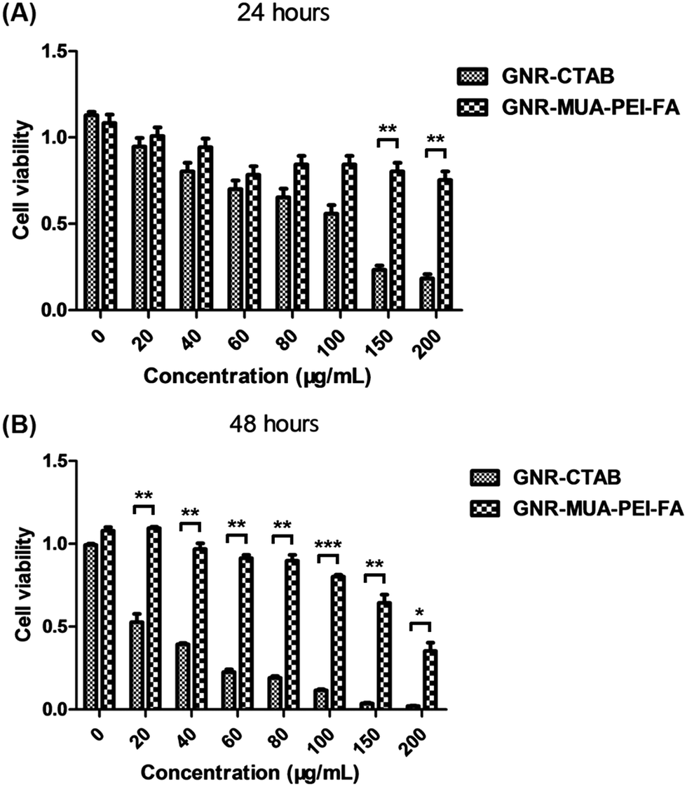 | ||
| Fig. 2 Assessment of cell viabilities. B16-BL6 cells were incubated with a medium containing different amount of GNR-CTAB or GNR-MUA-PEI-FA, and the final concentration of GNR-CTAB or GNR-MUA-PEI-FA was 0 μg mL−1, 20 μg mL−1, 40 μg mL−1, 60 μg mL−1, 80 μg mL−1, 100 μg mL−1, 150 μg mL−1, and 200 μg mL−1. After incubation for 24 hours (A) or 48 hours (B), the cell viabilities were measured by the MTT assay. (*, P < 0.05; **, P < 0.01; ***, P < 0.001). | ||
3.3. Assessment of apoptosis
To assess the capacity of causing apoptosis of GNR-CTAB and GNR-MUA-PEI-FA, we repeated above experiments by analyzing apoptosis of the B16-BL6 cells after incubation with various amounts of GNR-CTAB and GNR-MUA-PEI-FA, and then stained apoptotic cells with Annexin V-FITC/PI apoptosis detection kit and analyzed by flow cytometry (Fig. 3). Compared to GNR-CTAB (Fig. 3A and C), GNR-MUA-PEI-FA (Fig. 3B and D) caused few cells to apoptosis even at high dose of 200 g mL−1 for 48 hours. These results suggested that when GNRs-CTAB had been modified with MUA-PEI-FA, GNR-MUA-PEI-FA had much lower cytotoxicity and better biocompatibility than GNRs-CTAB.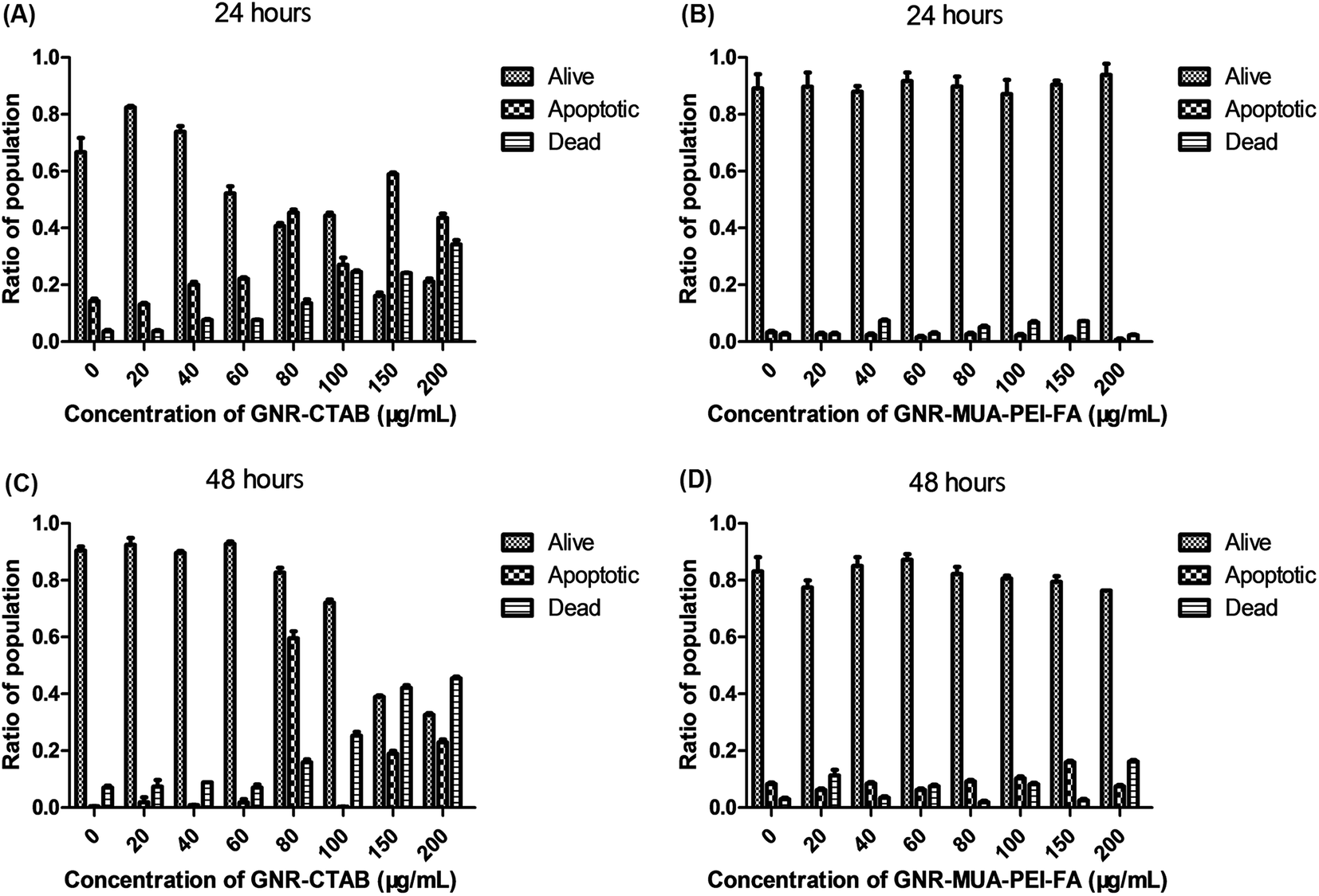 | ||
| Fig. 3 Apoptosis assay. The B16-BL6 cells were incubated with a series amount of GNR-CTAB (A and C) or GNR-MUA-PEI-FA (B and D) and t GNR-CTAB or GNR-MUA-PEI-FA at indicated concentrations. After 24 hours (A and B) or 48 hours (C and D), the ratios of alive, apoptotic and dead cells were determined at 24 hours by Annexic V-FITC/PI Apoptosis Detection Kit and analyzed by flow cytometry. The Annexin V-FITC and PI both positive or only Annexin V-FITC positive cells were regarded as apoptotic; the only PI positive ones were calculated as dead; the double negative ones were alive. Error bars represent the standard deviation of 3 experiments. | ||
3.4. Tumor-targeted delivery of siRNA
One desirable function of nano-vectors is to condense DNA/RNA to form stable complexes against enzymatic degradation.34 We thus tested the capacity of GNR-MUA-PEI-FA to condense siRNA electrostatically using a gel shift assay. In this assay, free siRNA migrates relatively unimpeded through the gel, while the migration of siRNA bound to GNR-MUA-PEI-FA is slowed down or stopped.35 The result showed that, for GNR-MUA-PEI-FA, the ratio of GNR-MUA-PEI-FA:siRNA required to fully inhibit migration of siRNA was more than 8:1 (wt:wt) (Fig. 4A). Therefore, when the ratio (wt:wt) of GNR-MUA-PEI-FA to siRNA is more than 8:1, GNR-MUA-PEI-FA could effectively adsorb all the siRNA molecules.
 | ||
| Fig. 4 Tumor-targeted delivery of siRNA by GNR-MUA-PEI-FA. (A) Gel shift assay for assessing the siRNA-loading capacity of GNR-MUA-PEI-FA. Equal volumes containing 0.5 μg of siRNA and the desired amount of GNR-MUA-PEI-FA were mixed and incubated for 30 minutes at the indicated weight ratios of siRNA to GNR-MUA-PEI-FA. Electrophoresis was done and the gel was taken out for visualizing the bands. (B) Tumor-targeted delivery by GNR-MUA-PEI-FA. Cy3-GAPDH was used to assess the cellular uptake of LIP2000, GNR-MUA-PEI, GNR-MUA-PEI-FA as the vehicle for siRNA delivery. B16-BL6 (B) and HUVEC (C) were incubated with LIP2000/Cy3-GAPDH, GNR-MUA-PEI/Cy3-GAPDH, or GNR-MUA-PEI/Cy3-GAPDH-FA for 4 hours. The population of cell that had up-taken vehicle/Cy3-GAPDH was determined by flow cytometry. Error bars represent the standard deviation of 3 experiments. (**, P < 0.01). | ||
To assess the enhancement of cellular uptake due to folic acid, we compared the percentages of metastatic melanoma cell B16-BL6 which had up-taken Cy3-labelled siRNA (Cy3-siRNA) bound to different vehicles: GNR-MUA-PEI-FA and GNR-MUA-PEI in vitro for 4 hours. The transfection efficacy was determined by the Cy3 positive cells using flow cytometry. As shown in Fig. 4B, GNR-MUA-PEI-FA achieved high transfection rate of 80.8 ± 0.50%, which is similar to when using LIP2000 (a commercial standard siRNA transfection reagent) that showed 85.0 ± 1.60% positively transfected cells. We also analyzed the influence of FA on the cellular uptake of GNRs and siRNA by B16-BL6 in Fig. 4B. Compared to GNR-MUA-PEI-FA, GNR-MUA-PEI without FA dropped down the transfection rate to 42.4 ± 1.75% (Fig. 4B), which indicated FA could promote the cellular uptake of siRNA and GNRs by B16-BL6 significantly. Taken together, these results suggested that GNR-MUA-PEI-FA could be a comparative vehicle for siRNA transfecting B16-BL6, while FA is capable of enhancing cellular uptake of siRNA carried by GNRs.
Folic acid (FA, the synthetic form of folate) is a water-soluble B vitamin, which is essential for de novo synthesis and one carbon transfer reactions.36 Folate receptors (FRs), also known as folate-binding proteins (FBP), are N-glycosylated proteins with high binding affinity to folate in vivo. Functional FR expression is low or absent in most non-malignant tissues, in contrast, neoplasias, especially epithelial carcinomas, consistently and uniformly express high levels of FRs.36 The frequent overexpression within tumors and highly restricted distribution in normal tissues suggest that FRs can potentially be exploited as a tumor-specific cell surface marker that can be used in the targeted delivery of cancer therapeutics.37 Since B16-F10 melanoma consistently express folate receptors,38 it an ideal modal for investigating the potential therapeutic application of GNR-MUA-PEI-FA. Indeed, addition of folic acid (GNR-MUA-PEI-FA) significantly enhanced the siRNA delivery to melanoma cell B16-BL6 (Fig. 4B). To further confirm the tumor-specific targeting by FA, in a parallel experiment, we also performed quantitative measurements on the count of HUVEC as a non-tumor targeting experiments. We observed that as shown in Fig. 4C, the correspondent percentages of HUVEC that had been transfected with Cy3-siRNA by GNR-MUA-PEI was 51.8 ± 2.75%.The addition of folic acid using GNR-MUA-PEI-FA failed to increase transfection rate (47.8 ± 1.80%) in HUVEC cells (Fig. 4C), which was also much less than that observed in B16-BL6 (Fig. 4B). These results suggested that folic acid is capable of facilitating GNR-MUA-PEI-FA targeting of melanoma cells (B16-BL6), and transfecting siRNA more efficiently in tumor cells than in vascular endothelial system cells (HUVEC). Thus, GNR-MUA-PEI-FA probably was an ideal carrier for delivery of siRNA to melanoma in vivo. Our results are in agreement with other studies, which have utilized gold nanorods for selective delivery of agents to cancer cells using enzyme activated release of payload selectively intracellularly.39
3.5. Validation of gene silencing by GNR-MUA-PEI/siRNA-FA
Next, we attempted to validate the gene silencing ability of GNR-MUA-PEI/siRNA-FA, using the house-keeping gene glyceraldehyde 3-phosphate dehydrogenase (GAPDH) as a reference and IDO2 as the siRNA target. After treatment with GNR-MUA-PEI-FA loading with IDO2 siRNA for 24 hours, relative siIDO2 mRNA levels were reduced by about 77.8 ± 3.72% compared to the cells treated with a scrambled sequence non-targeting siRNA (siGL2) (Fig. 5A). In Fig. 5B and C, WB analysis was further confirmed that significant knockdown of IDO2 expression was successful after gene silencing. These results indicated that the siRNA could be released from GNR-MUA-PEI-FA and retain the biological functions of gene silencing. In support of this notion, Dzamukova et al. created nanotube carriers with dextrin end stoppers to allow for selective intercellular release of encased payload. Specifically, the payload-filled nanotubes penetrate through the cellular membranes and intercellular glycosyl hydrolases-cause degradation of the dextrin tube-end stoppers triggers the release of the payload from inside the nanorod.39 Future research may include incorporation of an enzyme-based activation of siRNA release using a nanoparticle delivery system and targeting enzymes associated with neoplasia. | ||
| Fig. 5 Gene silencing of GNR-MUA-PEI/siRNA-FA. (A) B16-BL6 cells were incubated with a final concentration of 16 μg mL−1 of GNR-MUA-PEI/siIDO2-FA (wt(GNR-MUA-PEI-FA):wt(siIDO2) = 15:1) or GNR-MUA-PEI/siGL2-FA (wt(GNR-MUA-PEI-FA):wt(siGL2) = 15:1) (as the non-specific siRNA control) or PBS for 24 hours, and then the mRNA expressions of IDO2 in the samples were quantified with qRT-PCR using GAPDH as a reference; (B) B16-BL6 cells were transfected with GNR-MUA-PEI/siIDO2-FA or GNR-MUA-PEI/siGL2-FA or PBS at the same concentrations as described above. 24 hours later, the amount of IDO2 or GAPDH was determined by western blot (WB) protein assays; (C) the calculation results of western blot of (B). Error bars represent the standard deviation of 3 experiments. (*, P < 0.05; **, P < 0.01). | ||
3.6. Assessment of tumor-killing capacity of GNR-MUA-PEI-FA
The photothermal therapeutic effects of GNR-MUA-PEI-FA in B16-BL6 cells was examined in vitro upon NIR laser irradiation. Tumor-killing of B16-BL6 cells was evaluated by MTT assay at 24 hours after incubating GNR-MUA-PEI-FA followed by laser irradiation. As shown in Fig. 6A, when the power density of irradiation with 808 nm wavelength laser was 2 W cm−2 for 0, 0.5, 1, 2, 5 and 10 minutes, initial tumor killing (19%) of B16-BL6 cells by GNR-MUA-PEI-FA occurred as early as 1 minute of irradiation. The increased killing of tumor cells was observed, up to 57%, 73% and 73%, when the laser irradiation time extended to 2, 5, and 10 minutes, respectively, as compared with control cells that received GNR-MUA-PEI-FA but without the dose of irradiation (Fig. 6B). These results suggested that photothermal effects of GNRs using GNR-MUA-PEI-FA could be efficiently achieved to kill melanoma cells, and 5 minute irradiation showed an optimal killing capacity to tumor cells, therefore, which was applied for the next study.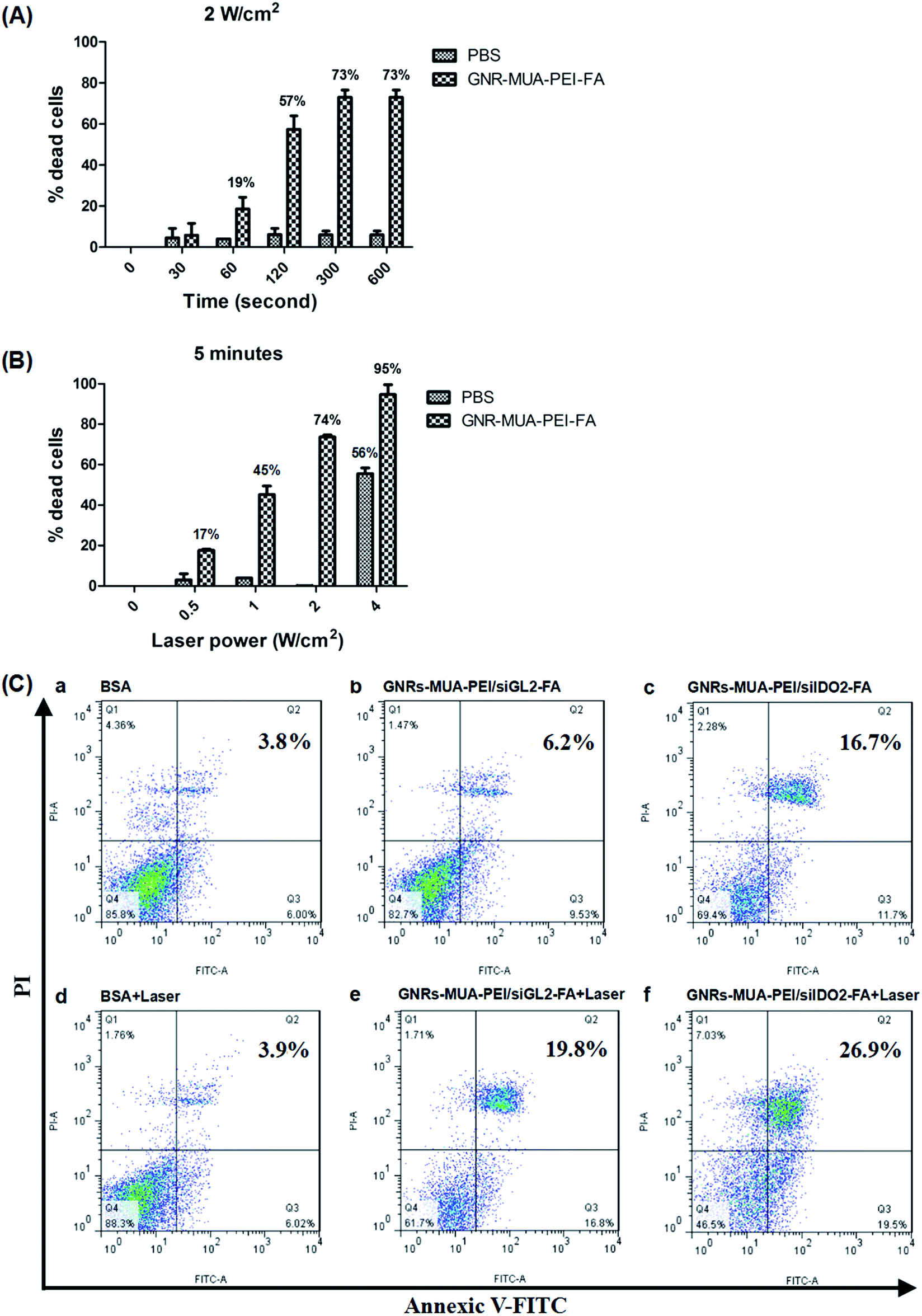 | ||
| Fig. 6 In vitro photothermal effects of GNR-MUA-PEI-FA and GNR-MUA-PEI/siIDO2-FA. B16-BL6 cells were incubated with a final concentration of 15 μg mL−1 of GNR-MUA-PEI-FA or PBS for overnight. Subsequently, the cells were irradiated at (A) 2 W cm−2 for various times (0 minutes, 0.5 minutes, 1 minute, 2 minutes, 5 minutes, 10 minutes) or at (B) 5 minutes for different power density (0 W cm−2, 0.5 W cm−2, 1 W cm−2, 2 W cm−2, 4 W cm−2). After 24 hours, the cell viabilities were measured by the MTT assay and the percentages of dead cells = (cell viabilities before laser irradiation − cell viabilities after laser irradiation)/(cell viabilities before laser irradiation). (C) Apoptosis of tumor cells induced by GNR-MUA-PEI/siIDO2-FA. B16-BL6 cells were incubated with PBS (a) or GNR-MUA-PEI/siGL2-FA (b), or GNR-MUA-PEI-FA-siIDO2 (c), at the final concentration of 16 μg mL−1 of wt(GNR-MUA-PEI-FA):wt(siIDO2) = 15:1) for overnight. Cells were remained no irradiation (a–c) or were irradiated (d–f) at 2 W cm−2 for 5 minutes with the wavelength of laser was 808 nm. The apoptotic and necrotic cell populations were determined at 24 hours by Annexic V-FITC/PI Apoptosis Detection Kit and analyzed by flow cytometry. Error bars represent the standard deviation of 3 experiments. | ||
We also measured the photothermal therapeutic effects of GNR-MUA-PEI-FA in B16-BL6 cells at different laser power density when the duration of irradiation was constant on 5 minutes (Fig. 6B). The results suggested that 1 W cm−2 is the minimum laser density necessary to kill tumor cells; when the laser power was added to 2 W cm−2, killing of tumor cells by GNR-MUA-PEI-FA was significantly increased to 74% (Fig. 6B). An increase (95%) of tumor cell killing by GNR-MUA-PEI-FA was observed when the laser power was set up at higher power (4 W cm−2), however, control cells that was treated with PBS also induced up to 56% cell death, suggesting the threshold of laser power should not exceed this maximum dose that may be harmful to the GNRs-untreated/normal cells in PTT. Thus the condition for the further in vivo experiments was optimized to be 1 W cm−2 of the power density and 2 minutes of the irradiation duration, which could effectively achieve photothermal effects of GNRs without non-specific injury to the non-malignant control cells.
To investigate apoptosis of B16-BL6 induced by IDO2 knockdown, we carried out the comparison experiments of the treatments of GNR with or without presence of IDO2 siRNA. According to the flow cytometry results (Fig. 6C(a–c)), the percentages of apoptotic cell population in the GNR-MUA-PEI/siIDO2-FA treated cells was 16.7%, which was significantly higher than the cells treated with control GNR-MUA-PEI/siGL2-FA (6.2%). Addition of IDO2 siRNA remarkably increased tumor cells to apoptosis, which is also consistent to our earlier discovery of silencing IDO2 causing tumor cell apoptosis.16 To clarify whether this increased apoptosis may enhance the killing ability of GNRs in PTT, we repeated above experiments under the condition of laser irradiation. As shown in Fig. 6C(d–f), apoptosis (19.8%) occurs in the GNR-MUA-PEI/siGL2-FA treatment along the combination of laser irradiation (808 nm, 2 W cm−2, 5 minutes), while GNR-MUA-PEI/siIDO2-FA, which incorporated with IDO2 siRNA, dramatically increased tumor cell apoptosis to 26.9%. These data suggested that IDO2 knockdown by GNR-MUA-PEI/siIDO2-FA induces tumor B16-BL6 cells to apoptosis and enhances the tumor cell killing ability of photothermal effects of GNRs, highlighting that GNR-MUA-PEI/siIDO2-FA could exert synergy of photothermal effects of GNRs and the IDO2 knockdown inducing tumor cell apoptosis.
In our studies the use of gold nanoparticles to deliver immune stimulatory siRNA, while concurrently inducing killing of cancer cells via laser induced hyperthermia was accomplished. A similar approach to ours was developed by Chen et al.,40 who utilized gold nanoparticles generated in a multi-layered manner by stabilization with hexadecyltrimethylammonium bromide and layering with polyelectrolytes and BSA. This approach was developed for selective release of doxorubicin subsequent to photothermal activation. Comparison between this approach and the approach developed by us for delivery of siRNA will require experimentation.
3.7. Transdermal delivery of siRNA to melanoma by GNR-MUA-PEI-FA
Cutaneous melanoma is a highly invasive disease. It would be ideal if there were a topical drug delivery system to suppress the growth of melanoma or even induce tumor regression in the case of immediate surgical removal is not possible. Therefore, we next explored the in vivo transdermal delivery of siRNA to melanoma by GNR-MUA-PEI-FA. The tumors were generated by subcutaneous injection of B6-BL6 cells on the back of C57BL/6 mice. After two weeks, the H&E staining histological section evidenced that the melanoma models were successfully prepared (Fig. 8A). Mixtures of Cy3-labeled siGAPDH, GNR-MUA-PEI/Cy3-labeled siGAPDH or GNR-MUA-PEI/Cy3-labeled siGAPDH-FA with glycerin and DMSO were topically applied on the skins above the inoculated tumors for 4 hours before analysis of fluorescent microscopy images of melanoma tissues (Fig. 7). The Cy3-labeled siGAPDH siRNA alone was not able to enter the cells and showed almost no fluorescence (Fig. 7A and B), while siRNA delivered by GNR-MUA-PEI only showed light fluorescence (Fig. 7C and D). In contrast, targeted delivery of siRNA using GNR-MUA-PEI-FA displayed the most intense fluorescence (Fig. 7E and F), suggesting that GNR-MUA-PEI-FA is as an effective transdermal reagent to reach the melanoma tissue for delivering siRNA to melanoma and promoting the cellular uptake of GNR-MUA-PEI-FA/siRNA by the melanoma cells in vivo. | ||
| Fig. 7 In vivo topical tumor-specific delivery of siRNA by GNR-MUA-PEI-FA. B16-BL6 melanoma tumor cells were inoculated on to C57BL/6 mice as described in Materials and methods. After 2 weeks, mice were anesthetized and the following reagents were applied on the skins above inoculated tumors: (A) and (B) Cy3-siGAPDH, (C) and (D) GNR-MUA-PEI/Cy3-siGAPDH, and (E) and (F) GNR-MUA-PEI/Cy3-siGAPDH-FA (all containing 6 μg of Cy3-siRNA, wt(GNR-MUA-PEI or GNR-MUA-PEI-FA):wt(Cy3-siRNA) = 15:1). 4 hours after above topical application of Cy3-labeled siRNA, and then the mice were scarified and the tumors were collected. The tumors tissues were frozen sectioned and the images were visualized with a fluorescence microscope. | ||
Nevertheless, the clinical development of the current approach will need to take into consideration the shape of the nanoparticle. As reviewed by Troung et al., shape influences biodistribution, selective uptake into cells, as well as possible selectivity of tumor uptake versus uptake into the tumor microenvironment. While advantages of nanorods to other shapes have been previously demonstrated in the cited review,20 future optimization studies will explore this questions.
3.8. Evaluation of anti-tumor efficacy of GNR-MUA-PEI/siIDO2-FA in photothermal therapy (PTT)
To investigate the in vivo anti-tumor efficacy of GNR-MUA-PEI/siIDO2-FA, which initiated a PTT of GNRs by NIR laser and enhanced anti-tumor therapy due to silencing IDO2, we tested therapeutic PTT in melanoma bearing mice. After 14 day treatment of melanoma in Fig. 8B, compared to PBS treating melanoma, GNR-MUA-PEI/siGL2-FA did not inhibit the tumor growth without the laser irradiation. In contrast, treatment of GNR-MUA-PEI/siGL2-FA with the laser irradiation (808 nm, 1 W cm−2, 2 minutes) significantly decreased the tumor volume (Fig. 8B), demonstrating the photothermal effects of GNRs. In addition, the treatment of GNR-MUA-PEI/siIDO2-FA, in combination of irradiation, showed the best therapeutic effects, implying the critical role of silencing IDO2 in inhibiting the tumor growth more effectively. In addition, after 14 day treatment of melanoma, the mice were sacrificed and the tumors were collected at the end point of observation. The size (Fig. 8C) and weight (Fig. 8D) of tumors were confirmed the PTT enhanced by silencing IDO2 as a promising treatment strategy for melanoma therapy using GNR-MUA-PEI/siIDO2-FA. Taken together, these data are the first demonstration that the enhanced anti-tumor therapy is achievable by knocking down IDO2 together with the photothermal effects of GNRs in melanoma therapy.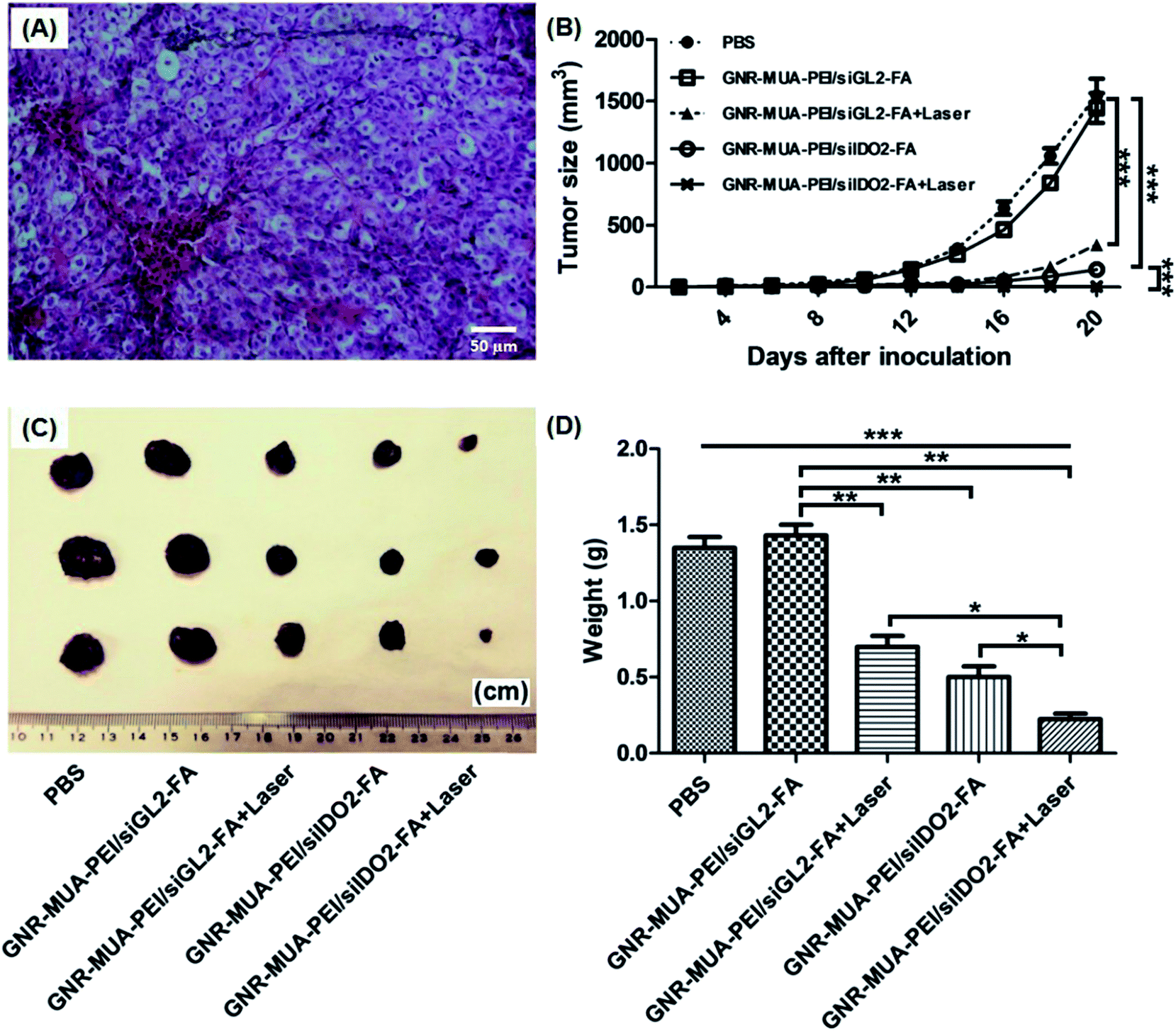 | ||
| Fig. 8 In vivo photothermal effects of GNR-MUA-PEI/siIDO2-FA for melanoma. (A) Melanoma models and histology sections. Female C57BL/6 mice were inoculated with B16-BL6 cells. After 2 weeks, mice were scarified. The tumors were collected and the hematoxylin and eosin (HE) staining was performed on paraffin-embedded sections. The prepared specimen was examined using a light microscope. Melanoma bearing mice were treated with PBS or GNR-MUA-PEI-FA/siRNA (wt(GNR-MUA-PEI-FA):wt(siRNA) = 15:1, 6 μg, siIDO2 or siScrambled), by a topical application onto the skins above the tumors on day 6, 10, 14, 18. Twelve hours after each application of above mixture, a laser irradiation (1 W cm−2, 2 minutes) was conducted. The tumor sizes of growth were measured with caliper (B). 20 days after tumor inoculation, the mice were sacrificed and tumors were collected. The images of tumor were displayed (C) and the tumor weights were measured (D). Error bars represent the standard deviation of 3 experiments. (*, P < 0.05; **, P < 0.01; ***, P < 0.001). | ||
4. Conclusion
In summary, we have designed and generated a novel nanoplatform GNR-MUA-PEI-FA/siIDO2, which possesses several integrated components: GNRs that exerts photothermal killing of melanoma cells upon NIR laser, FA that promotes tumor-specific delivery of siRNA to melanoma cells, and siIDO2 that can enhance inhibition of tumor growth upon knockdown of IDO2 gene. This new nanoplatform of GNR-MUA-PEI/siIDO2-FA represents a novel concept of GNR-based photothermal therapy and enhanced anti-tumor therapy by silencing IDO2 on the tumor cells. Encouragingly, this GNR-MUA-PEI/siIDO2-FA nanoplatform has displayed multiple advantages such as low cytotoxicity and improved biocompatibility, tumor specific targeting ability into B16-BL6 cells, and efficient induction of gene silencing. The feasibility of this new methods was evidenced by a robust therapeutic efficacy by inhibiting the growth of tumors in a murine melanoma model at a low laser power density (1 W cm−2, 2 minutes), which paves a novel way for cancer therapy by combining photothermia and gene silencing of IDO2.Conflict of interest
The authors of this manuscript have no conflicts of interest to disclose.Author contributions
All the authors are contributed to this manuscript. Planned experiments: Y. Z., W. M.; performed experiments Y. Z., N. S., J. F., Y. L., X. Z., S. P., Z. Y., Y. G., Z. W.; analyzed data: Y. Z., W. M., Q. S., Y. F., K. Y., N. Z.; wrote the paper: Y. Z., N. S., T. E. I, W. M. All authors are assured that we met the criteria for authorship and all reviewed the manuscript.Abbreviations
| GNA | Gold nanorods |
| FA | Folic acid |
| IDO2 | Indoleamine dioxygenase 2 |
| PTT | Photothermal therapy |
| TEM | Transmission electron microscope |
| CTAB | Cetyltrimethylammonium bromide |
| MUA | Mercaptoundecanoic acid |
| PEI | Poly ethylene imine |
| siRNA | Small interfering RNA |
| LSPR | Localized surface plasmon resonance |
| LIP2000 | Lipofactamine 2000 |
Acknowledgements
This study was supported by grants from Natural Science Foundation of China (NSFC, No. 81273303, 11574255), Jiangxi Provincial Natural Science Foundation (20151BAB205060, 20124ACB00800, 20133BBG70019), Nanchang University Doctoral Foundation (No. 06301097), Jiangxi Provincial Post-Doctoral Foundation (No. 00018422), and Fujian Provincial Science and Technology Plan (Cooperation) Key Project (No. 2014I0016).References
- I. Russo, F. Caroppo and M. Alaibac, Cancer, 2015, 7, 1371–1387 CrossRef PubMed.
- A. M. Eggermont, A. Spatz and C. Robert, Lancet, 2014, 383, 816–827 CrossRef CAS.
- National Cancer Institute, Surveillance, Epidemiology and End Results, http://www.seer.cancer.gov/statfacts/, accessed June 30, 2013.
- M. R. Middleton, P. Lorigan, J. Owen, L. Ashcroft, S. M. Lee, P. Harper and N. Thatcher, Br. J. Cancer, 2000, 82, 1158–1162 CrossRef CAS PubMed.
- A. A. Tarhini, J. M. Kirkwood, W. E. Gooding, S. Moschos and S. S. Agarwala, Cancer, 2008, 113, 1632–1640 CrossRef CAS PubMed.
- M. Singh and W. W. Overwijk, Cancer Immunol. Immunother., 2015, 64, 911–921 CrossRef CAS PubMed.
- M. Terabe and J. A. Berzofsky, Trends Immunol., 2007, 28, 491–496 CrossRef CAS PubMed.
- D. H. Munn, Curr. Opin. Immunol., 2006, 18, 220–225 CrossRef CAS PubMed.
- J. B. Katz, A. J. Muller and G. C. Prendergast, Immunol. Rev., 2008, 222, 206–221 CrossRef CAS PubMed.
- Y. Yan, G. X. Zhang, B. Gran, F. Fallarino, S. Yu, H. Li, M. L. Cullimore, A. Rostami and H. Xu, J. Immunol., 2010, 185, 5953–5961 CrossRef CAS PubMed.
- H. J. Ball, A. Sanchez-Perez, S. Weiser, C. J. Austin, F. Astelbauer, J. Miu, J. A. McQuillan, R. Stocker, L. S. Jermiin and N. H. Hunt, Gene, 2007, 396, 203–213 CrossRef CAS PubMed.
- X. Zheng, J. Koropatnick, M. Li, X. Zhang, F. Ling, X. Ren, X. Hao, H. Sun, C. Vladau, J. A. Franek, B. Feng, B. L. Urquhart, R. Zhong, D. J. Freeman, B. Garcia and W. P. Min, J. Immunol., 2006, 177, 5639–5646 CrossRef CAS.
- C. A. Blache, E. R. Manuel, T. I. Kaltcheva, A. N. Wong, J. D. Ellenhorn, B. R. Blazar and D. J. Diamond, Cancer Res., 2012, 72, 6447–6456 CrossRef CAS PubMed.
- X. Zheng, J. Koropatnick, D. Chen, T. Velenosi, H. Ling, X. Zhang, N. Jiang, B. Navarro, T. E. Ichim, B. Urquhart and W. Min, Int. J. Cancer, 2013, 132, 967–977 CrossRef CAS PubMed.
- S. Maleki Vareki, D. Chen, C. Di Cresce, P. J. Ferguson, R. Figueredo, M. Pampillo, M. Rytelewski, M. Vincent, W. Min, X. Zheng and J. Koropatnick, PLoS One, 2015, 10, e0143435 Search PubMed.
- Y. Liu, Y. Zhang, X. Zheng, X. Zhang, H. Wang, Q. Li, K. Yuan, N. Zhou, Y. Yu, N. Song, J. Fu and W. Min, Oncotarget, 2016 DOI:10.18632/oncotarget.8617.
- A. Fire, S. Xu, M. K. Montgomery, S. A. Kostas, S. E. Driver and C. C. Mello, Nature, 1998, 391, 806–811 CrossRef CAS PubMed.
- H. Borna, S. Imani, M. Iman and S. Azimzadeh Jamalkandi, Expert Opin. Biol. Ther., 2015, 15, 269–285 CrossRef CAS PubMed.
- D. D. Lasic and D. Papahajopoulos, Science, 1995, 267, 1275 CrossRef CAS PubMed.
- N. P. Truong, M. R. Whittaker, C. W. Mak and T. P. Davis, Expert Opin. Drug Delivery, 2015, 12, 129–142 CrossRef CAS PubMed.
- T. Lammers, F. Kiessling and W. E. Hennink, J. Controlled Release, 2012, 161, 175–187 CrossRef CAS PubMed.
- S. Venkataraman, J. L. Hedrick, Z. Y. Ong, C. Yang, P. L. Ee, P. T. Hammond and Y. Y. Yang, Adv. Drug Delivery Rev., 2011, 63, 1228 CrossRef CAS PubMed.
- X. Hu, J. Hu, J. Tian, Z. Ge, G. Zhang, K. Luo and S. Liu, J. Am. Chem. Soc., 2013, 135, 17617 CrossRef CAS PubMed.
- P. Wagstaff, A. Ingels, P. Zondervan, J. J. de la Rosette and M. P. Laguna, Curr. Opin. Neurol., 2014, 24, 474–482 CrossRef PubMed.
- T. J. Vogl, P. Farshid, N. N. Naguib and S. Zangos, Eur. J. Radiol., 2013, 23, 797–804 CrossRef PubMed.
- A. Poletti, G. Fracasso, G. Conti, R. Pilot and V. Amendola, Nanoscale, 2015, 7, 13702–13714 RSC.
- J. Song, P. Huang, H. Duan and X. Chen, Acc. Chem. Res., 2015, 48, 2506–2515 CrossRef CAS PubMed.
- R. K. Kannadorai, G. G. Chiew, K. Q. Luo and Q. Liu, Cancer Lett., 2015, 357, 152–159 CrossRef CAS PubMed.
- J. Song, L. Pu, J. Zhou, B. Duan and H. Duan, ACS Nano, 2013, 7, 9947–9960 CrossRef CAS PubMed.
- Z. Chen, X. Shan, Y. Guan, S. Wang, J. J. Zhu and N. Tao, ACS Nano, 2015, 9, 11574–11581 CrossRef CAS PubMed.
- A. Heber, M. Selmke and F. Cichos, Phys. Chem. Chem. Phys., 2015, 17, 20868–20872 RSC.
- B. Nikoobakht and M. A. El-Sayed, Chem. Mater., 2003, 15, 1957–1962 CrossRef CAS.
- E. J. Jin, Y. A. Choi, J. K. Sonn and S. S. Kang, Mol. Cells, 2007, 24, 139–147 CAS.
- D. Putnam, Nat. Mater., 2006, 5, 439–451 CrossRef CAS PubMed.
- K. S. Siu, D. Chen, X. Zheng, X. Zhang, N. Johnston, Y. Liu, K. Yuan, J. Koropatnick, E. R. Gillies and W. P. Min, Biomaterials, 2014, 35, 3435–3442 CrossRef CAS PubMed.
- X. Pan and R. J. Lee, Expert Opin. Drug Delivery, 2004, 1, 7–17 CrossRef CAS PubMed.
- J. Sudimack and R. J. Lee, Adv. Drug Delivery Rev., 2000, 41, 147–162 CrossRef CAS PubMed.
- T. Kurosaki, T. Morishita, Y. Kodama, K. Sato, H. Nakagawa, N. Higuchi, T. Nakamura, T. Hamamoto, H. Sasaki and T. Kitahara, Mol. Pharm., 2011, 8, 913–919 CrossRef CAS PubMed.
- M. R. Dzamukova, E. A. Naumenko, Y. M. Lvov and R. F. Fakhrullin, Sci. Rep., 2015, 5, 10560 CrossRef CAS PubMed.
- H. Chen, X. Chi, B. Li, M. Zhang, Y. Ma, S. Achilefu and Y. Gu, Biomater. Sci., 2014, 2, 996–1006 RSC.
| This journal is © The Royal Society of Chemistry 2016 |