Solvation, rotational relaxation and fluorescence correlation spectroscopic study on ionic liquid-in-oil microemulsions containing triple-chain surface active ionic liquids (SAILs)

Chiranjib Banerjee , Niloy Kundu, Arpita Roy, Debasis Banik, Mintu Halder and Nilmoni Sarkar*
Department of Chemistry, Indian Institute of Technology, Kharagpur 721302, WB, India. E-mail: nilmoni@chem.iitkgp.ernet.in; Fax: +91-3222-255303

Received 21st May 2016 , Accepted 26th July 2016

First published on 26th July 2016


Abstract

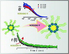
In this article, solvation dynamics and rotational relaxation approaches have been applied to explore the microheterogeneity of surface active ionic liquid (SAIL) containing microemulsions, i.e., [P13][Tf2N]/[BHD][AOT]/[IPM] and [N3111][Tf2N]/[BHD][AOT]/[IPM], using coumarin 480 (C-480) as a probe molecule. The average solvation time constants of Coumarin-480 (C-480) decreases with an increase in R value (R = [P13][Tf2N] or [N3111][Tf2N]/[BHD][AOT]) at a temperature of 25 °C for both the systems. This is attributed to the movement of the probe molecules from the interface to the pool of the microemulsion. However, the rotational relaxation time of the probe molecules (C-480) increases with increasing value of R as the pool of the microemulsion is more viscous compared to the interfacial region. Besides these, for the first time, we have measured the diffusion coefficients of these novel aggregates applying fluorescence correlation spectroscopic technique. The decrease in diffusion coefficients (Dt) with increase in R confirms the swelling of the microemulsions with addition of RTILs.


1. Introduction

The self-assembly of surfactant molecules, like microemulsion, is an interesting field due to their potential applications in separation science, environmental science, materials science and reaction engineering with certain unique advantages.1,2 Microemulsions are transparent (or translucent) systems of oil, water and surfactant, frequently in combination with a cosurfactant (having droplet sizes usually in the range of 5–100 nm) and are thermodynamically stable and isotropic in nature. They are generally classified as oil-in-water (o/w), water-in-oil (w/o) or bicontinuous systems depending on the structural features and are characterized by ultra-low interfacial tension between oil and water phases. In an o/w microemulsion, the dispersed oil is stabilized by the surfactant and co-surfactant molecules, whereas in a w/o microemulsion, the water droplet positions itself inside the resultant reverse micelle. The properties of microemulsions containing water cluster or highly polar organic solvent cores with dimensions in the order of nanometer have been studied extensively.3–6 Pileni et al. described different techniques to control the size of the reverse micelle (RM)/microemulsion, along with the probable reasons such as localization of solutes which causes structural perturbation of RM or microemulsion.7,8 The experiments performed by Kahlweit et al. provided a general idea on the behavior of microemulsions.9,10 Recently, attempts have been made for the preparation and study of reverse micelles where various polar solvents, having relatively high dielectric constants and which are immiscible in hydrocarbon solvents, have been used to replace the core water.11,12 These waterless microemulsion systems have been found to be more advantageous than the aqueous ones particularly because of their applicability as good reaction media. Nowadays, room-temperature ionic liquids (RTILs) are also being used as polar solvents because they constitute “green” substituents to classic (volatile) organic solvents.13,14 Recently, we have observed an increased stability of microemulsion at high temperature using a room temperature ionic liquid (RTIL) as polar core solvent, surface active ionic liquid (SAIL) as surfactant and isopropyl myristate (IPM) as organic solvent.15

Eastoe et al.16 investigated the structure of the system containing 1-butyl-3-methylimidazolium tetrafluoroborate ([bmim][BF4]) polar nanodroplets dispersed in cyclohexane (as a continuous phase) and it was first reported by Han and co-workers,17 by small-angle neutron scattering (SANS) and it showed a regular increase in droplet volume with gradual addition of [bmim][BF4]. These discoveries helped in the spectroscopic exploration of the formation mechanism of microemulsions that contain an IL with the electropositive imidazolium ring.18 Our group has also reported the preparation of IL-in-oil microemulsions with various RTILs.19 The modification of formation of microemulsion is not restricted to RTILs; surfactants which form microemulsions can also be modified. In our first approach towards such modifications, we prepared IL-in-oil microemulsion with SAILs.20 SAIL can be defined as functional ionic liquid with the combined properties of IL and surfactant; in other words, IL bearing long alkyl chain having amphiphilic character is termed as surface-active ionic liquid (SAIL).21 Subsequently, further modifications were made to prepare triple chain SAILs which have the ability to form IL-in-oil microemulsions as well as unilamellar vesicles.22

There is immense interest in understanding the nature of biological water, i.e., the water molecules near a biological system.23 In this regard; solvation dynamics and rotational relaxation studies are efficient ways to obtain fruitful information. These studies provide valuable information regarding the structure and dynamics of hydration layers and also, about the dynamics of self-assemblies and biomolecules themselves.

Fluorescence correlation spectroscopy (FCS) is emerging as an important technique for applications in biochemical field to study diffusion as well as conformational transitions in proteins,24–26 DNA,27 RNA,28 polypeptides,29 etc. on microseconds and longer timescales.

In this article, initially we have probed the microheterogeneity of [P13][Tf2N]/[BHD][AOT]/[IPM], and [N3111][Tf2N]/[BHD][AOT]/[IPM] microemulsions using solvation dynamics and rotational relaxation studies and it is further extended to single molecular level using FCS technique. The uniqueness of this study lies in the fact that the hydrophilic ionic liquid ([bmim][BF4]) used in our previous study has been replaced by hydrophobic ionic liquids such as N-methyl-N-propylpyrrolidinium bis(trifluoromethanesulfonyl)imide ([P13][Tf2N]), and N,N,N-trimethyl-N-propylammonium bis(trifluoromethanesulfonyl)imide, ([N3111][TF2N]).20 Also, the use of tripled-chain SAIL as well as biocompatible organic solvent (IPM, isopropyl myristate) makes this study more important. The diffusion time of IL-in-oil microemulsion has been determined through FCS which is first of its kind. It is believed that the findings would make the physicochemical and spectroscopic characterization of microemulsion systems more convenient and lead to an improved understanding of the microstructure of such systems. Moreover, the determination of diffusion coefficients using FCS technique makes the present article more significant.

2. Experimental section

2.1. Materials

Coumarin 480 (C-480) (laser grade), and 4-(dicyanomethylene)-2-methyl-6-(4-dimethylaminostyryl)-4H-pyran (DCM) (laser grade) were bought from Exciton. Sodium 1,4-bis(2-ethylhexyl)sulfosuccinate (NaAOT) and benzyl-n-hexadecyldimethylammonium chloride (BHDCl) were purchased from Sigma-Aldrich. AOT was dried in vacuum for 30 h before use. Isopropyl myristate (IPM) (SRL, India) was used as received. The ILs, N-methyl-N-propylpyrrolidinium bis(trifluoromethanesulfonyl)imide ([P13][Tf2N]) and N,N,N-trimethyl-N-propylammonium bis(trifluoromethanesulfonyl)imide ([N3111][TF2N]), procured from Kanto Chemicals (purity > 98%), were also used as received. All the structures are shown in Scheme 1. The detailed description of the sample preparation is included in the ESI. As water can affect the physical properties of RTILs, it is essential to report their water contents at experimental condition. The water content for both the ILs was measured using a digital automatic Karl Fischer Titrator (model VEEGO/MATIC-MD). The RTILs were clear, colorless and odorless with a moisture content below 1 ppm for [P13][Tf2N] and 25 ppm for [N3111][TF2N]. The concentration of C-480 was kept at 4 μM throughout the experiments and for FCS measurements; the concentration of DCM was maintained as 2.5 nM.
image file: c6ra13197b-s1.tif
Scheme 1 Structure of Surface Active Ionic Liquid (SAIL), ionic liquids, IPM, coumarin-480 and DCM.

2.2. Instrumentation

The detailed description of the instrumentation is included in the ESI.

3. Results and discussion

3.1. Steady-state studies

The absorption and emission spectra of C-480 were taken in isopropyl myristate (IPM) and both the microemulsions, i.e., [P13][Tf2N]/[BHD][AOT]/[IPM] and [N3111][Tf2N]/[BHD][AOT]/[IPM] systems (Fig. S1, ESI). In neat IPM, C-480 showed emission maximum at 425 nm. The addition of [BHD][AOT] shifted the emission maximum to 433 nm. Thus, the movement of the probe molecule from bulk IPM to the polar core of the [BHD][AOT] microemulsion is clearly indicated by this red shift of emission maximum (8 nm). Addition of ILs, [P13][Tf2N] and [N3111][Tf2N], to [BHD][AOT]/[IPM] microemulsion caused further red shifts in the emission maxima for both the systems (Fig. S2, ESI). The gradual red shift of emission maximum with the addition of ILs clearly indicates the transfer of probe molecules toward the polar IL pool in each microemulsion. The positions of the emission maxima were tabulated in our earlier publication where we have characterized the microemulsions.22 The emission spectra of C-480 in both the neat ionic liquids were taken and it is important to note that the emission maxima in neat [P13][Tf2N] IL (460 nm) is significantly different from that in [P13][Tf2N]/[BHD][AOT]/IPM microemulsion (438 nm at R = 0.61). Thus, it clearly indicates that the environment sensed by the C-480 inside the pool of the microemulsion is not similar to that in neat IL. The absorbance at the red end of the spectrum increased on addition of hydrophobic RTILs, i.e., [P13][Tf2N] and [N3111][Tf2N], to the C-480/[BHD][AOT]/[IPM] solution (Fig. S1, ESI). This provides an indication to the migration of a significant number of C-480 molecules from bulk solvent to the ionic liquid pool of microemulsion. The absorption and emission spectra of C-480 in microemulsions at different R values are illustrated in Fig. S1 and S2 (ESI), respectively. The emission maxima, monitored at R = 0.61, remained unchanged with increase in temperature (288–318 K) for both the systems, i.e., [P13][Tf2N]/[BHD][AOT]/[IPM] and [N3111][Tf2N]/[BHD][AOT]/[IPM], which indicates that a similar environment is sensed by the probe molecule at all temperatures or, in other words, variation of temperature does not lead to change in the partitioning of C480 between the two pseudophases (the organic and the micellar).

Thermochromism can alter the time resolved experimental results and earlier, we have showed that the temperature have substantial effect on the solvation and rotational dynamics of probe molecule in IL containing microemulsions.30 For this reason, the absorption and emission spectra of C-480 were taken at different temperatures in both the systems (data not shown). But, no changes in the absorption and emission spectra were observed. Thus, thermochromism does not take place for C-480 in microemulsion which is reflected in the absorption and emission spectra of C-480.

3.2. Time-resolved measurements

Time resolved anisotropy as well as solvation dynamics were monitored in the present systems which have been discussed below.
3.2.1. Time-resolved anisotropy measurements. Absorption and emission spectra were used to obtain information regarding the location as well as the partitioning of the probe molecules, C-480 in both the studied systems, i.e., [P13][Tf2N]/[BHD][AOT]/[IPM] and [N3111][Tf2N]/[BHD][AOT]/[IPM]. Now, to support the observation and monitor the effect of the movement of C-480 molecules on rotational relaxation with addition of RTILs, time resolved fluorescence anisotropy decays were recorded for both the systems. Fluorescence anisotropy decay is the most suitable technique to investigate the molecular dynamics near the site of the dye molecule because of its direct relation with the reorientation dynamics of excited molecule. The following equation has been used to calculate time resolved anisotropy, r(t):31
 
image file: c6ra13197b-t1.tif(1)
where, G is the correction factor for detector sensitivity to the polarization direction of emission. The value of G is 0.6 for our setup. I(t) and I(t) are the intensity of the fluorescence decays polarized parallel and perpendicular to the direction of polarization of the excitation light, respectively.

In neat IPM, the rotational time of C-480 is 0.34 ns and in [BHD][AOT]/IPM, at R = 0.0, the rotational relaxation time, τrot of C-480 is increased to 0.52 ns and the anisotropy decay is single exponential in nature (Table S1 and Fig. S3(a) of ESI). The increase in rotational relaxation time (τrot) of C-480 indicates the movement of probe molecules from IPM to [BHD][AOT]/IPM. Variation of anisotropy decay with increasing R value was observed and has been shown in Fig. 1. The average rotational relaxation time (τrot) of C-480 in [P13][Tf2N]/[BHD][AOT]/[IPM] microemulsions at R = 0.30 (at 298 K) was found to be 0.84 ns with components of 0.53 ns (80%) and 2.09 ns (20%) (Table 1) and the residuals of the fitted anisotropy decays are shown in Fig. S4, ESI. The probe molecule sensed two different regions inside the microemulsions: a relatively fast region near the head group of [BHD][AOT] surfactant and a slow region in the IL pool inside the microemulsion, which is indicated by the observed biexponential nature and the faster component in the rotational relaxation time of C-480 in IL microemulsion is close enough to the rotational time of C-480 at R = 0.00. The faster rotational relaxation time of C-480 is defined as τfast and the slower relaxation time is defined as τslow while, afast and aslow are their relative amplitudes, respectively.


image file: c6ra13197b-f1.tif
Fig. 1 Time-resolved fluorescence anisotropy decays of C-480 in (a) [P13][Tf2N]/[BHD][AOT]/[IPM], (b) [N3111][Tf2N]/[BHD][AOT]/[IPM] microemulsions with variation of R values at 298 K.
Table 1 Anisotropy decay parameters of C-480 in [P13][Tf2N]/[BHD][AOT]/[IPM] and [N311][Tf2N]/[BHD][AOT]/[IPM] microemulsions at different R valuesa
Systems R ([RTIL/[BHD][AOT]) afast aslow τfast (ns) τslow (ns) τrotb (ns) Viscosityc (cP)
a Experiments were performed at 298 K.b Error in experimental data of ±5%.c Error in experimental data of ±3%.
[P13][Tf2N]/[BHD][AOT]/[IPM] 0.30 0.80 0.20 0.53 2.09 0.84 11.07
0.46 0.74 0.26 0.52 2.23 0.96 13.69
0.61 0.68 0.32 0.53 2.29 1.09 15.16
[N3111][Tf2N]/[BHD][AOT]/[IPM] 0.30 0.77 0.23 0.54 1.72 0.81 8.64
0.46 0.73 0.27 0.54 1.88 0.90 11.21
0.61 0.72 0.28 0.52 2.09 0.96 13.69


On addition of IL, i.e., with increase in the value of R from 0.46 to 0.61, the average rotational relaxation time (τrot) of C-480 in [P13][Tf2N]/[BHD][AOT]/[IPM] microemulsion increased from 0.96 ns with components of 0.52 ns (74%) and 2.23 ns (26%) to 1.09 ns with components of 0.53 ns (68%) and 2.29 ns (32%) (Table 1). With increasing the amount of IL to the core of the microemulsion, the amplitude of the slower component is increased which confirms that the probe molecules are relocated towards the IL pool with addition of IL. The transfer of probe molecule towards IL-pool of the microemulsion can be the reason for this increment in average rotational relaxation time (τrot) with increasing value of R. Two main factors guiding the change in the anisotropy value with change of R are the restriction and the viscosity of the microenvironment around the probe molecules.

Now, with increase in R, the probe molecule moves to the pool of the microemulsion from the interfacial region. As a result, the constraint of the environment around the probe molecule is somewhat released, although the microviscosity of the environment increases drastically that led to an increase in time resolved anisotropy values. We have also measured the rotational time of C-480 in neat ILs (Fig. S3(b) of ESI) and the average rotational time of C-480 in neat IL is much higher than that in IL microemulsion. Hence, it can be said that the probe molecule sensed higher microviscosity on moving towards the IL-pool of the microemulsions (Table 1). To further support this observation, we have monitored the time resolved anisotropy value with increasing value of R for the other system also, i.e., [N3111][Tf2N]/[BHD][AOT]/[IPM]. The system showed an average anisotropy value of 0.81 ns with components of 0.54 ns (77%) and 1.72 ns (23%) at R = 0.30. On addition of ILs, an effect similar to that in case of [P13][Tf2N]/[BHD][AOT]/[IPM] microemulsion was observed, i.e., the rotational relaxation time (τrot) increased from 0.90 ns with components of 0.54 (73%) ns and 1.80 ns (27%) at R = 0.46 to 0.96 ns with components of 0.52 ns (72%) and 2.09 ns (28%) at R = 0.61 (Table 1). Hence, we can generalize our statement that with increase in the value of R, i.e., with addition of hydrophobic ILs the anisotropy value for both of our studied systems increases due to the increasing viscosity of the surroundings sensed by the probe molecule. Another notable point needs to be mentioned here is that in our previous study, we measured the rotational relaxation time on addition of hydrophilic IL, i.e., with increasing R value and ended up with similar observation.32 As a result, it can be said that the effect of R value in the rotational relaxation in the microemulsion is seems to be same irrespective of the nature of the ILs, i.e., hydrophilic or hydrophobic.

3.2.2. Solvation dynamics. The fluorescence decays of C-480 show marked dependence on the emission wavelengths. The representative decays of C-480 in [P13][Tf2N]/[BHD][AOT]/[IPM] microemulsion monitored at different wavelengths at 298 K are shown in Fig. 2. A fast decay is observed at shorter wavelength, while, at a longer wavelength, a growth precedes the emission spectra in the subnanosecond time scale. The solvent relaxation taking place in our present system results in the fast decay at the blue end and the slow rise at the red end of the emission spectrum.33,34 The red edge and blue edge decay profiles are best fitted by a biexponential function. The time-resolved emission spectra (TRES) are constructed following the procedure of Fleming and Maroncelli.35,36 The TRES at given time t, S(λ;t) is calculated by the fitted decays, D(t;λ), by relative normalization to the steady-state spectrum S0(λ), as follows:
 
image file: c6ra13197b-t2.tif(2)

image file: c6ra13197b-f2.tif
Fig. 2 Fluorescence decays of C-480 in [P13][Tf2N]/[BHD][AOT]/[IPM]microemulsion at R = 0.30 with different emission wavelengths.

Each TRES is fitted by a log-normal line shape function, which is defined as

 
image file: c6ra13197b-t3.tif(3)
where, g0, νp, b and Δ are the peak height, peak frequency, asymmetric parameter and width parameter, respectively.

The value of peak frequency is obtained by log-normal fitting of TRES. The respective TRES plot of C-480 in microemulsion is shown in Fig. S5(b) of ESI. The solvent response function defined below is used to monitor the solvation dynamics

 
image file: c6ra13197b-t4.tif(4)
where, ν(t), ν(0), and ν(∞) are the peak frequencies at time t, time zero, and time infinity, respectively. The solvent response function, C(t), is fitted using the following biexponential decay function,
 
C(t) = a1et/τ1 + a2et/τ2 (5)
where, τ1 and τ2 are the two relaxation times with respective amplitudes a1 and a2, following the relation a1 + a2 = 1. The C(t) versus time plots are given in Fig. 3. The decay parameters of C(t) are summarized in Table 2. The average solvation time was calculated using
 
τav = a1τ1 + a2τ2 (6)


image file: c6ra13197b-f3.tif
Fig. 3 Decays of solvent response functions, C(t) of C-480 in (a) [P13][Tf2N]/[BHD][AOT]/[IPM] microemulsion and (b) [N3111][Tf2N]/[BHD][AOT]/[IPM] microemulsion with variation of R values at 298 K.
Table 2 Decay parameters of C(t) of C-480 in [P13][Tf2N]/[BHD][AOT]/[IPM] and [N3111][Tf2N]/[BHD][AOT]/[IPM] microemulsions at different R values
Systems R ([RTIL/[BHD][AOT]) afast aslow τfast (ns) τslow (ns) τsa (ns) Sizeb (nm) R2
a Error in experimental data of ±0.08 ns.b Experimental error ±4%; R2 = coefficient of determination of decay parameters.
[P13][Tf2N]/[BHD][AOT]/[IPM] 0.30 0.13 0.87 0.45 10.51 9.20 3.61 0.992
0.46 0.10 0.90 0.51 9.88 8.94 5.27 0.997
0.61 0.12 0.88 0.57 9.76 8.65 7.00 0.997
[N3111][Tf2N]/[BHD][AOT]/[IPM] 0.30 0.20 0.80 0.48 11.00 8.89 3.55 0.997
0.46 0.19 0.81 0.53 9.84 8.07 5.00 0.997
0.61 0.14 0.86 0.58 8.80 7.64 6.50 0.997
[BHD][AOT]/IPM 0.00 0.00 1.00 14.7 14.7 2.80 0.991


The solvation dynamics in confined media is much more complex compared to the bulk media. For this reason, calculation of average solvation time is necessary to avoid the complexity of explaining individual components originating from different types of interactions and the locations of the probes. Thus, average solvation time is best to compare the retardation of solvation process in any confined system compared to bulk medium.

A detailed understanding of the structure of the microemulsion and the location of the probe within the microemulsion is necessary to perceive the solvation dynamics process. Inside microemulsion, the RTIL molecules face much more hindrance during movement than that in neat RTIL. Hence, the solvation process inside microemulsion is expected to be slower than that in neat RTIL. Earlier, our group have studied the solvation dynamics of C-480 in neat [N3111][Tf2N], where the average solvation time was found to be 0.21 ns with the time constant of 0.04 ns (67%) and 0.56 ns (33%) and in case of [P13][Tf2N], the reported average solvation time is 0.47 ns with the time constant of 0.35 ns (78%) and 0.92 ns (22%).19,37 Solvation dynamics in a RTIL occurs in a slow nanosecond time scale along with a sub-picosecond component.38,39 Several computer simulations have been carried out to understand the solvation process in RTILs, which suggest for the involvement of the collective motion of the cations and the anions for this process.40 To reveal the structural heterogeneity of RTIL, various studies have been reported.41,42

In either of our investigated systems, i.e., [P13][Tf2N]/[BHD][AOT]/[IPM] and [N3111][Tf2N]/[BHD][AOT]/[IPM] microemulsions, a bimodal solvation time is observed for all the R values. The solvent relaxation in RTILs is dependent on translational motion of ions of the RTILs. There are two types of RTILs in the microemulsion that exist in different environments, near and away from head groups of surfactant, i.e., [BHD][AOT]. The RTILs at the interfacial regions, i.e., near the surfactant molecule, are more restricted due to the translational motion of the ions compared to those away from surfactant molecules in microemulsion. So, RTIL molecules present at the interfaces of the microemulsion generate the slow components, while those away from surfactant molecules contribute to the fast component of solvation dynamics. They are defined as τslow and τfast and their corresponding amplitudes are termed as aslow and afast (Table 2). Two component model of the solvent relaxation in different confined systems is also reported earlier in the literature.43,44 Bhattacharyya and coworkers studied solvation dynamics in a neat ionic liquid, 1-pentyl-3-methyl-imidazolium tetra-fluoroborate ([pmim][BF4]) and its microemulsion in Triton X-100 (TX-100)/benzene using femtosecond up-conversion. They also observed more than one solvation components which originate from different regions of the microemulsion.43 Pandey and coworkers also observed two types of regions in water-in-ionic liquid (w/IL) microemulsions applying Fourier transform infrared (FTIR) spectroscopy.44

Interestingly, the increase in average solvation time of C-480 on going from neat IL to IL in microemulsion (almost 20 times) is much less compared to the solvation time on going from neat water to water containing microemulsion (almost 100 to 1000 times).45,46 The slight retardation of average solvation time suggests that the movement of cation and anion of the IL is slightly affected by the confinement.30

The average solvation time of C-480 in [BHD][AOT]/IPM solution (at R = 0.00) is found to be 14.7 ns and it is adequately fitted by single exponential decay function (Fig. S5(a), ESI). However, in our investigated system, [P13][Tf2N]/[BHD][AOT]/[IPM] microemulsion at R = 0.30, the average solvation time is found to be 9.20 ns with time constants of 0.45 ns (13%) and 10.51 ns (87%) (Table 2). Now, on addition of RTILs, i.e., [P13][Tf2N], the average solvation time is gradually decreased; at R = 0.46 the observed average solvation time is 8.94 ns with components of 0.51 ns (10%) and 9.88 ns (90%). With further addition of RTILs, i.e., at R = 0.61, it is decreased to 8.65 ns with components of 0.57 ns (12%) and 9.76 ns (88%). The results imply that with gradual addition of hydrophobic IL, i.e., [P13][Tf2N], the average solvation time decreases. At low concentration of IL, most of ILs are involved in solvating the surfactant and forming ion–dipole interaction with them. As the R value increases, the proportion of the IL in the interfacial region decreases and they are populated in the pool of the microemulsions which lead to an increase in the diffusional motion of the ions of the ILs and consequently, the average solvation time is decreased.19 On increasing the value of R from 0.30 to 0.61, the size of the microemulsion increases from 3.61 nm to 7.00 nm. The observation for the other microemulsion, i.e., [N3111][Tf2N]/[BHD][AOT]/[IPM] is also similar; the average solvation time is decreased from 8.89 ns with components of 0.48 ns (20%) and 11.00 ns (80%) at R = 0.30 to 7.64 ns with components of 0.58 ns (14%) and 8.80 ns (86%) at R = 0.61 (Table 2). The size of the microemulsion is also increased from 3.55 nm at R = 0.30 to 6.50 nm at R = 0.61. From the above results, one can conclude that with increasing value of R, the average solvation time decreases for both the systems, i.e., [P13][Tf2N]/[BHD][AOT]/[IPM] and [N3111][Tf2N]/[BHD][AOT]/[IPM]. However, close scrutiny of the data tabulated in Tables 1 and 2 reveals that the changes in solvation time is less in case of [P13][Tf2N]/[BHD][AOT]/[IPM] system compared to the other one, i.e., [N3111][Tf2N]/[BHD][AOT]/[IPM]. The results can be explained by considering the viscosity of the system. In case of [P13][Tf2N]/[BHD][AOT]/[IPM], the viscosity changes from 11.07 to 15.16 cP, with variation of R from 0.30 to 0.61. However, for [N3111][Tf2N]/[BHD][AOT]/[IPM] system, the viscosity changes from 8.64 to 13.69 cP with similar variation in R value. The relatively slow collective motion of the cations and anions in viscous medium are responsible for the larger solvation time. Another notable observation for both the investigated system is that the decrement in the solvation time with variation of R is small compared to the literature reports involving hydrogen-bonded solvents like water, methanol, glycerol etc.47,48 Shirota et al. have observed similar type of tiny R dependence on solvation time of aprotic solvent (dimethylformamide, DMF) in reverse micellar solution in compared to the protic solvent (formamide, FA) in reverse micelles where solvation time strongly depends on R.49

3.3. Fluorescence correlation spectroscopic (FCS) study

Although, FCS technique is widely applied in biology, however the utility of FCS in the field of colloidal chemistry has not yet been explored. Only a few reports on the application of FCS in microemulsions are available.50,51 The diffusion constant of w/o AOT reverse micelles prepared in a ternary mixture of water, sodium bis(2-ethylhexyl) sulfosuccinate (AOT), and isooctane, with sulforhodamine-B (SRhB) as a fluorescent marker was determined by Pal et al.50 In FCS measurements, photophysical effects of the fluorescent molecule, such as saturation and photobleaching, can severely influence the correlation data. Thus, the choice of fluorescent molecule is an important aspect. To avoid such phenomena, DCM was used as probe molecule. The concentration of DCM was kept at 2.5 nM in order to avoid any possibility of the presence of more than one DCM in a microemulsion droplet. Increase or decrease in the dye concentration only populates or depopulates the number of microemulsion droplets containing dye. Now, if the fluorophore strongly binds to the aggregates, the diffusional motion as analyzed from FCS can be ascribed to the motion of that self-assembly.52 For the neutral fluorophore DCM, Bhattacharyya et al. have showed that the diffusion coefficient of DCM is more or less the same in two differently charged micelles of identical size.52 Thus, DCM is located in the hydrophobic zone of the microemulsion because of its hydrophobic nature, and thus, leading to much accurate results in the determination of diffusion time of the droplets compared to C-480.

Fig. 4 shows the correlation curves of DCM in [P13][Tf2N]/[BHD][AOT]/[IPM] and [N3111][Tf2N]/[BHD][AOT]/[IPM] microemulsions with varying R. These curves are fitted assuming a two-component diffusion model, as the fits to a single-component diffusion model are found to be unsatisfactory (as judged by residuals and R2 values, Fig. 5, Table 3). The instrumentation and the analysis of FCS data are shown in ESI. It is, thus, evident that the investigated probe molecule, DCM, exhibits a bimodal diffusion behavior in both the microemulsions. The diffusion coefficients for the probe in these microemulsions, estimated from an average of 20 data sets, are tabulated in Table 3. The bimodal diffusion behavior of the probe can perhaps be attributed to the existence of the probe molecules in two distinct environments; inside the hydrophobic domain of the microemulsion, and in bulk solvent (IPM).


image file: c6ra13197b-f4.tif
Fig. 4 Fluorescence correlation curves of the DCM with variation of R, (a) [P13][Tf2N]/[BHD][AOT]/[IPM], (b) [N3111][Tf2N]/[BHD][AOT]/[IPM] microemulsions.

image file: c6ra13197b-f5.tif
Fig. 5 Double (a) and single (b) component fitting of [P13][Tf2N]/[BHD][AOT]/[IPM]microemulsion at R = 0.31.
Table 3 Diffusion parameters of the DCM dye in microemulsion at different R and different bulk solventsa
Systems R ([RTIL/[BHD][AOT]) D1b (μm2 s−1) D2b (μm2 s−1) R2
a R2 = coefficient of determination, D1 and D2 are the two diffusion coefficients.b Experimental error ±5%.
[P13][Tf2N]/[BHD][AOT]/[IPM] 0.30 33.61 179.26 0.99
0.46 29.88 175.38 0.99
0.61 28.00 166.01 0.98
[N3111][Tf2N]/[BHD][AOT]/[IPM] 0.30 38.41 182.19 0.99
0.46 32.79 177.40 0.99
0.61 29.87 170.12 0.99
[BHD][AOT]/[IPM] 0.00 40.00 185.03 0.98
IPM   176.00 0.98
[N3111][Tf2N]   127.61 9.68 0.99
[P13][Tf2N]   74.85 9.55 0.99


To confirm our assumption, we have also measured the diffusion time of DCM in neat IPM and the diffusion coefficient value of DCM in IPM is close enough to the faster component of diffusion coefficient (D2) in microemulsion. We have also measured the diffusion time of DCM in neat IL (Fig. S6, ESI) and the FCS traces were adequately fitted to a two component diffusion model. The diffusion time of DCM in neat [N3111][Tf2N] is slower than that of neat [P13][Tf2N] as the viscosity of [N3111][Tf2N] (69 cp) is higher than that of [P13][Tf2N] (51cp). However, the bimodal distribution in the diffusion behavior of fluorophores in IL has been reported earlier by several groups.53,54 The multiple values of diffusion coefficients suggest the presence of microheterogeneity or the nanoscale organization of the ILs. DCM experience different friction in the different regions of the IL, such as polar and nonpolar (cluster of alkyl chain) domains formed by the IL. Now, it is considered that the molecules do not change between two different environments while passing through the observation volume. In order to see the structural changes of the microemulsion droplets, the FCS measurements were extended with variation in the value of R. With increase in R value, the diffusion coefficient of the microemulsion droplets decreased supporting the swelling of these aggregates. For [P13][Tf2N]/[BHD][AOT]/[IPM] microemulsion, at R = 0.30, two diffusion coefficients have been obtained which are 33.61 μm2 s−1 and 179.26 μm2 s−1.

The two diffusion coefficients signify that the probe molecule sense two different regions inside the microemulsions: a region near the tail of [BHD][AOT] surfactant, which is responsible for slow diffusion coefficient, and bulk solvent, responsible for the fast diffusion coefficient. At R = 0.61, the value of the diffusion coefficient of the aggregates was 28.00 μm2 s−1. The decrease in diffusion coefficient from 33.61 to 28.00 μm2 s−1 with increase in R is a signature of the swelling of the microemulsion, as the larger particles diffuse slowly compared to the smaller ones. Similar effect is observed for the other microemulsion also. Beside these, microemulsions droplets are known to exchange, typically on millisecond to sub-microsecond time scale. However, for disperses emulsions with sufficiently small droplets (<500 Å), the tendency to coalesce will be counteracted by an energy barrier. Recently, McPhee et al. have investigated the intermicellar interaction by FCS using Cy-3 (Cyanine-3) as a probe molecule.55 Now, Cy-3 form different aggregate (dimer) in solution and the H-aggregate formation of Cy-3 induce the formation of a transient reverse micelle dimer. However, in our case, unlike Cy-3, DCM does not form any aggregate in solution and thus, it does not induce to form transient RM dimer.

Hence, we can say that the FCS technique has been successfully applied to determine the diffusion coefficient of the microemulsion droplets, which confirms the swelling of the aggregates with increasing concentration of the RTILs.

4. Conclusion

In conclusion, we have studied solvent and rotational relaxation processes in microemulsions composed of tripled-chain SAILs, hydrophobic ILs and biocompatible solvent, IPM. The average solvation and rotational relaxation time show dependence on R. The average solvation time decreases with increasing R values, which can be attributed to the transfer of probe molecule from the interfacial region to the polar core of both the microemulsions under investigation. The red shift in absorption and emission spectra, i.e., steady state results justify the movement of the probe molecules. However, the rotational relaxation time shows completely reversed results. The average rotational relaxation time increases with increasing value of R for both the systems. This could be due to the increase in viscosity of the microenvironment sensed by the probe molecules at the pool of the microemulsions. Most importantly, we have showed that it is possible to determine the diffusion coefficient of the microemulsion droplets applying FCS technique. Indeed, we observed that the diffusion coefficient of the microemulsion droplets decreases with increase in the concentration of RTILs, confirming the swelling of the aggregates.

Acknowledgements

N. S. is grateful to SERB, Department of Science and Technology and Council of Scientific and Industrial Research (CSIR), Government of India for generous research grants. C. B. is thankful to UGC; N. K. and D. B. are thankful to IIT Kharagpur and A. R. is thankful to CSIR for the research fellowships.

References

  1. M. J. Schwuger, K. Stickdorn and R. Schomaecker, Chem. Rev., 1995, 95, 849–864 CrossRef CAS.
  2. T. F. Jaramillo, S. H. Baeck, B. R. Cuenya and E. W. McFarland, J. Am. Chem. Soc., 2003, 125, 7148–7149 CrossRef CAS PubMed.
  3. E. J. Beckman, Science, 1996, 271, 613–614 CrossRef CAS.
  4. V. Challa, K. Kuta, S. Lopina, H. M. Cheung and E. V. Meerwall, Langmuir, 2003, 19, 4154–4161 CrossRef CAS.
  5. D. S. Venables, K. Huang and C. A. Schmuttenmaer, J. Phys. Chem. B, 2001, 105, 9132–9138 CrossRef CAS.
  6. N. Kundu, A. Roy, D. Banik and N. Sarkar, J. Phys. Chem. B, 2016, 120, 1106–1120 CrossRef CAS PubMed.
  7. M. P. Pileni, T. Zemb and C. Petit, Chem. Phys. Lett., 1985, 118, 414–420 CrossRef CAS.
  8. M. P. Pileni, J. Phys. Chem., 1993, 97, 6961–6973 CrossRef CAS.
  9. M. Kahlweit, R. Strey and P. Firman, J. Phys. Chem., 1986, 90, 671–677 CrossRef CAS.
  10. M. Kahlweit, R. Strey and G. Busse, J. Phys. Chem., 1990, 94, 3881–3894 CrossRef CAS.
  11. N. Li, Y. A. Gao, L. Q. Zheng, J. Zhang, L. Yu and X. W. Li, Langmuir, 2007, 23, 1091–1097 CrossRef CAS PubMed.
  12. Y. A. Gao, N. Li, L. Q. Zheng, X. T. Bai, L. Yu, X. Y. Zhao, J. Zhang, M. W. Zhao and Z. Li, J. Phys. Chem. B, 2007, 111, 2506–2513 CrossRef CAS PubMed.
  13. Z. Qiu and J. Texter, Curr. Opin. Colloid Interface Sci., 2008, 13, 252–262 CrossRef CAS.
  14. J. Kuchlyan, N. Kundu and N. Sarkar, Curr. Opin. Colloid Interface Sci., 2016, 25, 27–38 CrossRef CAS.
  15. V. G. Rao, C. Banerjee, S. Ghosh, S. Mandal, J. Kuchlyan and N. Sarkar, J. Phys. Chem. B, 2013, 117, 7472–7480 CrossRef CAS PubMed.
  16. J. Eastoe, S. E. Gold, A. Rogers, T. Paul, R. K. Welton and I. G. Heenan, J. Am. Chem. Soc., 2005, 127, 7302–7303 CrossRef CAS PubMed.
  17. H. X. Gao, J. C. Li, B. X. Han, W. N. Chen, J. L. Zhang, R. Zhang and D. D. Yan, Phys. Chem. Chem. Phys., 2004, 6, 2914–2916 RSC.
  18. Y. A. Gao, J. Zhang, H. Y. Xu, X. Y. Zhao, L. Q. Zheng, X. W. Li and L. Yu, ChemPhysChem, 2006, 7, 1554–1561 CrossRef CAS PubMed.
  19. R. Pramanik, S. Sarkar, C. Ghatak, V. G. Rao and N. Sarkar, J. Phys. Chem. B, 2011, 115, 2322–2330 CrossRef CAS PubMed.
  20. V. G. Rao, S. Ghosh, C. Ghatak, S. Mandal, U. Brahmachari and N. Sarkar, J. Phys. Chem. B, 2012, 116, 2850–2855 CrossRef CAS PubMed.
  21. P. D. Galgano and O. A. El Seoud, J. Colloid Interface Sci., 2010, 345, 1–11 CrossRef CAS PubMed.
  22. C. Banerjee, S. Mandal, S. Ghosh, J. Kuchlyan, N. Kundu and N. Sarkar, J. Phys. Chem. B, 2013, 117, 3927–3934 CrossRef CAS PubMed.
  23. B. Bagchi, Chem. Rev., 2005, 9, 3199 Search PubMed.
  24. K. Chattopadhyay, S. Saffarian, E. L. Elson and C. Frieden, Proc. Natl. Acad. Sci. U. S. A., 2002, 99, 14171–14176 CrossRef CAS PubMed.
  25. K. Chattopadhyay, E. L. Elson and C. Frieden, Proc. Natl. Acad. Sci. U. S. A., 2005, 102, 2385–2389 CrossRef CAS PubMed.
  26. D. K. Sasmal, T. Mondal, S. S. Majumdar, A. Choudhury, R. Banerjee and K. Bhattacharyya, J. Phys. Chem. B, 2011, 115, 13075–13083 CrossRef CAS PubMed.
  27. L. Edman, U. Mets and R. Rigler, Proc. Natl. Acad. Sci. U. S. A., 1996, 93, 6710–6715 CrossRef CAS.
  28. H. D. Kim, G. U. Nienhaus, T. Ha, J. W. Orr, J. R. Williamson and S. Chu, Proc. Natl. Acad. Sci. U. S. A., 2002, 99, 4284–4289 CrossRef CAS PubMed.
  29. H. Neuweiler, S. Doose and M. A. Sauer, Proc. Natl. Acad. Sci. U. S. A., 2005, 102, 16650–16655 CrossRef CAS PubMed.
  30. V. G. Rao, S. Mandal, S. Ghosh, C. Banerjee and N. Sarkar, J. Phys. Chem. B, 2012, 116, 8210–8221 CrossRef CAS PubMed.
  31. J. R. Lakowicz, Principles of Fluorescence Spectroscopy, Kluwer, Plenum, Dordrecht, NY, 2nd edn, 1999 Search PubMed.
  32. D. Chakrabarty, D. Seth, A. Chakraborty and N. Sarkar, J. Phys. Chem. B, 2005, 109, 5753 CrossRef CAS PubMed.
  33. S. Pal, S. Balasubramanian and B. Bagchi, J. Chem. Phys., 2002, 117, 2852–2859 CrossRef CAS.
  34. S. Pal, S. Balasubramanian and B. Bagchi, J. Phys. Chem. B, 2003, 107, 5194–5202 CrossRef CAS.
  35. M. Maroncelli and G. R. Fleming, J. Chem. Phys., 1987, 86, 6221–6239 CrossRef CAS.
  36. R. S. Fee and M. Maroncelli, Chem. Phys., 1994, 183, 235–247 CrossRef CAS.
  37. R. Pramanik, S. Sarkar, C. Ghatak, V. G. Rao, P. Setua and N. Sarkar, J. Phys. Chem. B, 2010, 114, 7579–7586 CrossRef CAS PubMed.
  38. S. K. Das and M. Sarkar, Chem. Phys. Lett., 2011, 515, 23–28 CrossRef CAS.
  39. S. K. Das and M. Sarkar, J. Phys. Chem. B, 2012, 116, 194–202 CrossRef CAS PubMed.
  40. H. Shirota and E. W. Castner Jr, J. Phys. Chem. A, 2005, 109, 9388–9392 CrossRef CAS PubMed.
  41. H. K. Kashyap and R. Biswas, J. Phys. Chem. B, 2010, 114, 16811–16823 CrossRef CAS PubMed.
  42. S. Daschakraborty and R. Biswas, J. Phys. Chem. B, 2014, 118, 1327–1339 CrossRef CAS PubMed.
  43. A. Adhikari, K. Sahu, S. Dey, S. Ghosh, U. Mandal and K. Bhattacharyya, J. Phys. Chem. B, 2007, 111, 12809–12816 CrossRef CAS PubMed.
  44. R. Rai and S. Pandey, Langmuir, 2014, 30, 10156–10160 CrossRef CAS PubMed.
  45. K. Bhattacharyya and B. Bagchi, J. Phys. Chem. A, 2000, 104, 10603–11061 CrossRef CAS.
  46. K. Bhattacharyya, Acc. Chem. Res., 2003, 36, 95–101 CrossRef CAS PubMed.
  47. N. Sarkar, K. Das, A. Datta, S. Das and K. Bhattacharyya, J. Phys. Chem., 1996, 100, 10523–10527 CrossRef CAS.
  48. H. Shirota and K. Horie, J. Phys. Chem. B, 1999, 103, 1437–1443 CrossRef CAS.
  49. H. Shirota and H. Segawa, Langmuir, 2004, 20, 329–335 CrossRef CAS PubMed.
  50. N. Pal, S. D. Verma, M. K. Singh and S. Sen, Anal. Chem., 2011, 83, 7736–7744 CrossRef CAS PubMed.
  51. M. F. Khan, M. K. Singh and S. Sen, J. Phys. Chem. B, 2016, 120, 1008–1020 CrossRef PubMed.
  52. S. Dey, U. Mandal, S. S. Mojumdar, A. K. Mandal and K. Bhattacharyya, J. Phys. Chem. B, 2010, 114, 15506–15511 CrossRef CAS PubMed.
  53. S. Patra and A. Samanta, J. Phys. Chem. B, 2012, 116, 12275–12283 CrossRef CAS PubMed.
  54. D. K. Sasmal, A. K. Mandal, T. Mondal and K. Bhattacharyya, J. Phys. Chem. B, 2011, 115, 7781–7787 CrossRef CAS PubMed.
  55. J. T. McPhee, E. Scott, N. E. Levinger and A. V. Orden, J. Phys. Chem. B, 2011, 115, 9585–9592 CrossRef CAS PubMed.

Footnotes

Electronic supplementary information (ESI) available: Sample preparation, general theory of fluorescence correlation spectroscopy, table of rotational time of C-80 in different ILs and IPM, UV-visible and fluorescence spectra, time resolved anisotropy decays, decays of solvent response function of C-480 in microemulsion at R = 0.00, FCS traces of DCM in different solvents. See DOI: 10.1039/c6ra13197b
C. B. and N. K. have equal contributions.

This journal is © The Royal Society of Chemistry 2016
Click here to see how this site uses Cookies. View our privacy policy here.