Deposition of a hydrophilic nanocomposite-based coating on silicone hydrogel through a laser process to minimize UV exposure and bacterial contamination

Guobang Huanga, Wai Hei Tseb and Jin Zhang*ab
aDepartment of Chemical and Biochemical Engineering, University of Western Ontario, London, Ontario N6A 5B9, Canada. E-mail: jzhang@eng.uwo.ca
bDepartment of Medical Biophysics, University of Western Ontario, London, ON N6A3K7, Canada

Received 19th May 2016 , Accepted 5th July 2016

First published on 5th July 2016


Abstract

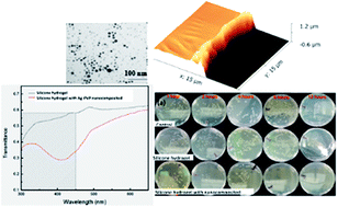
To prevent contact lens from biofouling and to minimize UV exposure to human eyes, a nanocomposite-based coating made of silver (Ag) nanoparticles and polyvinylpyrrolidone (PVP) is deposited on synthetic silicone hydrogels through matrix assisted pulsed laser evaporation (MAPLE) with a pulsed Nd:YAG laser at 532 nm. The average diameter of Ag NPs that have undergone the MAPLE process for 60 min is 11.61 ± 3.58 nm. The thickness of the Ag–PVP nanocomposite coating with a deposition time of 60 min is around 930 ± 15 nm. Our results demonstrate that the oxygen permeability of silicone hydrogel with a nanocomposite coating is similar to that of commercialized contact lenses; over 60% of UV light in the range of 300–450 nm can be blocked. Moreover, the silicone hydrogel with the nanocomposite coating can reduce over 65.4 ± 1.6% of protein (human lysozyme) absorption as compared to silicone hydrogel-based contact lens, and kill all cultured bacteria in 8 hours. This research work demonstrates a new way to deposit biocompatible nanocomposite coatings on silicone hydrogels used as contact lens to efficiently minimize UV exposure and biofouling.


1. Introduction

Extended exposure to solar ultraviolet (UV) radiation including UVA (400–320 nm), UVB (320–290 nm), and UVC (290–200 nm) may increase the risk of macular degeneration and the development of cataracts.1,2 According to the American Optometric Association, the majority of sunglasses cannot block sun radiation reaching the eyes from the side or around the glasses.3 Quite recently, UV-blocking agents have been cross-linked into the silicone hydrogel network to minimize UV absorption through the cornea.4 Silicone hydrogels with siloxane groups have been used extensively for contact lenses due to their transparent properties, high oxygen permeability, and good biocompatibility.5–7 Unfortunately most globular proteins tend to stick onto the hydrophobic surface of the silicone hydrogel, which provides a conditioning layer for microbial colonization and subsequent biofilm formation. Such biofouling on the surface of silicone-based medical devices are serious problems causing inflammation and/or infection of the eyes.8 Electrical neutrality has been applied to form hydrogen bonds on polymers to realize low/non-biofouling surfaces.9 Recently, drug-eluting contact lenses have attracted attention because of the capability for incorporating therapeutics into the silicone hydrogel matrix.10 No feasible method has been reported to treat silicone hydrogels to minimize both UV exposure and biofouling.

Polyvinylpyrrolidone (PVP) has been used to improve the hydrophilicity of the surface of polymer materials for various fields including the chemical industry, food, biomedical field, etc.11,12 In addition, it is noted that silver nanoparticles (Ag NPs) and silver ions have been recognized as effective antimicrobial agents, and have been used in wound or burn dressings, catheters, and bone cement.13 The antibacterial mechanism of Ag NPs is still unclear. One popular theory is that the Ag NPs release silver ions into bodily fluids, which are known to cause damage to bacterial DNA, proteins, enzymes, and cell walls. Some studies indicate the Ag NPs will interact with the cell wall and then destroy the metabolic response.14 In addition, the special surface plasmon resonance absorbance of Ag nanostructures could replace UV blocking agents in contact lenses to minimize UV exposure. Most methods in modifying the surface are wet chemical processes, dip-coating, and spin-coating, where organic chemicals are involved and require additional steps to remove them.15 Moreover, few works have been done to deposit Ag NPs physically incorporated with PVP on silicone hydrogels.

Compared to other coating techniques, matrix assisted pulsed laser evaporation (MAPLE) is an environmental-friendly method without the addition of chemical reagents. Fig. 1 displays the MAPLE process.


image file: c6ra12991a-f1.tif
Fig. 1 Illustration of the deposition of Ag–PVP nanocomposite coating on a silicone hydrogel by the MAPLE process.

The target material is suspended in a volatile solvent to be frozen with liquid nitrogen (LN2).16,17 A pulsing laser beam is introduced into the vacuum chamber to irradiate the frozen target. The energy absorbed by the chosen solvent is converted into thermal energy to vaporize the solvent, which minimizes the photochemical decomposition of proteins and polymers.18–20 The MAPLE process has demonstrated the capability for retention of the properties and structures of deposited organic molecules.16 In the MAPLE process, excimer lasers or Nd:YAG lasers with second or third harmonics are normally used; infrared laser sources are utilized in particular cases.21,22 Quite recently, we reported the deposition of a layer of polyethylene glycol (PEG) onto silicone hydrogel through MAPLE with a pulsed Nd:YAG laser of 532 nm.23 Few studies have demonstrated a nanocomposite coating produced by MAPLE.24,25 The MAPLE technique could be a suitable tool for the surface modification of biomaterials due to a lack of solvent and oxygen contamination. In this paper, Ag NPs and PVP were deposited at the same time onto silicone hydrogels by MAPLE. The performance of the silicone hydrogel contact lenses with nanocomposite coating was studied. We hope the nanocomposite coating is capable of minimizing UV exposure and bacterial contamination concurrently.

2. Experimental

All chemicals used in this paper are purchased from Sigma-Aldrich Canada.

2.1 Deposition of nanocomposite coating on silicone hydrogel by MAPLE

The silicone hydrogel was synthesized through a photo-polymerization.26 N,N-Dimethylacrylamide, 3-methacryloxypropyltris(trimethylsiloxy)silane, and bis-alpha,omega-(methacryloxypropyl)polydimethylsiloxane were mixed by the volume ratio of 2[thin space (1/6-em)]:[thin space (1/6-em)]4[thin space (1/6-em)]:[thin space (1/6-em)]1 to yield 3 ml, followed by the addition of 15 μl of ethylene glycol dimethacrylate and 0.3 ml of ethanol. Nitrogen was purged into the mixture for 15 min before 8 mg of the photo-initiator (diphenyl(2,4,6-trimethylbenzoyl)phosphine oxide) was added and stirred for 5 min. After that, the mixture was photo-polymerized under UV irradiation for 50 min. 30% ethanol was applied to wash the hydrogel after photo-polymerization. The thickness of all silicone hydrogel samples was 200 μm.

Ag NPs were synthesized from silver nitrate by photochemical reduction.20,21 100 ml of ethylene glycol was added into a 250 ml flask followed by 10 min of nitrogen purging. 1.5 g of PVP (MW = 10[thin space (1/6-em)]000) was used as a structure directing agent, which was dissolved in ethylene glycol under stirring for 30 min. 1 g of silver nitrate (AgNO3) was then added to the above solution. The mixture was under UVA irradiation for 24 hours. The produced Ag NPs were separated from the solution, and washed with a solution of ethanol and acetone.

The coating of Ag–PVP nanocomposites on the surface of silicone hydrogel was fabricated by MAPLE deposition as shown in Fig. 1. Ag NPs and PVP in a weight ratio of 1.5[thin space (1/6-em)]:[thin space (1/6-em)]1 were dissolved in isopropanol at a concentration of 0.5 wt%, followed by freezing with liquid nitrogen. A Nd:YAG laser with a wavelength (λem) of 532 nm was used in the MAPLE deposition. The deposition was conducted for 30 min with a background pressure of 1 × 10⁻⁶ Torr. The substrate-to-target distance was 6 cm.

The Ag NPs produced by the photochemical reduction and the Ag NPs that had undergone the MAPLE process for 60 min were characterized by transmission electron microscopy (TEM) and FTIR (Fourier Transform Infrared spectroscopy, Bruker FTIR-IFS 55). The thickness of the coating was approximately 930 ± 15 nm, as measured by atomic force microscopy (Dimension 3100, Veeco Inc.). The nanocomposite coating was further studied by EDX (Energy-dispersive X-ray spectroscopy, Hitachi 3400s).

2.2 Performance of silicone hydrogel-based contact lens with/without the nanocomposite coating

UV-visible spectrophotometry (UV-vis, UV-3600 Shimadzu) was employed to study the light transmission of the silicone hydrogel with/without the nanocomposite coating. In addition, silicone hydrogel samples 1 cm × 1 cm were mounted on the BioTester 5000 test system (CellScale Biomaterials Testing). The samples were stretched uniaxially with a pre-loading of 10 mN as per a previously reported method.7 The stress and strain were calculated from the data and the stress–strain curves of different samples were plotted to determine Young’s modulus (E) by using the equation below:
 
image file: c6ra12991a-t1.tif(1)
where E is in Pascal (Pa), F is the force applied in Newton (N), A is the area perpendicular to the force vector (m2), δL is the displacement of the materials (m), and Lo is the original length of the materials (m). Measurements in triplicate were carried out to obtain the statistic Young’s modulus of the samples.

Fig. 2 shows our designed system for measuring oxygen permeability. Argon gas was purged into the distilled water for 10 minutes (lower chamber), then the hydrogel sample (diameter = 10 mm × thickness = 1 mm) was placed into the connector. Oxygen gas flowed into the upper chamber for 10 minutes (T). The pressure of oxygen in the upper chamber was 1.013 × 10⁵ Pa. A 550A Dissolved Oxygen Meter (YSI Inc.) was used to measure the dissolved oxygen concentration before (C1) and after oxygen flow (C2). The volume (V) of the lower chamber was 250 ml. The oxygen transport for control (P0) was 0.04525 ± 0.00263 mg min−1; which was obtained without a sample in the connector. The relative oxygen permeability through the hydrogel was calculated using eqn (2).

 
image file: c6ra12991a-t2.tif(2)


image file: c6ra12991a-f2.tif
Fig. 2 Illustration of the system for measuring oxygen permeability of the silicone hydrogel samples.

2.3 Protein adhesion test

Human lysozyme (LSZ) was used here to study protein adhesion. Silicone hydrogel samples (1 cm × 1 cm) with/without Ag–PVP nanocomposite coating were immersed in PBS for 24 hours and then soaked in 0.5 mg ml−1 of LSZ in PBS solution for 3 hours at 37 °C to facilitate protein adhesion. PBS was used to rinse the samples 3 times to remove the non-absorbed LSZ on the surface of the hydrogel. The samples were then immersed in 1 wt% SDS–PBS solution under sonication for 20 minutes to completely detach LSZ from the hydrogel surface and into the solution. A BCA protein assay kit (Micro BCATM Protein Assay Kit, Thermo Scientific, U.S.A.) was used to determine the protein concentration in the SDS–PBS solution with a UV-visible plate reader at the wavelength of 562 nm.

2.4 Antibacterial efficiency and cytotoxicity of the nanocomposite coating

The antibacterial efficiency of the nanocomposite coatings was tested by a drop-test method.27,28 The samples (1 cm × 1 cm) were placed into sterilized 90 mm Petri dishes. Glass cover slips were used as control samples. Then 100 μl PBS solutions with non-pathogenic E. coli at a concentration of 106 CFU ml−1 were dropped onto the surface of each sample. The samples were laid at ambient temperature for different time periods (i.e. 1, 2, 4, 8 and 12 hours). After each time interval, the bacteria containing drops were washed from the sample surfaces using 5 ml of PBS into the sterilized Petri dish. Then 10 μl of each bacteria suspension was spread on a LB agar plate. The number of surviving bacteria on the Petri dishes was counted after a 24 h incubation at 37 °C. The relative viability of E. coli was calculated: the number of colonies in the sample divided by the number of colonies in the control. Meanwhile the response of the silicone hydrogel with/without the Ag–PVP coating to the Gram-positive bacterium, S. aureus (ATCC), was also tested. All experiments were run in triplicate.

In addition, NIH/3T3 mouse fibroblast cells (ATCC) were used to study the cytotoxicity of the silicone hydrogel samples with the Ag–PVP nanocomposite coating. Approximately 102[thin space (1/6-em)]000 3T3 mouse fibroblast cells, determined using a haemocytometer, were seeded in a 24-well plate and incubated overnight to ensure good cell adhesion onto the plate. Silicone hydrogel samples with/without the nanocomposite coatings were then added into the cultured cells and incubated for 24 hours under sterile conditions. The positive control sample was cultured cells without hydrogel samples. After 24 hours, the samples were removed and the media aspirated. As per the protocol of the Vybrant® MTT Cell Proliferation Assay Kit, cells were then labeled using formazan dissolved in DMSO. Controls and treated cells in microplates were incubated for 10 min followed by spectrophotometric analysis with the absorbance read at 540 nm.

3. Results and discussion

3.1 Silver nanoparticles before and after deposition by MAPLE

Fig. 3a demonstrates the TEM micrograph of Ag NPs synthesized by photochemical reduction. Fig. 3b is the TEM micrograph of the Ag NPs deposited on Cu grids by MAPLE for 60 min. The insert small figures of Fig. 3a and b depict the size distribution. The average size of Ag NPs without MAPLE is estimated at 11.29 nm ± 1.88 nm, while the average size of Ag NPs that have undergone the 60 min’s of MAPLE is 11.61 nm ± 3.58 nm. The size distribution of Ag NPs that have undergone MAPLE is broader although most of the energy of laser irradiation at λ = 532 nm with 300 mJ cm−2 of fluence may be absorbed by the solvent, isopropanol. The 532 nm laser could incur a photon–electron interaction in Ag NPs, which causes a rapid temperature rise in the Ag NPs.23 When the temperature of the silver particle reaches the boiling point, atomic sand or small particles are ejected through vaporization into the surrounding solvent.29,30 As a result, the reduction of particle size occurs. The small particles are very unstable in the solution and tend to aggregate onto the surface of other silver particles, resulting in an increase in size. Therefore, the size of Ag NPs becomes non-uniform after the MAPLE process.
image file: c6ra12991a-f3.tif
Fig. 3 TEM micrograph of (a) Ag NPs produced by photochemical reduction; (b) Ag NPs deposited on a Cu grid through the MAPLE process. The scale bar refers to 100 nm.

3.2 Chemical analysis of the nanocomposite coating

FTIR spectra of samples can be observed in Fig. 4. The spectrum of (a) is the FTIR spectrum of pure PVP, with respective bands at1287 cm−1, 1072 cm−1 and 1017 cm−1 indicating the C–N vibration band from PVP.31 Fig. 4b is the FTIR spectrum of Ag–PVP NPs, in which the C–N vibration bands red-shift to 1290 cm−1, 1075 cm−1, and 1019 cm−1 compared with pure PVP. This confirms that the silver atom is coordinated with the N atoms of PVP.32 In Fig. 4, the vibration band of C[double bond, length as m-dash]O as shown in the spectrum of (b) also red shifts from 1651 cm−1 to 1655 cm−1, which indicates the coordination between silver atoms and C units of PVP.32 Fig. 4c and d are the respective FTIR spectra of silicone hydrogel with Ag–PVP nanocomposite coating and bare silicone hydrogel. The bands at 1723 cm−1, 1644 cm−1, 1250 cm−1, and 1038 cm−1 refer to the C[double bond, length as m-dash]O vibration of the silicone hydrogel shown in the spectrum of (d). After MAPLE, most of the C[double bond, length as m-dash]O vibration bands of the silicone hydrogel coating with the Ag–PVP nanocomposite coating remains the same, only the band of 1644 cm−1 shifts to 1651 cm−1 compared to bare silicone hydrogel. This is due to the overlap of C[double bond, length as m-dash]O vibration bands of the Ag–PVP nanocomposite (1655 cm−1) and bare silicone (1644 cm−1). The spectrum of (c) indicates that silicone–Ag–PVP also has the C–N (1289 cm−1) vibration band which does not exist on the bare silicone hydrogel. In addition, the O–H stretching vibration band can be observed in the samples of PVP, the colloidal Ag NPs with the stabilizer of PVP. The MAPLE produced the Ag–PVP nanocomposite coating as shown in Fig. 4 because of the presence of the hydrophilic PVP. The FTIR spectra of silicone hydrogel with the Ag–PVP coating by MAPLE and the drop-air-dried method have no significant difference as shown in the ESI, which further indicates that the structure of PVP is maintained in the MAPLE process.
image file: c6ra12991a-f4.tif
Fig. 4 FTIR spectra of (a) PVP, (b) colloidal Ag NPs with the stabilizer of PVP produced by the photochemical process, (c) Ag–PVP nanocomposite coating on silicone hydrogel by the MAPLE process and (d) silicone hydrogel.

3.3 EDX analysis of the silicone hydrogel with the Ag–PVP nanocomposite coating

Fig. 5 depicts the EDX mapping and EDX spectra of the Ag–PVP nanocomposite coating deposited on silicone hydrogel by MAPLE. The presence of elemental silver on the silicone hydrogel is observed in Fig. 5a and e. It also indicates that the Ag NPs are homogenously distributed on the silicone hydrogel.
image file: c6ra12991a-f5.tif
Fig. 5 EDX analysis. EDX mapping of (a) Ag (b) C (c) O and (d) Si; (e) EDX spectrum of Ag–PVP nanocomposites deposited on the silicone hydrogel through the MAPLE process.

3.4 Mechanical properties and oxygen permeability of the silicone hydrogel with the nanocomposite coating

The thickness of the Ag–PVP nanocomposite coating measured by AFM is approximately 930 ± 15 nm, as shown in Fig. 6.
image file: c6ra12991a-f6.tif
Fig. 6 Cross-section of the Ag–PVP nanocomposite coating measured by AFM.

The Young’s modulus of the silicone hydrogel increases from 0.708 ± 0.005 MPa to 0.811 ± 0.005 MPa after coating of the Ag–PVP nanocomposite by MAPLE. It is noted that Young’s modulus of human skin can range between 0.42 MPa and 0.85 MPa depending on ages.33 Young’s modulus of silicone hydrogel with/without the nanocomposite coating are similar to human skin, which is an important factor to be considered prior to applications with body implants and contact lens.

In addition, the synthetic silicone hydrogels demonstrate high oxygen permeability due to oxygen transporting through the siloxane-phase.26 Table 1 shows the result of O2 passing through different samples.

Table 1 Relative O2 permeability of silicone hydrogels with/without the Ag–PVP coating
Samples Silicone hydrogel (thickness = 200 μm) Silicone hydrogel (thickness = 200 μm) with the Ag–PVP nanocomposite coating
Relative O2 permeability 79.6 ± 0.5% 78.8 ± 0.5%


The results indicate that the oxygen permeability of the nanocomposite-coating silicone hydrogels has a similar value to synthetic silicone hydrogels; with thickness of 200 μm which is similar to those of contact lenses.

3.5 Light transmittance of the silicone hydrogel with the Ag–PVP nanocomposite coating

UV-visible spectrophotometry was employed to study the light transmittance of the silicone hydrogel with/without the Ag–PVP nanocomposite coating. In Fig. 7, the light transmittance of silicone hydrogel with the Ag–PVP nanocomposite coating was able to reduce by over 60% in the range of 300 nm to 450 nm and therefore reducing the side effect of UV exposure on human eyes.
image file: c6ra12991a-f7.tif
Fig. 7 Light transmittance of the silicone hydrogels with/without the Ag–PVP nanocomposite coating.

3.6 Protein adsorption

The protein adsorption of the bare silicone hydrogel and silicone hydrogel with the Ag–PVP nanocomposite coating, as confirmed by FTIR, was tested by the micro BCA method. LSZ adsorption of the bare silicone and Ag–PVP coated silicone is 7.92 μg cm−2 and 2.74 μg cm−2, respectively. The protein adsorbed on the silicone hydrogel with the Ag–PVP nanocomposite coating decreased 65.4 ± 1.6% compared to that of the silicone hydrogel. PVP has demonstrated the capability for reducing non-specific protein adsorption as it provides a more hydrophilic surface, to reduce irreversible protein adsorption compared to the bare silicone hydrogel.18 Lysozyme is one of major common proteins found on worn contact lenses.34 Its hydrophobic amino acids can protect the inside of the protein molecule and its hydrophilic amino acids side chain are held outside to interact with the environment.34,35 When a protein interacts with a hydrophobic surface, the protein core attempts to interact with the hydrophobic surface in order to reach lower entropy. As a result, this may denature the protein structure. Thus, the MAPLE produced Ag–PVP nanocomposite coating improves the hydrophilicity of the silicone hydrogel and can reduce irreversible protein adsorption.

3.7 Antibacterial efficiency and cytotoxicity of the Ag–PVP nanocomposite coating

Ag NPs demonstrate an efficient antibacterial property due to the interaction between releasing silver ions from Ag NPs and the bacteria cell.36 Fig. 8a shows the surviving bacterial colonies on agar plates cultured separately on the surface of the control sample, silicone hydrogel, and silicone hydrogel with the Ag–PVP nanocomposite coating. It is evident that the viability of bacteria cultured on the silicone hydrogel with the Ag–PVP nanocomposite coating decreases with time. Fig. 8b and c indicates the relative viabilities of E. coli and S. aureus, respectively, on the silicone hydrogel with and without the Ag–PVP nanocomposite coating over different culture periods. The relative number of surviving bacterial colonies (both E. coli and S. aureus) on the bare silicone remains around 85% even when the incubation time increases, indicating that the bare silicone hydrogel does not have the ability to inhibit the bacterial growth. On the other hand, the relative number of surviving bacteria on silicone–Ag–PVP decreases with increasing incubation time; from 1 hour to 12 hours. After 8 hours, there is almost no bacteria growth from the silicone hydrogel with the Ag–PVP nanocomposite coating sample.
image file: c6ra12991a-f8.tif
Fig. 8 Antimicrobial efficiency. (a) Plate counting of E. coli from control, silicone hydrogels and silicone hydrogels with Ag–PVP nanocomposite coating; (b) antibacterial test (E. coli) of silicone hydrogels with/without the Ag–PVP nanocomposite coating. (c) Antibacterial test (S. aureus) of silicone hydrogels with/without Ag–PVP the nanocomposite coating.

NIH/3T3 mouse fibroblast cells were used for the cell viability test. The results indicate that the cell viability of silicone hydrogel and silicone hydrogel with the Ag–PVP nanocomposite coating is 146.5 ± 2.5% and 81.1 ± 0.5%, respectively. It is noted that the relative cell viability for materials with good biocompatibility is normally beyond 80%.37 Meanwhile, the relative decrease of cell viability of the silicone hydrogel with the nanocomposite coating could be caused by many factors, e.g. the increased thickness, the change of the surface roughness, the slightly decreased oxygen permeability, etc. We are working on the effect of thickness of the nanocomposite coating deposited on silicone hydrogel on cell viability, which will be reported in a different manuscript.

4. Conclusions

The Ag–PVP nanocomposite coating was deposited on a silicone hydrogel by MAPLE. The average diameter of Ag NPs that have undergone MAPLE for 60 min is 11.61 ± 3.58 nm which is a broader size distribution compared to the Ag NPs produced by photochemical reduction with a diameter of 11.29 ± 1.88 nm. MAPLE did not influence the polymer (PVP) structure according to results of the FTIR spectra. Silicone hydrogel with the Ag–PVP nanocomposite coating can block over 60% of the light in the UVA region. Other physical performances of the silicone hydrogel with the Ag–PVP nanocomposite coating in terms of oxygen permeability and mechanical properties are retained. In addition, the silicone hydrogel with the Ag–PVP nanocomposite coating shows high resistance to protein-sticking, which can reduce about 65.4 ± 1.6% of the LSZ adsorption compared to bare silicone hydrogel. The antimicrobial test assay further demonstrates that the silicone hydrogel with the Ag–PVP nanocomposite coating is able to inhibit bacterial growth. After an 8 hour incubation, the Ag–PVP nanocomposite coating can eliminate almost all E. coli, while E. coli keeps growing on the bare silicone hydrogel. It is expected that the Ag–PVP nanocomposite coating produced by MAPLE can add additional value on silicone hydrogel-based contact lens to minimize UVA exposure, and to prevent bacterial contamination.

Acknowledgements

Authors are grateful the Natural Sciences and Engineering Research Council of Canada (NSERC) for financial support.

Notes and references

  1. H. R. Taylor, S. West, B. Muñoz, F. S. Rosenthal, S. B. Bressler and N. M. Bressler, Arch. Ophthalmol., 1992, 110, 99–104 CrossRef CAS PubMed.
  2. S. Wittenberg, Optom. Vis. Sci., 1986, 63, 676–689 CrossRef CAS.
  3. H. L. Chandler, K. S. Reuter, L. T. Sinnott and J. J. Nichols, Invest. Ophthalmol. Visual Sci., 2010, 51, 172–178 Search PubMed.
  4. L. Buratto, L. Werner, M. Zanini and D. Apple, Phacoemulsification: principles and techniques, Slack Incorporated, New Jersey, 2003, p. 666 Search PubMed.
  5. N. Annabi, A. Tamayol, J. A. Uquillas, M. Akbari, L. E. Bertassoni, C. Cha, G. Camci-Unal, M. R. Dokmeci, N. A. Peppas and A. Khademhosseini, Adv. Mater., 2014, 26, 85–124 CrossRef CAS PubMed.
  6. J. Pozuelo, V. Compañ, J. M. González-Méijome, M. González and S. Mollá, J. Membr. Sci., 2014, 452, 62–72 CrossRef CAS.
  7. P. C. Nicolson, Eye Contact Lens, 2003, 29, S30–S32 Search PubMed.
  8. I. Banerjee, R. C. Pangule and R. S. Kane, Adv. Mater., 2011, 23, 690–718 CrossRef CAS PubMed.
  9. X. Liu, Y. Xu, Z. Wu and H. Chen, Macromol. Biosci., 2013, 13, 147–154 CrossRef CAS PubMed.
  10. J. Jiang, L. Zhu, L. Zhu, H. Zhang, B. Zhu and Y. Xu, ACS Appl. Mater. Interfaces, 2013, 5, 12895–12904 CAS.
  11. H. Yu, X. Xu, X. Chen, T. Lu, P. Zhang and X. Jing, J. Appl. Polym. Sci., 2007, 103, 125–133 CrossRef CAS.
  12. M. Matsuda, K.-i. Yamamoto, T. Yakushiji, M. Fukuda, T. Miyasaka and K. Sakai, J. Membr. Sci., 2008, 310, 219–228 CrossRef CAS.
  13. S. Siddhartha, B. Tanmay, R. Arnab, S. Gajendra, P. Ramachandrarao and D. Debabrata, Nanotechnology, 2007, 18, 225103 CrossRef.
  14. Q. Wei, B. Li, C. Li, J. Wang, W. Wang and X. Yang, J. Mater. Chem., 2006, 16, 3606–3608 RSC.
  15. J. J. Lin, W.-C. Lin, S.-D. Li, C.-Y. Lin and S.-h. Hsu, ACS Appl. Mater. Interfaces, 2013, 5, 433–443 CAS.
  16. R. Cristescu, D. Mihaiescu, G. Socol, I. Stamatin, I. N. Mihailescu and D. B. Chrisey, Appl. Phys. A, 2004, 79, 1023–1026 CrossRef CAS.
  17. D. B. Chrisey, A. Pique, R. A. McGill, J. S. Horwitz, B. R. Ringeisen, D. M. Bubb and P. K. Wu, Chem. Rev., 2003, 103, 553–576 CrossRef CAS PubMed.
  18. D. M. Bubb, P. K. Wu, J. S. Horwitz, J. H. Callahan, M. Galicia, A. Vertes, R. A. McGill, E. J. Houser, B. R. Ringeisen and D. B. Chrisey, J. Appl. Phys., 2002, 91(4), 2055–2058 CrossRef CAS.
  19. A. P. Caricato, A. Luches, G. Leggieri, M. Martino and R. Rella, Vacuum, 2012, 86, 661–666 CrossRef CAS.
  20. V. Grumezescu, A. M. Holban, F. Iordache, G. Socol, G. D. Mogoşanu, A. M. Grumezescu, A. Ficai, B. Ş. Vasile, R. Truşcă, M. C. Chifiriuc and H. Maniu, Appl. Surf. Sci., 2014, 306, 16–22 CrossRef CAS.
  21. Y. Guo, A. Morozov, D. Schneider, J. W. Chung, C. Zhang, M. Waldmann, N. Yao, G. Fytas, C. B. Arnold and R. D. Priestley, Nat. Mater., 2012, 11, 337–343 CrossRef CAS PubMed.
  22. D. M. Bubb, A. O. Sezer, J. Gripenburg, B. Collins and E. Brookes, Appl. Surf. Sci., 2007, 253, 6465–6470 CrossRef CAS.
  23. P. Yin, G. B. Huang, W. H. Tse, Y. G. Bao, J. Denstedt and J. Zhang, J. Mater. Chem. B, 2015, 3, 3234–3241 RSC.
  24. Z. Wu, H. Chen, X. Liu, Y. Zhang, D. Li and H. Huang, Langmuir, 2009, 25, 2900–2906 CrossRef CAS PubMed.
  25. L. E. Smith, S. Rimmer and S. MacNeil, Biomaterials, 2006, 27, 2806–2812 CrossRef CAS PubMed.
  26. J. Kim, A. Conway and A. Chauhan, Biomaterials, 2008, 29, 2259–2269 CrossRef CAS PubMed.
  27. S. Q. Sun, B. Sun, W. Zhang and D. Wang, Bull. Mater. Sci., 2008, 31, 61–66 CrossRef CAS.
  28. C. C. Trapalis, P. Keivanidis, G. Kordas, M. Zaharescu, M. Crisan, A. Szatvanyi and M. Gartner, Thin Solid Films, 2003, 433, 186–190 CrossRef CAS.
  29. H. Kurita, A. Takami and S. Koda, Appl. Phys. Lett., 1998, 72, 789–791 CrossRef CAS.
  30. A. Takami, H. Kurita and S. Koda, J. Phys. Chem. B, 1999, 103, 1226–1232 CrossRef CAS.
  31. F. Mafuné, J.-y. Kohno, Y. Takeda, T. Kondow and H. Sawabe, J. Phys. Chem. B, 2000, 104, 9111–9117 CrossRef.
  32. H. Wang, X. Qiao, J. Chen, X. Wang and S. Ding, Mater. Chem. Phys., 2005, 94, 449–453 CrossRef CAS.
  33. D. Luensmann and L. Jones, Cont. Lens Anterior Eye, 2012, 35, 53–64 CrossRef PubMed.
  34. N. B. Omali, Z. J. Zhao, H. Zhu, D. Tilia and M. D. P. Willcox, Mol. Vision, 2013, 19, 390–399 CAS.
  35. P. G. Agache, C. Monneur, J. L. Leveque and J. De Rigal, Arch. Dermatol. Res., 1980, 269, 221–232 CrossRef CAS PubMed.
  36. S. Eckhardt, P. S. Brunetto, J. Gagnon, M. Priebe, B. Giese and K. M. Fromm, Chem. Rev., 2013, 113, 4708–4754 CrossRef CAS PubMed.
  37. S. H. Bae, J.-H. Che, J. M. Seo, J. Jeong, E. T. Kim, S. W. Lee, K. Koo, G. J. Suaning, N. H. Lovell, D. D. Cho, S. J. Kim and H. Chung, Invest. Ophthalmol. Visual Sci., 2012, 53, 2653–2657 CAS.

Footnote

Electronic supplementary information (ESI) available. See DOI: 10.1039/c6ra12991a

This journal is © The Royal Society of Chemistry 2016
Click here to see how this site uses Cookies. View our privacy policy here.