DOI:
10.1039/C6RA12986B
(Paper)
RSC Adv., 2016,
6, 67481-67487
Cobalt(II) ions detection using carbon dots as an sensitive and selective fluorescent probe
Received
19th May 2016
, Accepted 3rd July 2016
First published on 4th July 2016
Abstract
A simple method was designed for detecting cobalt ions (Co2+) based on the analyte-induced fluorescence quenching of carbon dots (CDs). CDs with a quantum yield of 38.7% were synthesized by hydrothermal treatment of Carbopol 934 and diethylenetriamine. Through the metal–ligand interaction, the prepared CDs can allow highly sensitive and selective detection of Co2+. The color change (from transparent to brown) of the solution can be clearly seen with the naked eye. This effective sensing platform shows high sensitivity and selectivity towards Co2+. Moreover, the CDs are also successfully utilized for monitoring the Co2+ content of natural water.
1. Introduction
Cobalt (Co) uptake commonly occurs in our daily life by breathing air, eating food or drinking water. Meanwhile excessive Co uptake is associated with various diseases such as asthma, decreased pulmonary function, thyroid damage and so on.1 Thus there is a need to design selective and sensitive methods for Co2+ detection.2–4 Various methods such as atomic absorption spectrometry, ICP-AES, chemiluminescence and fluorescence spectroscopy have been developed for Co2+ detection.5–8 However, most of the methods have the disadvantages of crucial pH requirements, lengthy and tedious processes, complicated operation and high costs which restrict their application. Meanwhile, fluorescent probes show excellent high sensitivity and simplicity and are becoming promising analytical methods.
Carbon-based nanomaterials, graphene quantum dots (GQDs) and carbon dots (CDs) have been the most popular nanomaterials in many applications such as bioimaging, fluorescent sensors and theranostics.9–12 Ju's group synthesized GQDs for the determination of Fe3+, glutathione and H2O2 through different methods.13–16 Lin et al. reported GQDs as effective probes for the detection of Cu2+, L-cysteine, glucose and H2O2.17,18 CDs with size below 10 nm show strong fluorescence with tunable emission, good photostability, excellent biocompatibility and easy functionalization.19–21 Because of these properties, various researchers have investigated their applications.22–24 Chen et al. synthesized CDs for label-free detection of Hg2+ and Ag+.25,26 Along with the development of sensitive ion studies, CDs can also serve as effective probes for dopamine, H2O2 and theranostic applications.27–30
Recently, Shi's group reported CDs as a probe for Co2+ through a Fenton-like reaction.31 However, this approach included complex processing including electronic spinning resonance and chemiluminescence measurements which limited its application. Li's team synthesized CDs from cysteine for the detection of Co2+ through the reaction of Co2+ and cysteine molecules or residues on the surfaces of CDs.32 This kind of specific identification, however, restricted the reactive raw materials of CDs. Herein, we reported that CDs can serve as an effective and simple probe for recognizing and detecting Co2+ (Scheme 1). CDs were synthesized from Carbopol 934, which is widely used in commercial applications.33 On the surface of the prepared CDs, the carboxyl moieties can provide coordination sites for metal ions. In the present study, CDs show high selectivity and sensitivity towards Co2+. Besides, the sensing platform has been successfully used for sensing Co2+ in real water samples.
 |
| | Scheme 1 Schematic illustration for the possible synthetic mechanism and Co2+ detection mechanism of CDs. | |
2. Experimental
2.1 Chemicals and apparatus
Carbopol 934, diethylenetriamine and ethanol were purchased from Tianjin Guangfu chemical reagents Co., Ltd. (Tianjin, China). Deionized water distilled by our own method was used throughout the experiments. The solutions of metal ions were prepared from Ba(NO3)2, La(NO3)3, Ce(NO3)3, Mn(NO3)2, Fe(NO3)3, Co(NO3)2, Ni(NO3)2, PdCl2, CuCl2, AgNO3, Zn(NO3)2, Cd(NO3)2, Hg(NO3)2, Al(NO3)3 and Pb(NO3)2 (purchased from Tianjin Kemiou Chemical Reagent Co., Ltd). All the chemicals were analytical grade and used as received.
Transmission electron microscopy (TEM) measurements were performed on a H-7650 electronic microscope from HITACHI, Japan. The Fourier transform infrared spectroscopy (FTIR) spectra were recorded on a TENSOR37 Fourier-transform infrared spectrometer from Tianjin Gangdong Scientific and Technological Development Co., Ltd. Wide-angle (10-808, 40 kV/100 mA) X-ray powder diffraction (XRD) data were recorded on a Rigaku D/max 2550 VB/PC diffractometer using nickel-filtered Cu Kα radiation with a wavelength of λ = 1.5406 Å. X-Ray photoelectron spectroscopy (XPS) measurements were performed on an X-ray photoelectron spectroscope (EDAX, GENESIS 60S). UV-vis absorption spectra of the samples were recorded on a Purkinje General TU-1901 UV-vis spectrophotometer from Beijing Purkinje General Instrument Co., Ltd. Fluorescence emission spectroscopy was carried out on an F-380 fluorescence spectrophotometer from Tianjin Gangdong Scientific and Technological Development Co., Ltd. A PHS-3W pH meter (Shanghai Lida instrument factory) was utilized to measure the pH values of aqueous solutions.
2.2 CDs synthesis
CDs were synthesized by a one-step hydrothermal method according to the reported procedure with a small modification.34 Carbopol 934 (0.3 g) and diethylenetriamine (0.46 g) were dissolved in deionized water (20 mL) and heated at 200 °C for 5 h in a poly(tetrafluoroethylene) autoclave (50 mL). After cooling to room temperature, the obtained solution was first neutralized with a Na2CO3 solution. The solid was obtained by rotary evaporation and then re-dissolved in ethanol. The insoluble precipitate in the solution was removed by centrifugation and the supernatant liquid was then used. To remove the impurities, the solution was given a dialysis treatment with a dialysis bag (Mw = 3000) for 4 days. At last, the obtained solution changed to a transparent yellow color. Eventually, pure CDs were obtained by rotary evaporation.
2.3 Quantum yield measurement
The fluorescence quantum yield was determined by the slope method by the reference of quinine sulfate: comparing the integrated photoluminescence intensity (λex = 360 nm) and the absorbance value (several values gave the curve) of the CDs samples with that of the references. Then, we used the equation: Q = Qst(Kx/Kst)(ηx/ηst)2, where Q is the fluorescence quantum yield, K is the slope determined by the curves and η is the refractive index.35 The subscript “st” refers to quinine sulfate and “x” refers to the CDs. For these aqueous solutions, ηx/ηst = 1. So, the equation was simplified to: Q = Qst(Kx/Kst).
2.4 Detection of Co2+
The detection of Co2+ was investigated at room temperature. CDs were dispersed in doubly distilled water to obtain a 1 mg mL−1 aqueous solution. The required concentration of Co2+ solution was 2 mM, which was prepared by dissolving Co(NO3)2 in doubly distilled water. In a typical assay, 0.2 mL CDs stock solution and a known volume of standard Co2+ solution were added to a 10 mL standard flask. Then, the solutions were diluted with water to form different concentrations of Co2+. After mixing completely and maintaining this for 30 minutes, fluorescence measurements were carried out at the excitation wavelength of 340 nm with excitation and emission slit widths of 5 and 10 nm. The selectivity and competitiveness for Co2+ were confirmed by adding some common metal ions (2 mL of a concentration of 2 mM) under the same conditions. All the samples were prepared at room temperature.
3. Results and discussion
3.1 Preparation and characterization of CDs
Citric acid, sodium citrate or adipic acid with amine or ammonium have been used for the syntheses of CDs.35–37 Inspired by the reported studies, Carbopol 934 and diethylenetriamine were applied to synthesize CDs through a hydrothermal method. In the procedure, both amide and ester bonds were formed through carbonization of the polymers, which were polymerized by carboxyl groups with amine (Scheme 1). The as-prepared CDs are well separated from each other with near spherical morphologies (Fig. 1a). As shown in Fig. 1b, the average size of CDs is about 5.6 nm, by counting 100 random nanoparticles from various spots of the TEM image. In addition, the XRD pattern exhibits a sharp diffraction peak at 21° corresponding to the (002) lattice spacing of carbon materials (Fig. 1c).38 Then, the surface functional groups of CDs were studied by FTIR (Fig. 1d). The obvious absorption bands at 3130 and 1128 cm−1 are ascribed to the stretching vibration of C–OH and N–H. The weak absorption band at 3020 cm−1 corresponds to the stretching vibration of C–H. The FTIR spectrum also reveals the stretching vibration of COO− at 1629 and stretching peak of –NHCO– at 1407 cm−1, respectively.39
 |
| | Fig. 1 TEM image (a), size distribution (b), XRD (c) and FTIR (d) spectrum of CDs. | |
To study further the components of CDs, XPS was carried out (Fig. 2). Results reveal three main peaks of C 1s at 284 eV, N 1s at 398 eV and O 1s at 532 eV with the percentage compositions of 60.34%, 8.68% and 22.07%, demonstrating that carbon is the major component of the prepared CDs. The high resolution C 1s are fitted into three main peaks centered at 284 eV, 285 eV and 288 eV, which are attributed to C–C, C–N and C![[double bond, length as m-dash]](https://www.rsc.org/images/entities/char_e001.gif) O bands (Fig. 2b). The N 1s spectrum reveals the presence of C–N–C (399 eV) and N–H (401 eV) groups (Fig. 2c). From the O 1s spectrum, it can be seen that there are two oxygen states of CO at 530 eV and C–OH at 531 eV (Fig. 2d). All the results discussed above demonstrate that the prepared CDs are nitrogen and oxygen-containing nanoparticles.
O bands (Fig. 2b). The N 1s spectrum reveals the presence of C–N–C (399 eV) and N–H (401 eV) groups (Fig. 2c). From the O 1s spectrum, it can be seen that there are two oxygen states of CO at 530 eV and C–OH at 531 eV (Fig. 2d). All the results discussed above demonstrate that the prepared CDs are nitrogen and oxygen-containing nanoparticles.
 |
| | Fig. 2 XPS survey scan (a), enlarged regions for C 1s (b), N 1s (c), O 1s (d) of CDs. | |
The UV-vis and fluorescence spectra of CDs were then also investigated. The UV-vis absorption around 320 nm reveals the n–π* transition of CO.40 The CD fluorescence shows the strongest emission at 430 nm with the excitation wavelength of 340 nm. As shown in Fig. 3b, the broad fluorescence emission spectra are dependent on excitation wavelength. With quinine sulfate (54% in 0.1 M H2SO4, λex = 360 nm) as a reference material, the quantum yield was measured to be about 38.7%.
 |
| | Fig. 3 Absorption and fluorescence emission spectra (λex = 340 nm) of CDs (a); fluorescence spectra (excitation wavelength from 310 to 380 nm in 10 nm increments) of CDs (b). | |
3.2 Sensitivity and selectivity of CDs for the detection of Co2+
In this work, CDs with carboxylic groups were synthesized for sensing metal ions. Significantly, it was found that the prepared CDs show high recognition for Co2+. Fig. 4a shows that the increasing concentration of Co2+ leads to the gradual fluorescence quenching of CDs. The inset in Fig. 4a reveals that the fluorescence changes begin to occur more slowly at 100 μM Co2+. This result indicates that the concentration of Co2+ has reached saturation and no spare ligand sites on the surface of CDs were available for Co2+. The quenching efficiency can be expressed as the Stern–Volmer equation: F0/F = 1 + KC. Here F0 and F are the fluorescent intensities of CDs in the absence and presence of Co2+, K is the quenching constant and C is the concentration of Co2+. As shown in Fig. 4b, a good linear relationship is obtained in the Co2+ concentration range of 0 to 40 μM. The detection limit for Co2+ was calculated to be 0.45 μM at a signal-to-noise ratio of 3. The good linear relationship and high sensitivity is comparable to and even better than the reported literature (Table 1).
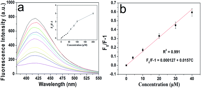 |
| | Fig. 4 Fluorescence quenching spectra (excited at 340 nm) in the presence of different concentrations of Co2+ (from top to bottom: 0 to 200 μM); the inset shows the plot of the relative fluorescence intensity (a); the linear relationship between the relative fluorescence intensity of CDs and Co2+ concentration (0 to 40 μM) (b). | |
Table 1 Comparison of different methods for the determination of Co2+
| Methods |
Materials |
Linear range/μM |
Detection limit/μM |
Ref. |
| Chemosensor |
Coumarin |
0–10 |
0.31 |
41 |
| Luminescence |
Lanthanide |
0.02–0.1 |
0.33 |
42 |
| Potentiometry |
p-(4-n-Butylphenylazo)calix[4]arene contained polymer membranes electrode |
9.2–100 |
4.00 |
43 |
| Colorimetry |
Silver nanoparticles |
5–100 |
7.00 |
44 |
| Colorimetry |
Peptide-modified gold nanoparticles |
2–10 |
2.00 |
45 |
| Fluorescence |
Carbon dots |
0–40 |
0.45 |
This work |
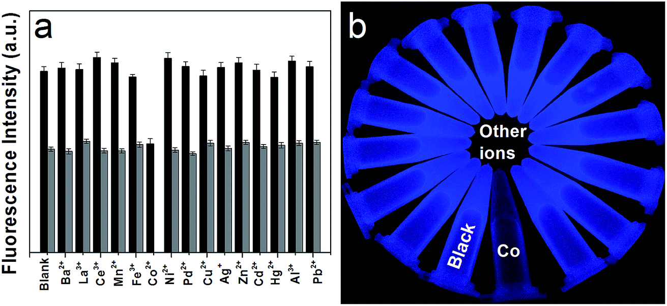
High selectivity is also an important factor for a sensitive probe. Therefore, the fluorescence responses of CDs in the presence of representative metal ions under the same conditions were studied, including Ba2+, La3+, Ce3+, Mn2+, Fe3+, Co2+, Ni2+, Pd2+, Cu2+, Ag+, Zn2+, Cd2+, Hg2+, Al3+, Pb2+. It can be seen that only Co2+ causes an obvious fluorescence quenching of CDs (Fig. 5a). Besides, it can also be visibly recognized in the presence of other ions under UV light (Fig. 5b). The competitive tests were also investigated by adding Co2+ to CDs in the presence of other metal ions (Fig. 5a). These show that other cations have no significant effect on the determination of Co2+.
 |
| | Fig. 5 Selectivity (black) and interference (gray) of CDs by Co2+ (10 μM) in the presence of different metal ion species (20 μM) (a); the color changes of the CD solution to different cations under 365 nm UV light irradiation (the concentration of all the metals was 100 μM) (b). | |
3.3 Possible mechanism for detecting Co2+ by CDs
It has been reported that carboxylic groups can bind with metal ions by metal–ligand interaction.46 Fig. 6a reveals the images of CDs in the presence of different concentrations of Co2+. The gradual color change (from transparent to brown) demonstrates that metal–ligand interaction between CDs and Co2+ may occur. Moreover, the UV-vis absorption spectra of CDs at 300 nm increase with the increasing concentration of Co2+, which is attributed to the metal–ligand interaction (Fig. 6b) (Scheme 1). On the other hand, it has been reported that thiol group can interact with metal ions through a heavy metal–thiol bond.47 As shown in Fig. 6c, the quenched fluorescence can be recovered after the addition of glutathione (GSH). The enhanced fluorescence may be due to the formation of Co2+–S between Co2+ and GSH which weakens the metal–ligand interaction between CDs and Co2+. In addition, other biothiols such as cysteine (Cys) and homocysteine can also recover the fluorescence of this system. The enhancement of the quenched fluorescence further demonstrates that this mechanism is feasible.
 |
| | Fig. 6 Photo images of CDs with different concentrations of Co2+ (0 to 200 μM) (a); UV-vis absorption spectra of CDs with various concentrations of Co2+ (0 to 200 μM) (b); fluorescence responses of CDs in the presence of Co2+ (100 μM) towards 100 μM Cys, Hcys and GSH (c). | |
3.4 Application of CDs for detecting Co2+ in real water samples
As the prepared CDs can be used as an effective fluorescent probe for Co2+, this method was used in environmental water samples to investigate its practical application. Lake water and tap water samples collected from our university campus were used as real water samples. The lake water samples were firstly filtered to remove suspended matter and then centrifuged for 15 min at 9000 rpm. No Co2+ was detected in lake water and tap water samples. Samples spiked with Co2+ concentration of 5, 10 and 20 μM were investigated through the standard addition method. The results shown in Table 2 reveal that the amounts of Co2+ are in great agreement with the added contents. The results indicate that the present method is capable of detecting Co2+ in practical samples.
Table 2 Determination of Co2+ in water samples (n = 3)
| Samples |
Added/μM |
Found/μM |
Recovery (%) |
RSD (%) |
| Lake water |
5 |
4.85 |
97.00 |
5.82 |
| 10 |
9.80 |
98.00 |
2.78 |
| 20 |
20.17 |
100.85 |
3.35 |
| Tap water |
5 |
5.14 |
102.80 |
1.58 |
| 10 |
10.10 |
101.00 |
2.93 |
| 20 |
20.05 |
100.25 |
1.75 |
4. Conclusions
In this study, CDs were synthesized through a simple method and served as an effective fluorescent probe for Co2+. Co2+ can induce the fluorescence quenching of CDs because of the metal–ligand interactions, which can be visibly seen through the color change of the solution (from colorless to brown). It shows a good linear response to Co2+ in the range of 0–40 μM with a detection limit of 0.45 μM. Besides, the probe also exhibits high selectivity and has been successfully applied to real water sample analyses. Therefore, this proposed method provides new feasibility for monitoring Co2+ in water samples without complicated sample pretreatment.
Acknowledgements
The work described in this manuscript was supported by the National Natural Science Foundation of China (No. 21374078, 51303132) and the Tianjin Research Program of Application Foundation and Advanced Technology (No. 15JCYBJC18100, 15JCTPJC59200).
References
- M. Gharehbaghi, F. Shemirani and M. D. Farahani, J. Hazard. Mater., 2009, 165, 1049–1055 CrossRef CAS PubMed.
- S. Okamoto and L. D. Eltis, Metallomics, 2011, 3, 963–970 RSC.
- A. F. Ngomsik, A. Bee, J. M. Siaugue, D. Talbot, V. Cabuil and G. Cote, J. Hazard. Mater., 2009, 166, 1043–1049 CrossRef CAS PubMed.
- R. Zein, R. Suhaili, F. Earnestly, Indrawati and E. Munaf, J. Hazard. Mater., 2010, 181, 52–56 CrossRef CAS PubMed.
- M. R. Awual, T. Yaita and Y. Okamoto, Sens. Actuators, B, 2014, 203, 71–80 CrossRef CAS.
- C. M. Sakamoto-Arnold and K. S. Johnson, Anal. Chem., 1987, 59, 1789–1794 CrossRef CAS.
- J. M. O. Souza and C. R. T. Tarley, Int. J. Environ. Anal. Chem., 2009, 89, 489–502 CrossRef CAS.
- L. Zi, Y. Huang, Z. Yan and S. Liao, J. Lumin., 2014, 148, 359–363 CrossRef CAS.
- L. P. Lin, M. C. Rong, F. Luo, D. M. Chen, Y. R. Wang and X. Chen, TrAC, Trends Anal. Chem., 2014, 54, 83–102 CrossRef CAS.
- Q. H. Yao, L. P. Lin, T. T. Zhao and X. Chen, J. Org. Chem., 2015, 27, 1523–1530 Search PubMed.
- Q. T. Huang, X. F. Lin, F. M. Li, W. Weng, L. P. Lin and S. R. Hu, J. Org. Chem., 2015, 27, 1604–1614 Search PubMed.
- V. Georgakilas, J. A. Perman, J. Tucek and R. Zboril, Chem. Rev., 2015, 115, 4744–4822 CrossRef CAS PubMed.
- J. Ju and W. Chen, Biosens. Bioelectron., 2014, 58, 219–225 CrossRef CAS PubMed.
- J. Ju, R. Z. Zhang, S. J. He and W. Chen, RSC Adv., 2014, 4, 52583–52589 RSC.
- J. Ju and W. Chen, Anal. Chem., 2015, 87, 1903–1910 CrossRef CAS PubMed.
- J. Ju, R. Z. Zhang and W. Chen, Sens. Actuators, B, 2016, 228, 66–73 CrossRef CAS.
- L. P. Lin, X. H. Song, Y. Y. Chen, M. C. Rong, Y. R. Wang, L. Zhao, T. T. Zhao and X. Chen, Anal. Chim. Acta, 2015, 891, 261–268 CrossRef CAS PubMed.
- L. P. Lin, X. H. Song, Y. Y. Chen, M. C. Rong, T. T. Zhao, Y. R. Wang, Y. Q. Jiang and X. Chen, Anal. Chim. Acta, 2015, 869, 89–95 CrossRef CAS PubMed.
- X. H. Gao, C. Du, Z. H. Zhuang and W. Chen, J. Mater. Chem. C, 2016 10.1039/C6TC02055K.
- A. Diac, M. Focsan, C. Socaci, A. M. Gabudean, C. Farcau, D. Maniu, E. Vasile, A. Terec, L. M. Veca and S. Astilean, RSC Adv., 2015, 5, 77662–77669 RSC.
- A. M. Craciun, A. Diac, M. Focsan, C. Socaci, K. Magyari, D. Maniu, I. Mihalache, L. M. Veca, S. Astilean and A. Terec, RSC Adv., 2016, 6, 56944–56951 RSC.
- Y. P. Sun, C. Shen, J. Wang and Y. Lu, RSC Adv., 2015, 5, 16368–16375 RSC.
- Y. P. Sun, C. Shen, J. Wang and Y. Lu, RSC Adv., 2015, 5, 20691 RSC.
- X. Wang, X. Shen, B. Z. Li, G. Y. Jiang, X. M. Zhou and H. J. Jiang, RSC Adv., 2016, 6, 18326–18332 RSC.
- R. Z. Zhang and W. Chen, Biosens. Bioelectron., 2014, 55, 83–90 CrossRef CAS PubMed.
- X. H. Gao, Y. Z. Lu, R. Z. Zhang, S. J. He, J. Ju, M. M. Liu, L. Li and W. Chen, J. Mater. Chem. C, 2015, 3, 2302–2309 RSC.
- Q. T. Huang, X. F. Lin, C. Q. Lin, Y. Zhang, S. R. Hu and C. Wei, RSC Adv., 2015, 5, 54102–54108 RSC.
- Q. T. Huang, H. Q. Zhang, S. R. Hu, F. M. Li, W. Weng, J. H. Chen, Q. X. Wang, Y. S. He, W. X. Zhang and X. X. Bao, Biosens. Bioelectron., 2014, 52, 277–280 CrossRef CAS PubMed.
- J. Y. Zhou, J. J. Gu, C. X. Tian, D. C. Jiang, Y. Chen and K. Xi, RSC Adv., 2016, 6, 39480–39483 RSC.
- A. S. Krishna, C. Radhakumary, S. S. Priya, R. M. Ramesan and K. Sreenivasan, RSC Adv., 2016, 6, 56313–56318 RSC.
- J. X. Shi, C. Lu, D. Yan and L. N. Ma, Biosens. Bioelectron., 2013, 45, 58–64 CrossRef CAS PubMed.
- C. L. Li, C. C. Huang, A. P. Periasamy, P. Roy, W. C. Wu, C. L. Hsu and H. T. Chang, RSC Adv., 2015, 5, 2285–2291 RSC.
- D. S. MacLean-McDavitt, J. D. Robertson and M. Jay, Pharm. Res., 2003, 20, 435–441 CrossRef CAS.
- Y. Zou, F. Y. Yan, L. F. Dai, Y. M. Luo, Y. Fu, N. Yang, J. Y. Wun and L. Chen, Carbon, 2014, 77, 1148–1156 CrossRef CAS.
- S. J. Zhu, Q. N. Meng, L. Wang, J. H. Zhang, Y. B. Song, H. Jin, K. Zhang, H. C. Sun, H. Y. Wang and B. Yang, Angew. Chem., Int. Ed., 2013, 52, 3953–3957 CrossRef CAS PubMed.
- Y. M. Guo, Z. Wang, H. W. Shao and X. Y. Jiang, Carbon, 2013, 52, 583–589 CrossRef CAS.
- F. Y. Yan, D. P. Kong, Y. M. Luo, Q. H. Ye, J. J. He, X. F. Guo and L. Chen, Microchim.Acta, 2016, 183, 1611–1618 CrossRef CAS.
- Y. Liu, Y. N. Liu, S. J. Park, Y. F. Zhang, T. Kim, S. Chae, M. Park and H. Y. Kim, J. Mater. Chem. A, 2015, 3, 17747–17754 CAS.
- W. Lu, X. Qin, S. Liu, G. Chang, Y. Zhang, Y. Luo, A. M. Asiri, A. O. Al-Youbi and X. Sun, Anal. Chem., 2012, 84, 5351–5357 CrossRef CAS PubMed.
- S. L. Hu, K. Y. Niu, J. Sun, J. Yang, N. Q. Zhao and X. W. Du, J. Mater. Chem., 2009, 19, 484–488 RSC.
- Z. D. Liu, W. L. Wang, H. J. Xu, L. Q. Sheng, S. S. Chen, D. Q. Huang and F. Sun, Inorg. Chem. Commun., 2015, 62, 19–23 CrossRef CAS.
- C. H. Zeng, X. T. Meng, S. S. Xu, L. J. Han, S. L. Zhong and M. Y. Jia, Sens. Actuators, B, 2015, 221, 127–135 CrossRef CAS.
- P. Kumar and Y. B. Shim, Talanta, 2009, 77, 1057–1062 CrossRef CAS PubMed.
- Y. Yao, D. M. Tian and H. B. Li, ACS Appl. Mater. Interfaces, 2010, 2, 684–690 CAS.
- M. Zhang, Y. Q. Liu and B. C. Ye, Analyst, 2012, 137, 601–607 RSC.
- Y. Kim, R. C. Johnson and J. T. Hupp, Nano Lett., 2001, 1, 165–167 CrossRef CAS.
- Z. Li, Y. Wang, Y. N. Ni and S. Kokot, Sens. Actuators, B, 2015, 207, 490–497 CrossRef CAS.
|
| This journal is © The Royal Society of Chemistry 2016 |
Click here to see how this site uses Cookies. View our privacy policy here.