Silymarin nanoparticles through emulsion solvent evaporation method for oral delivery with high antioxidant activities, bioavailability, and absorption in the liver

Xiuhua Zhao, Yiping Deng, Ying Zhang, Yuangang Zu*, Bolin Lian, Mingfang Wu, Chang Zu and Weiwei Wu
Key Laboratory of Forest Plant Ecology, Northeast Forestry University, Ministry of Education, Harbin 150040, Heilongjiang, China. E-mail: yuangangzu@163.com; Fax: +86-451-82102082; Tel: +86-451-82191517

Received 18th May 2016 , Accepted 16th September 2016

First published on 21st September 2016


Abstract

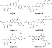
Silymarin (SM), a well-known hepatoprotective drug, is widely used to treat liver disorders. However, its application was seriously restricted because of its poor aqueous solubility. In this study, silymarin nanoparticles (SMNs) were prepared through emulsion solvent evaporation and freeze-drying methods to improve their solubility. SM nanoemulsion was optimized through a single-factor experiment. SMNs with MPS of 107.1 ± 13.7 nm were produced under optimum conditions. These SMNs were characterized by scanning electron microscopy, transmission electron microscopy, X-ray diffraction, differential scanning calorimetry, and thermal gravimetric analysis to determine their solvent residue, equilibrium solubility, dissolution rate, stability, antioxidant activity, bioavailability, and absorption in the liver. The results showed a smaller particle size and lower crystallinity in SMNs than those in raw SM. The solubility of SMNs was 4.87 times higher in simulated gastric fluid and 3.70 times higher in simulated intestinal fluid than that of raw silymarin. In addition, the residual amounts of chloroform and ethanol were separately less than the amount indicated by the international conference on harmonization limit for class II. The antioxidant activity after incubation with plasma in vitro of SMNs was higher than that of raw SM. Moreover, the oral bioavailability of SMNs was 3.66 times higher than that of raw SM. The silybin absorption of SMNs was also significantly higher than that of raw SM in the other organs, and very high silybin concentrations were observed in the liver with a long retention time. Overall, the results concluded that SMNs can be applied in oral delivery formulations and show potential application value for liver disease therapy.


Introduction

Silymarin (SM) is a polyphenolic component isolated from the fruits and seeds of the milk thistle plant Silybum marianum (family Asteraceae).1 SM is a mixture of several flavonolignans, including silybin A, silybin B, isosilybin A, isosilybin B, silychristin, and silydianin (structure shown in Fig. 1).2 Silybin constitutes the principal chemical component in the purified extract of SM.3 SM has been widely used to treat various acute and chronic liver toxicities, inflammation, fibrosis, and oxidative stress.4 However, the bulky multi-ring structure and poor oral bioavailability of SM lead to its low aqueous solubility; hence, the clinical role of SM is limited.4 To solve this problem, several approaches, such as formation of silybin–phospholipid complex,5 solid dispersions,6 encapsulated liposomes,7 and self-microemulsifying drug delivery system,8 have been used to improve the dissolution rate of SM. The dissolution and bioavailability of treated SM have been improved relative to raw SM. Nonetheless, many disadvantages were encountered in different SM preparations. For example, the silybin–phospholipid complex and the silybin solid dispersions exhibited a large particle size and poor stability. The encapsulated liposomes underwent a complex preparation method and demonstrated poor stability. Moreover, the self-microemulsifying drug delivery system presented inconvenience in storage and transportation.
image file: c6ra12896c-f1.tif
Fig. 1 Chemical structure of SM.

In accordance with the Noyes–Whitney equation,9 the dissolution rate of poorly water-soluble drugs can be increased by reducing the particle size to micro- or nano-scale and increasing the interfacial surface area. In the past decade, bottom-up techniques, such as supercritical fluid,10 liquid precipitation,11 and emulsion solvent evaporation (ESE) methods,12 have been widely applied to obtain ultrafine drug particles.

Nanoparticles with small size, spherical form, narrow distribution, and easy storage and transportation can be prepared by ESE.13 In the present study, we prepared silymarin nanoparticles (SMNs) by using the ESE technique, which had not been reported in the literature. First, we selected an appropriate organic phase with high solubility of raw SM. Afterward, the organic phase containing SM and water phase was mixed and fully emulsified. Subsequently, the emulsion was processed through high-pressure homogenization. Furthermore, the organic solvent was removed by nanoemulsion through evaporation. Finally, the processed SM was dried by lyophilization. Single-factor experiments were conducted to determine the optimum conditions. Eight main factors, i.e., the drug concentration, proportion of water to organic phase, concentration of poloxamer 188, homogenate speed and time, homogenization pressure (HP) and cycles, and proportion of SM to mannitol, were selected to optimize the prepared SMNs. The physical and chemical properties of the obtained SMN powder were characterized by scanning electron microscopy (SEM), transmission electron microscopy (TEM), X-ray diffraction (XRD), differential scanning calorimetry (DSC), thermal gravimetric analysis (TGA), dissolving capability test, stability test and solvent residual determination. The antioxidant activity of SM including 2,2-diphenyl-1-picrylhydrazyl (DPPH) free radical scavenging and reducing power was assessed. Moreover, the bioavailability and biodistribution of silybin in SMNs in different organs were assessed.

Materials and methods

Materials

Silymarin (containing 33% of silybin) was purchased from Jiamusi Sanjiang Silymarin Co., Ltd. (Heilongjiang, PR China). Poloxamer 188 was obtained from Hubei Hong Qi Chemical Co., Ltd. (Hubei, PR China). β-Glucuronidase, mannitol, methanol, chloroform, ethanol, acetic acid, n-hexane, hydrochloric acid, ether, NaOH, KH2PO4, DPPH, potassium ferricyanide, trichloroacetic acid, FeCl3, Na2HPO4·12H2O, NaH2PO4·2H2O, H3BO3, and NaB4O7·10H2O were purchased from Sigma-Aldrich Co., LLC. (St. Louis, MO, USA). Deionized water was prepared with Hitech-K flow water purification system (Hitech Instruments Co., Ltd., Shanghai, PR China).

Preparation of SMNs

SMNs were prepared through ESE followed by freeze-drying method. Chloroform containing 30% (v/v) ethanol was used as an organic phase of emulsion. In the organic phase, a certain amount of SM solution was slowly added dropwise to deionized water with a certain concentration of poloxamer 188 as surfactant. Afterward, the two liquid phases were mixed using a high-speed homogenizer (FSH-II, Jiangsu, China) at room temperature and homogenized using a nano-homogenizer machine (AH-100D, ATS Engineering Inc., Canada) to obtain the SM nanoemulsion. The organic phase was removed through rotary evaporation by using a R201BL rotary evaporator (SENCO, Shanghai, China) at an evaporation temperature of 45 °C. Subsequently, the SM nanosuspension was obtained. A certain amount of mannitol was added as lyoprotectant into the SM nanosuspension and mixed evenly. The mixture was pre-frozen at −40 °C for 2 h and then freeze dried at −50 °C for 48 h to obtain SMNs.

Single-factor experiments were performed to determine the optimum conditions. The following seven factors were selected to prepare the SM nanoemulsion through the preliminary experiment: drug concentration, proportion of water to organic phase, concentration of poloxamer 188, homogenate speed, time, HP, and cycles. One factor was changed at a time, whereas the other factors were kept constant. The range of drug concentration tested was 0.5–15 mg mL−1. The volume ratios of water to organic phase tested ranged from 6[thin space (1/6-em)]:[thin space (1/6-em)]1 to 38[thin space (1/6-em)]:[thin space (1/6-em)]1. The concentration of poloxamer 188 ranged from 0.5 mg mL−1 to 4.5 mg mL−1. Homogenate speed ranged from 3000 rpm to 11[thin space (1/6-em)]000 rpm, and homogenization time was set from 0.5 min to 15 min. HP was set from 300 bar to 1100 bar, and the frequency was set from 3 times to 11 times. All the specific parameters are listed in Table 1. During drug concentration testing, the proportion of water to organic phase was 22[thin space (1/6-em)]:[thin space (1/6-em)]1, the concentration of poloxamer 188 was 2.5 mg mL−1, the homogenate speed was 7000 rpm, the homogenization time was 7 min, the homogenization pressure was 700 bar, and the homogenate cycle was seven times. The same method as above was applied to study the other factors.

Table 1 Factors and levels of the single-factor experiments
Levels Factor
(A) (B) (C) (D) (E) (F) (G) (H)
Drug concentration (mg mL−1) Proportion of water to organic phase (v/v) Concentration of poloxamer 188 (mg mL−1) Homogenate speed (rpm) Homogenate time (min) Homogenization pressure (bar) Homogenization cycles (times) Proportion of silymarin to mannitol (m/m)
1 0.5 6[thin space (1/6-em)]:[thin space (1/6-em)]1 0.5 3000 0.5 300 3 1[thin space (1/6-em)]:[thin space (1/6-em)]1
2 1 10[thin space (1/6-em)]:[thin space (1/6-em)]1 1 4000 1 400 4 1[thin space (1/6-em)]:[thin space (1/6-em)]3
3 3 14[thin space (1/6-em)]:[thin space (1/6-em)]1 1.5 5000 3 500 5 1[thin space (1/6-em)]:[thin space (1/6-em)]5
4 5 18[thin space (1/6-em)]:[thin space (1/6-em)]1 2 6000 5 600 6 1[thin space (1/6-em)]:[thin space (1/6-em)]7
5 7 22[thin space (1/6-em)]:[thin space (1/6-em)]1 2.5 7000 7 700 7 1[thin space (1/6-em)]:[thin space (1/6-em)]9
6 9 26[thin space (1/6-em)]:[thin space (1/6-em)]1 3 8000 9 800 8 1[thin space (1/6-em)]:[thin space (1/6-em)]11
7 11 30[thin space (1/6-em)]:[thin space (1/6-em)]1 3.5 9000 11 900 9 1[thin space (1/6-em)]:[thin space (1/6-em)]13
8 13 34[thin space (1/6-em)]:[thin space (1/6-em)]1 4 10[thin space (1/6-em)]000 13 1000 10 1[thin space (1/6-em)]:[thin space (1/6-em)]15
9 15 38[thin space (1/6-em)]:[thin space (1/6-em)]1 4.5 11[thin space (1/6-em)]000 15 1100 11 1[thin space (1/6-em)]:[thin space (1/6-em)]17


Mannitol significantly affected the mean particle size (MPS) in SMNs through preliminary experiments. A certain proportion of mannitol was added in the SM nanosuspension during the lyophilization step. The proportion of SM to mannitol (mass/mass) ranged from 1[thin space (1/6-em)]:[thin space (1/6-em)]1 to 1[thin space (1/6-em)]:[thin space (1/6-em)]17. All the levels are also listed in Table 1.

Characterization of SMNs

Surface morphology of silymarin. The morphology of raw silymarin and SMNs was examined through SEM (Quanta 200, FEI, The Netherlands) and TEM (Hitachi, H-7650, Tokyo, Japan). The suitable amount of raw SM powder was mounted on the surface of an aluminum stub by using a carbon tape. SMNs dispersed in pure water were dropped on a silver paper, and then the blow-dried silver paper was fixed on a SEM stub by using a double-sided adhesive tape. Before analysis, the samples were sputter coated with gold under an argon atmosphere. The raw SM was detected at 12.5 kV with the spot size was 4.5 nm and the SMNs were detected at 20 kV with the spot size was 5 nm. TEM was used to detect the morphology of a single SMN. The SMN samples were mounted on a microgrid carbon polymer supported on a copper grid by placing a few drops of SMN aqueous solution, followed by drying under ambient conditions, all in an air glovebox. The samples were then transferred to a microscope under exclusion of air by a special vacuum-transfer sample holder. The sample was detected at 100 kV and magnified 30[thin space (1/6-em)]000 times.
MPS analysis. The MPS activity of the obtained nanoparticles was detected by laser light scattering by using a ZetaPALS analyzer (Brookhaven Instruments, Holtsville, NY, USA). The obtained samples of SM nanoemulsion and nanosuspension were analyzed directly. SMN powder samples were prepared by dispersion in deionized water under an ultrasonic bath. Every measurement was repeated thrice.
XRD analysis. XRD analyses were performed to evaluate the changes in crystal structure. Mannitol, poloxamer 188, raw SM, SMNs, physical mixture of SM, poloxamer 188, and mannitol at the same mass ratios as SMNs were detected using a Cu target tube at 30 mA and 40 kV with an X-ray diffractometer (Philips, X'pert-Pro, Amsterdam, The Netherlands) with a rotating anode. The XRD patterns were obtained in the 2θ range of 5–50° by using a 0.02° step and 5° min−1 scan speed.
DSC analysis. To confirm the decreased crystallinity SMNs, DSC (TA Instruments, DSC 204, Woodland, CA, USA) was examined for mannitol, poloxamer 188, raw SM, SMNs, physical mixture of SM, poloxamer 188, and mannitol at the same mass ratios as SMNs. Up to 5 mg of each sample was investigated at a temperature heating rate of 5 °C min−1 from 25 °C to 300 °C by using a differential scanning calorimeter.
HPLC condition. The drug concentration was determined using a Waters HPLC (Waters Corporation, Milford, MA, USA) containing a pump (Waters 1525 binary) and UV detector (Waters 2478 Tunable Absorbance Detector) equipped with the DIKMA Diamonsil C18 column (5 μm, 4.6 mm × 150 mm). The integrator system is Breeze 2. The mobile phase, which consisted of 48% methyl alcohol, 51% deionized water, and 1% acetic acid (v/v), was delivered at 1 mL min−1. The samples were detected at 287 nm. The main components of SM were silybin and isosilybin with a peak time of 13.9 and 16.1 min, respectively. Thus, the concentration of SM was the total concentration of silybin and isosilybin. The experiment was conducted in triplicate.
Equilibrium solubility and dissolution rate test. The dissolution rates of raw SM and SMNs were compared using an USP apparatus (II) paddle method. Simulated gastric fluid (SGF) comprised 5 mL of 37% hydrochloric acid and 1000 mL of deionized water,14 and simulated intestinal fluid (SIF) consisted of 6.8 g L−1 KH2PO4, which was adjusted to pH 6.8 by NaOH.15 SGF and SIF were used as the dissolution medium. The stirring speed was set at 100 rpm at a solution temperature of 37.0 ± 0.5 °C. Raw SM (50 mg) and SMNs (containing 50 mg of SM) were added to 100 mL of the dissolution medium and then dissolved for 48 h. Afterward, the samples (1 mL) were withdrawn and centrifuged at 7000 rpm for 8 min. The supernatant (10 μL) was directly injected into the HPLC system and then assayed for SM concentration. The experiment was conducted in triplicate.

The dissolution study of raw SM and SMNs was performed by paddle method. The stirring speed was 100 rpm and kept at 37.0 ± 0.5 °C temperature. SGF and SIF were used as the dissolution medium. Raw SM (13 mg) and SMNs (containing 13 mg SM) were immersed in 200 mL of the dissolution medium. Subsequently, 5 mL of the release medium was collected after 1, 3, 5, 10, 15, 30, and 60 min and then centrifuged at 8000 rpm for 5 min. The mixture was immediately supplemented with the same volume of the dissolution medium. The filtrate samples were directly injected into the HPLC system, and then the SM concentration was assayed.

Stability of SMNs in SGF and SIF. The stability of SMNs in SGF and SIF was detected by laser light scattering by using a ZetaPALS analyzer. SMNs (containing 3.25 mg raw SM) were dispersed in 50 mL SGF or SIF. Then, 2.5 mL of SMNs dispersion was analyzed directly after 1, 3, 5, 10, 15, 30 and 60 min. Every measurement was repeated thrice.

Antioxidant activity after incubation with plasma

SM is a powerful antioxidant herbal drug, which can protect biological systems against oxidative stress. This study was conducted to evaluate the in vitro free radical scavenging activity and antioxidant properties of SM. The antioxidant properties of SM after incubation with plasma were evaluated by two methods, namely, DPPH radical scavenging assay and measurement of reducing power.

Raw SM (20 mg) and SMNs (containing 20 mg SM) were dispersed in 20 mL of SGF. The stirring speed was set at 100 rpm at a solution temperature of 37.0 ± 0.5 °C. After 0.5 h, 228 mg K2HPO4·3H2O were separately added in the solution, which was adjusted to pH 6.8 with NaOH,16 and the mixed solution was considered as SIF system. Meanwhile, 20 mL of SGF was treated in the same way as blank solution. At 1 h, the suspension of each sample was centrifuged for 10 min at 10[thin space (1/6-em)]000 rpm. The supernatant was obtained and diluted into different SM concentrations (1–0.05 mg mL−1) by blank solution. 0.2 mL different concentrations of raw SM and SMNs solution and blank solution were added in 1.8 mL rat plasma to incubate. After incubation at 37.0 ± 0.5 °C for 30 min, the plasma samples were ready for testing.

The scavenging activity of DPPH radicals of SM was described by the paper of Asghar.17 The inhibitory effect of different concentrations of raw SM and SMNs on DPPH was measured by spectrophotometry. Up to 0.5 mL of each plasma sample was added to 0.5 mL of 10 mg L−1 DPPH–ethanol solution. After mixing, all the solutions were incubated in the dark for 30 min, centrifuged for 3 min at 10[thin space (1/6-em)]000 rpm, and the absorbance was measured at 517 nm. The blank sample was the supernatant of blank plasma solution and ethanol mixture, and the control sample was the supernatant of blank plasma solution and 10 mg L−1 DPPH–ethanol solution mixture. The experiment was repeated thrice. IC50 represents the level at which 50% of the radicals were scavenged by test samples. The percentage of scavenging activity was calculated as follows:

DPPH radical scavenging activity (%) = [(Abscontrol − Abssample)/(Abscontrol)] × 100
where Abscontrol is the total absorbance of the centrifugated control sample, and Abssample is the total absorbance of centrifugated DPPH radicals and samples.

The reducing power of the samples was determined using the method of Oyaizu (1986) with some modifications.18 Up to 0.5 mL of each plasma sample was mixed with 0.5 mL of 0.2 M phosphate buffer (pH 6.6) and 0.5 mL of 1% potassium ferricyanide. After incubation at 50 °C for 20 min, 0.5 mL of 10% trichloroacetic acid was added to the mixture followed by centrifugation at 3000 rpm for 10 min. Up to 1 mL of the supernatant was mixed with 1 mL of deionized water and 0.2 mL of 0.1% ferric chloride, and the absorbance of the resultant solution was read at 700 nm after 10 min. The experiment was repeated thrice.

Bioavailability study

All animal experiments were performed in compliance with relevant laws and institutional guidelines following the approval of the ethics committee of the Harbin Medical University (Harbin, China). Animals were housed under standard conditions of temperature, humidity and light with food and water provided freely and allowed to acclimatize in the laboratory for at least 1 week prior to the experiment. Six male Sprague-Dawley rats (weight 200–250 g) were used in this study. The rats were randomly divided into two groups, each with three animals. Prior to all experiments, the animals were fasted overnight with free access to water. Two groups of rats were correspondingly given with raw SM and SMNs by gavage at a dose of 30 mg kg−1. Blood samples were collected from the orbital venous sinus into heparinized tubes at 0, 10, 20, 30, 60, 120, 240, 360, 480, 600, and 720 min after oral administration. The samples were immediately centrifuged at 3000 rpm for 10 min, and the supernatant plasma was stored at −20 °C until further extraction and analysis.

Frozen samples, after being thawed at room temperature, were treated as follows. Each 200 μL plasma sample was combined with 200 μL of PBS (pH 5.0) and 50 μL of β-glucuronidase solution (enzyme activity is equivalent to 15 μm). After vortexing for 3 min, the samples were placed in a water bath at 37 °C for 16 h. Up to 1 mL of borate buffer solution (pH 8.5) was added to the sample and vibrated for 30 s, then mixed with 2 mL of ether for extraction procedure centrifuged at 8000 rpm for 10 min. Eventually, 2 mL of the supernatant was transferred and evaporated by water bath at 45 °C. The residue was redissolved by 100 μL mobile phase and centrifuged at 8000 rpm for 10 min. Afterward, 10 μL of the supernatant was injected for HPLC analysis. The oral bioavailability of the samples was represented by the area under the plasma concentration–time curve (AUC).

Biodistribution study

A total of 18 male Sprague-Dawley rats (weight 200–250 g) were randomly divided into two groups, each with nine animals. Before administration, the animals were starved overnight with free access to water. Two groups of rats were correspondingly given with raw SM and SMNs by gavage at a dose of 30 mg kg−1, and they were killed after 0.5, 1, 2, 4, 8, and 12 h. The heart, liver, spleen, lung, kidney, and brain were collected immediately and placed in ice-cold 0.1 M phosphate buffer (pH 7.4) and then dried with tissue paper. Tissue samples were frozen at −20 °C until analysis.

The tissue samples were weighed accurately and homogenized using a glass tissue homogenizer after addition of physiologic saline. The samples were immediately centrifuged at 3000 rpm for 10 min, and 200 μL supernatant tissue homogenates were collected. The tissue homogenates were processed similarly as the plasma samples and analyzed by HPLC.

Results and discussion

Optimization study

The optimum conditions were determined using a single-factor test. The production efficiency (production under the condition of unit volume and energy consumption) and particle size played a key role in selecting the optimum conditions. During microemulsion optimization, the maximum production efficiency and particle size less than 300 nm were selected as the optimum conditions. The MPS was also chosen as indicator during the optimization of nanoemulsion and cryoprotectant. During preliminary experiments, the following factors, namely, the concentration of SM, volume ratio of water to organic phase, concentration of surfactants, homogenate speed and time, homogenization pressure and cycles, and proportion of SM to mannitol exhibited an effect on the production efficiency and particle size (Fig. 2), and detailed in the ESI.
image file: c6ra12896c-f2.tif
Fig. 2 Effect of each parameter on the MPS of SMNs.

In accordance with the above results of single-factor experiments, the optimum conditions were set as follows: 15 mg mL−1 SM, 6[thin space (1/6-em)]:[thin space (1/6-em)]1 volume ratio of water to organic phase, 0.5 mg mL−1 poloxamer 188, 3000 rpm of homogenate speed for 0.5 min, a homogenization pressure of 600 bar for six times, and 1[thin space (1/6-em)]:[thin space (1/6-em)]7 proportion of SM to mannitol. The experiment was repeated thrice under the optimum conditions, and SMNs with MPS of 107.1 ± 13.7 nm were obtained. The subsequent characteristics of the optimum sample were all obtained under these conditions.

The drug purity of SMNs was 3% studied by HPLC, and the HPLC figure of silybin and isosilybin is shown in Fig. S1. As shown in Fig. S2, the limits of residual ethanol and chloroform were less than the ICH limit of 5000 ppm (0.5%) and 60 ppm (0.006%) for solvents, respectively. Therefore, the SMNs met the ICH requirements and were suitable for pharmaceutical use.

Characterization of SMNs

Morphology and particle size. The morphology of the samples is shown in Fig. 3. The raw SM appeared as irregular blocks, with a particle size of 1 μm to 200 μm (Fig. 3a). The SMNs were nearly spherical and exhibited a smaller particle size than raw SM (Fig. 3b). The TEM image of SMNs is shown in Fig. 3c; the SMNs were nearly spherical with a particle size of about 100 nm. This finding was consistent with the result of the SEM image shown in Fig. 3b. Fig. 4 shows the normal distribution curve of SM nanoemulsion, SM nanosuspension, and aqueous dispersion of SMNs under optimum condition. Their MPS values were 45.9, 103.5, and 107.1 nm, correspondingly, and the particle-size distributions are 33.56–63.84 nm, 11.24–266.21 nm, and 9.39–273.15 nm, respectively. The possible reason for the increasing MPS is the particle agglomeration during the freeze-drying process.
image file: c6ra12896c-f3.tif
Fig. 3 Morphology of samples: (a) SEM result of raw SM (1000×); (b) SEM result of SMNs (70[thin space (1/6-em)]000×); (c) TEM result of SMNs (30[thin space (1/6-em)]000×).

image file: c6ra12896c-f4.tif
Fig. 4 Normal distribution curve of samples: (a) SM nanoemulsion under optimum condition; (b) SM nanosuspension under optimum condition; (c) SMNs.
Physical structure characterization. XRD and DSC analyses were performed to evaluate the occurrence of eventual structural changes at the crystal level. Fig. 5 shows the XRD results of raw SM, SMNs, poloxamer 188, mannitol and physical mixture of SM, poloxamer 188, and mannitol at the same mass ratio. The diffraction angles of the characteristic peaks of raw SM (Fig. 5a) were 14.583°, 19.626°, 24.449°, and 22.312°. The angles of poloxamer 188 (Fig. 5d) were 19.063° and 23.216°, and the angles of mannitol (Fig. 5e) were 23.444°, 18.860°, 14.718°, and 21.218°. Mannitol has three polymorphic crystalline forms α, β and δ. Compared with the data in the previous literature,19 it can be concluded that raw mannitol is β-form. All the characteristic peaks of raw SM, poloxamer 188, and mannitol can be found in the spectrogram of the physical mixture (Fig. 5c), which showed that the crystal form of raw SM did not change after physical mixing. The characteristic peaks of SMNs (Fig. 5b) were 9.631°, 20.307°, 21.093°, and 25.206°, which is consistent with δ-form mannitol. These suggested that mannitol in SMNs was polymorphic δ-form from β during the lyophilization step, and the SM in SMNs had transformed into amorphous form. The DSC curves of samples are displayed in Fig. 6. As shown in Fig. 6a, the curve of raw SM showed two endothermic peaks. The peaks at 143 °C and 256 °C were close to the melting point of the SM crystal. The peak at 56 °C was the melting point of the poloxamer 188 crystal, as shown in Fig. 6d. The curve of raw β-form mannitol showed an endothermic peak at 170 °C, as shown in Fig. 6e. In Fig. 6b, the peak of SMNs at 166 °C is attributed to δ-form mannitol. Raw SM showed two endothermic peaks at 143 °C and 256 °C, and the peak disappeared in the thermogram of SMNs. This evidence confirmed that SM in SMNs exhibited amorphous form, and this finding was in accordance with the XRD results. In many studies, the amorphous form can enhance dissolution. Hence, SMNs presented a higher dissolution rate than raw SM.
image file: c6ra12896c-f5.tif
Fig. 5 XRD patterns of raw SM, SMNs, poloxamer 188, mannitol, physical mixture of SM, and poloxamer 188 and mannitol. (a) Raw SM; (b) SMNs; (c) physical mixture of SM, poloxamer 188 and mannitol; (d) poloxamer 188; and (e) mannitol.

image file: c6ra12896c-f6.tif
Fig. 6 DSC curves of raw SM, SMNs, poloxamer 188, mannitol, physical mixture of silymarin, and poloxamer 188 and mannitol. (a) Raw SM; (b) SMNs; (c) physical mixture of SM, poloxamer 188 and mannitol; (d) poloxamer 188; and (e) mannitol.

The results of TGA (Fig. S3) showed that the smaller particle size of SMNs indicated a higher specific surface and physical mixture, leading to easier vaporization and a faster thermal decomposition rate than raw SM.

Equilibrium solubility and dissolution rate. The equilibrium solubility of raw SM and SMNs is shown in Fig. 7a. The terminal solubility of raw SM and SMNs was 28.01 μg mL−1 and 62.66 μg mL−1 in SGF and 29.50 μg mL−1 and 52.13 μg mL−1 in SIF, respectively. The equilibrium solubility of SMNs was increased 2.23 times in SGF and 1.76 times in SIF than that of raw SM. The aqueous dispersion of raw SM and SMNs is shown in Fig. 7b. In this photograph, raw SM cannot be completely dispersed in deionized water, and freeze-dried powder can disperse well in the solution at the same concentration of 5 mg mL−1 observable with a transparent opalescence and uniform state.
image file: c6ra12896c-f7.tif
Fig. 7 Solubility diagrams. (a) Results of equilibrium solubility of raw SM and SMNs; (b) photograph of the aqueous dispersion of raw SM at the concentration of 5 mg mL−1 (left), SMNs at the concentration of 5 mg mL−1 (right).

The dissolution profiles of raw SM and SMNs in two different dissolution media are shown in Fig. 8. At the beginning, SMNs showed a more rapid dissolution rate and solubility than raw SM in SGF. Approximately 7.41% and 86.88% of the drug were dissolved in 1 min from the raw SM and SMNs, respectively. As shown in Fig. 8a, 18.94% of the SM and 92.33% of the SMNs dissolved within 60 min. Thus, the dissolution rate of SMNs was 4.87 times higher than that of the raw drug at 60 min dissolution. Fig. 8b shows the dissolution profiles of raw SM and SMNs in SIF. At 1 min, 6.12% of the raw drug and 60.31% of the SMNs dissolved. Up to 20.95% of the raw SM and 77.66% of the SMNs dissolved within 60 min. Hence, the dissolution rate of SMNs within 60 min exhibited 3.70 times faster than that of the raw drug in SIF. In accordance with the Noyes–Whitney equation, the drug dissolution rate is in linear relationship with the surface area exposed to the dissolution medium.20 The increased dissolution rate and solubility of SMNs can be mainly ascribed to the greater surface area because of the large reduction in particle size. The amorphous form of the SMNs also contributed to the increased dissolution rate.21 Hence, the amorphous SMNs exhibited a higher dissolution rate and solubility than the raw SM. In addition, the SMNs can exert their antioxidant property efficiently because of their higher dissolution rate and solubility than the raw SM.


image file: c6ra12896c-f8.tif
Fig. 8 Dissolution profiles of raw SM and SMNs in (a) SGF and (b) SIF.

The stability results of SMNs in SGF and SIF were shown in Fig. 9. At 1 min, the MPS of SMNs was 111.3 nm in SGF and 89.2 nm in SIF. The MPS decreased quickly within 10 min and then reached the steady state. This phenomenon can be attributed to the nanoparticles dissolving into molecular, which is also agreed with the results of the dissolution rate.


image file: c6ra12896c-f9.tif
Fig. 9 Stability result of SMNs in SGF and SIF.

In vitro antioxidant activity of SM after incubation with plasma

The proton-radical scavenging action has been known as an important mechanism of antioxidation. As shown in Fig. 10, raw SM exerted significant scavenging effects on the DPPH radicals by decreasing the absorbance at 517 nm and increasing the effects with the increasing concentrations in the range of 0.005–0.1 mg mL−1. The IC50 value of SMNs on DPPH radical scavenging assay was found to be 0.052 mg mL−1 compared with 0.097 mg mL−1 of raw SM. To reach a similar extent of the DPPH-scavenging effect, the concentration required for raw SM was significantly higher than that required for SMNs. The results showed that SMNs achieved a higher DPPH-scavenging activity than raw SM. SMNs can exert their antioxidant property efficiently because of their higher dissolution rate and solubility than the raw SM. SMNs demonstrated a proton-donating ability and can serve as free radical inhibitors or scavengers, thereby potentially acting as primary antioxidants.
image file: c6ra12896c-f10.tif
Fig. 10 Scavenging effects of raw SM and SMNs on DPPH radical.

In the reducing power assay, the presence of reductants (antioxidants) in the samples would result in the reduction of the Fe3+/ferricyanide complex to its ferrous form.22 The amount of Fe2+ complex can then be monitored by measuring the formation of Perl's Prussian blue at 700 nm. Fig. 11 shows the dose–response curves for the reducing powers of the raw SM and SMNs. The reducing powers of all drugs also increased with the increase of their concentrations. These results revealed that the SMs were the electron donors and can react with free radicals, converting them to more stable products and terminating the radical chain reaction. From a comparison of the absorbance at 700 nm, the reducing powers of SMNs were found to be significantly more pronounced than raw SM. The maximum absorbance of SMNs in plasma at a dosage of 0.1 mg mL−1 showed that SMNs exhibited high reducing power values of 0.138, and raw SM only exhibited a low reducing power value of 0.067. In addition, the results of reducing power are consistent with the findings of DPPH radical-scavenging activity. Hence, the SMNs could be a promising antioxidant effect after absorbed into plasma and distributed to the organ.


image file: c6ra12896c-f11.tif
Fig. 11 Reducing power of raw SM and SMNs.

In vivo bioavailability of SMNs in rats

The blood concentration–time curves of raw SM and SMNs suspension after oral administration in rats are shown in Fig. 12. The absorption of the SMN group was significantly faster than that of the raw SM group in vivo. The silybin concentration in rat plasma of the SMNs group was also higher than that of the raw SM. The silybin concentration in rat plasma of the SMNs and the raw SM group reached the maximum of 2.49 and 0.63 μg mL−1 after 20 min of taking drugs, respectively. By comparing the corresponding AUC values of the two groups, the results demonstrated that the AUC values of the SMNs were 3.66 times higher than those of the raw SM. This result can be attributed to the particle-size reduction. Particle size reduction can help improve the efficiency of SMN absorption, which was beneficial to improve the bioavailability of SM. Thus, the oral bioavailability of the SMNs was improved significantly compared with the raw SM. The significant enhancement of oral bioavailability is also in accordance with the result of the dissolution test.
image file: c6ra12896c-f12.tif
Fig. 12 Silybin concentration–time curves of raw SM and SMNs.

Biodistribution of silybin

The silybin concentration in the organs after administration of raw SM and SMNs was studied at 0.5, 1, 2, 4, 8, and 12 h. The biodistribution results at 0.5 h are shown in Fig. 13a. The silybin concentration of SMNs group in the other organs except brain was expected to be higher than that of the raw SM. The sequence of silybin concentration of SMNs group was presented as follows: liver, heart, kidney, spleen, lung, and brain. The sequence of biodistribution of the raw SM group was at the heart, liver, spleen, lung, kidney, and brain. At 1 h (Fig. 13b), the liver and kidneys of SMNs group exhibited a relatively high drug concentration, and the concentration in the other organs significantly decreased. At 4 h (Fig. 13d), the liver showed a relatively high drug concentration as before with a value of 1.37 μg g−1. After 12 h (Fig. 13f), the concentration in the liver was the highest with a concentration of 0.21 μg g−1, which was 10 times higher than that of the raw SM. This result can be attributed to the high plasma absorption of silybin in SMNs with high dissolution rate and bioavailability. The high bioavailability SMNs can keep the drug concentration in the liver at a high level with a long retention time. SM is a well-known hepatoprotective drug and a powerful antioxidant, and the SMNs with high liver distribution are important for enhancing the therapeutic effect of SM. Thus, the obtained SMNs were concentrated in the liver, which is beneficial for liver disease therapy.
image file: c6ra12896c-f13.tif
Fig. 13 Silybin concentration of raw SM and SMNs in the organs: (a) 0.5 h; (b) 1 h; (c) 2 h; (d) 4 h; (e) 8 h; and (f) 12 h.

Conclusions

In this work, SMNs were successfully prepared by emulsion solvent evaporation, followed by freeze-drying method. The optimal conditions were set as follows: 15 mg mL−1 SM, 6[thin space (1/6-em)]:[thin space (1/6-em)]1 volume ratio of water to organic phase, 0.5 mg mL−1 poloxamer 188, 3000 rpm homogenate speed for 0.5 min, a homogenization pressure of 600 bar for six times, and proportion of SM to mannitol of 1[thin space (1/6-em)]:[thin space (1/6-em)]7. Under optimum conditions, SMNs with an MPS of 107.1 ± 13.7 nm were obtained. SMNs were nearly ellipsoid with uniform particle-size distribution. The analysis results indicated that the prepared SMNs were less crystalline. The residual amount of ethanol and chloroform was 0.0017% and 0.0010%, respectively, which were less than the ICH limit for class II. The antioxidant activity after incubation with plasma in vitro of SMNs was higher than that of raw SM. The oral bioavailability of silybin in SMNs group was improved significantly compared with the raw SM. Furthermore, the absorption of silybin in SMNs was significantly higher than that of raw SM in the organs, and very high silybin concentrations were observed in the liver with a long retention time. Therefore, the SMNs prepared by emulsion solvent evaporation may offer a great potential value to become a new oral SM formulation.

Acknowledgements

The authors are grateful for the precious comments and careful corrections made by anonymous reviewers. The authors would also like to acknowledge the financial support from the National Key Technology R&D Program (2012BAD21B0501), the National Natural Science Foundation of China (No. 21473023).

References

  1. G. Hahn, H. D. Lehmann, M. Kürten, H. Uebel and G. Vogel, On the pharmacology and toxicology of silymarin, an antihepatotoxic active principle from Silybum marianum (L.) Gaertn, Arzneim. Forsch., 1968, 18(6), 698–704 CAS.
  2. W. C. Hsu, L. T. Ng, T. H. Wu, L. T. Lin, F. L. Yen and C. C. Lin, Characteristics and antioxidant activities of silymarin nanoparticles, J. Nanosci. Nanotechnol., 2012, 12(3), 2022–2027 CrossRef CAS PubMed.
  3. F. Kvasnička, B. BíBa, R. ŠevčíK, M. Voldřich and J. Krátká, Analysis of the active components of silymarin, J. Chromatogr. A, 2003, 990(1–2), 239–245 CrossRef.
  4. G. Boigk, L. Stroedter, H. Herbst, J. Waldschmidt, E. O. Riecken and D. Schuppan, Silymarin retards collagen accumulation in early and advanced biliary fibrosis secondary to complete bile duct obliteration in rats, Hepatology, 1997, 26(3), 643–649 CrossRef CAS PubMed.
  5. Y. Song, J. Zhuang, J. Guo, Y. Xiao and Q. Ping, Preparation and properties of a silybin–phospholipid complex, Die Pharmazie, 2008, 63(1), 35–42 CAS.
  6. N. Sun, X. Wei, B. Wu, C. Jian, L. Yi and W. Wei, Enhanced dissolution of silymarin/polyvinylpyrrolidone solid dispersion pellets prepared by a one-step fluid-bed coating technique, Powder Technol., 2008, 182(1), 72–80 CrossRef CAS.
  7. M. S. El-Samaligy, N. N. Afifi and E. A. Mahmoud, Increasing bioavailability of silymarin using a buccal liposomal delivery system: preparation and experimental design investigation, Int. J. Pharmacol., 2006, 308(s 1–2), 140–148 CrossRef CAS PubMed.
  8. W. Wei, W. Yang and Q. Li, Enhanced bioavailability of silymarin by self-microemulsifying drug delivery system, Eur. J. Pharm. Biopharm., 2006, 63(3), 288–294 CrossRef PubMed.
  9. A. Dokoumetzidis, V. Papadopoulou and P. Macheras, Analysis of dissolution data using modified versions of Noyes–Whitney equation and the Weibull function, Pharm. Res., 2006, 23(2), 256–261 CAS.
  10. S. K. Kumar and K. P. Johnston, Modelling the solubility of solids in supercritical fluids with density as the independent variable, J. Supercrit. Fluids, 1988, 1(1), 15–22 CrossRef CAS.
  11. R. N. Jones, R. J. Rando, H. W. Glindmeyer, T. A. Foster, J. M. Hughes and C. E. O'Neil, et al. Abnormal lung function in polyurethane foam producers. Weak relationship to toluene diisocyanate exposures, Am. Rev. Respir. Dis., 1992, 146(4), 871–877 CrossRef CAS PubMed.
  12. P. B. O'Donnell and J. W. Mcginity, Preparation of microspheres by the solvent evaporation technique, Adv. Drug Delivery Rev., 1997, 28(1), 25–42 CrossRef.
  13. G. Cavallaro, E. F. Craparo, C. Sardo, G. Lamberti, A. A. Barba and A. Dalmoro, PHEA-PLA biocompatible nanoparticles by technique of solvent evaporation from multiple emulsions, Int. J. Pharm., 2015, 495(2), 719–727 CrossRef CAS PubMed.
  14. J. Hu, W. K. Ng, Y. Dong, S. Shen and R. B. H. Tan, Continuous and scalable process for water-redispersible nanoformulation of poorly aqueous soluble APIs by antisolvent precipitation and spray-drying, Int. J. Pharm., 2011, 404(1–2), 198–204 CrossRef CAS PubMed.
  15. E. K. Wasan, K. Bartlett, P. Gershkovich, O. Sivak, B. Banno and W. Zhao, et al. Development and characterization of oral lipid-based amphotericin B formulations with enhanced drug solubility, stability and antifungal activity in rats infected with Aspergillus fumigatus or Candida albicans, Int. J. Pharm., 2009, 372(1–2), 76–84 CrossRef CAS PubMed.
  16. C. Cheng, P. C. Wu, H. Y. Lee and K. Y. Hsu, Development and validation of an in vitroin vivo correlation (IVIVC) model for propranolol hydrochloride extended-release matrix formulations, J. Food Drug Anal., 2014, 22(2), 257–263 CrossRef CAS.
  17. Z. Asghar and Z. Masood, Evaluation of antioxidant properties of silymarin and its potential to inhibit peroxyl radicals in vitro, Pak. J. Pharm. Sci., 2008, 21(3), 249–254 CAS.
  18. M. Oyaizu, Antioxidative activities of browning products of glucosamine fractionated by organic solvent and thin-layer chromatography, J. Jpn. Soc. Food Sci. Technol., 1988, 35(11), 771–775 CrossRef CAS.
  19. T. Yoshinari, R. T. Forbes, P. York and Y. Kawashima, The improved compaction properties of mannitol after a moisture-induced polymorphic transition, Int. J. Pharm., 2003, 258(1–2), 121–131 CrossRef CAS PubMed.
  20. Y. Dong, W. K. Ng, J. Hu, S. Shen and R. B. H. Tan, A continuous and highly effective static mixing process for antisolvent precipitation of nanoparticles of poorly water-soluble drugs, Int. J. Pharm., 2010, 386(1–2), 256–261 CrossRef CAS PubMed.
  21. J. L. Italia, M. M. Yahya, D. Singh and M. N. V. R. Kumar, Biodegradable nanoparticles improve oral bioavailability of amphotericin B and show reduced nephrotoxicity compared to intravenous Fungizone, Pharm. Res., 2009, 26(6), 1324–1331 CrossRef CAS PubMed.
  22. Y. C. Chung, S. J. Chen, C. K. Hsu, C. T. Chang and S. T. Chou, Studies on the antioxidative activity of Graptopetalum paraguayense E. Walther, Food Chem., 2005, 91(3), 419–424 CrossRef CAS.

Footnote

Electronic supplementary information (ESI) available. See DOI: 10.1039/c6ra12896c

This journal is © The Royal Society of Chemistry 2016
Click here to see how this site uses Cookies. View our privacy policy here.