A marine algal polyphenol, dieckol, attenuates blood glucose levels by Akt pathway in alloxan induced hyperglycemia zebrafish model

Eun-A Kima, Seung-Hong Leeb, Ji-Hyeok Leea, Nalae Kanga, Jae-Young Oha, Seun-heuic, Ginnae Ahnd, Seok Chun Koe, Shanura P. Fernandoa, Seo-Young Kima, Sun-Joo Parkf, Young-Tae Kimg and You-Jin Jeon*a
aDepartment of Marine Life Sciences, Jeju National University, Jeju 690-756, Republic of Korea. E-mail: youjinj@jejunu.ac.kr; Fax: +82 64 756 3493; Tel: +82 64 754 3475
bDivision of Food Bioscience, Konkuk University, Chungju, Chungbuk 380-701, Republic of Korea
cDepartment of Pharmacology, School of Medicine, Ajou University, Suwon 443-749, Republic of Korea
dDepartment of Marine Bio-Food Sciences, Chonnam National University, Yeosu 550-749, Republic of Korea
eMarine-Integrated Bionics Research Center, Pukyoung National University, Busan 608-737, Republic of Korea
fDepartment of Chemistry, Pukyoung National University, Busan 608-737, Republic of Korea
gDepartment of Food Science and Biotechnology, Kunsan National University, Busan 608-701, Republic of Korea

Received 16th May 2016 , Accepted 4th July 2016

First published on 4th August 2016


Abstract

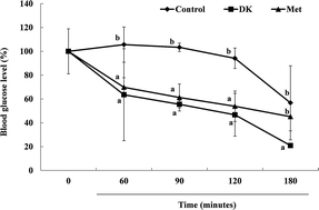
Recently, zebrafish (Danio rerio) have been used as a powerful model for human disease. The anti-diabetes activity of the anti-diabetic compound, dieckol (DK), one of the marine algal polyphenols isolated from Ecklonia cava, was confirmed in the zebrafish model. The zebrafish were divided to four groups, the normal (alloxan-untreated), alloxan-induced diabetic zebrafish without (control) and with DK, as well as those treated with metformin, a commercial drug. The blood glucose levels of the DK group of the hyperglycemic zebrafish at 90 min were decreased more than 3.3 times compared with the control group. Furthermore, reduced glucose-6-phosphate and phosphoenolpyruvate carboxykinase were observed with the treatments of DK and metformin in the liver tissues, and there was increased phosphorylation of protein kinase B (Akt) in the muscle tissue in the zebrafish model. Akt activation was involved in mediating the effect of DK on glucose transport activation and insulin sensitivity. These results prove that DK exerts a strong anti-diabetic effect by improving blood glucose regulation, hepatic glucose metabolic regulation and Akt up-regulation in alloxan induced hyperglycemic zebrafish.


1. Introduction

Diabetes mellitus, a disease of known significance to world health, is now taking its place as one of the main diseases of humans in the 21st century.1 Diabetes is characterized by either insufficient insulin production or by cellular resistance to insulin. When the pancreas is destroyed, there are high levels of glucose in the body, which in turn, damages many of the body's systems.2 The synthetic drugs used for the treatment of diabetes have several side effects, such as blindness, kidney disease, nerve damage, heart disease, amputation of feet, and stroke.3 Accordingly, recent natural products, especially those derived from herbs, have been in the limelight for use as drugs to treat diabetes because of their lower toxicity and fewer side effects than synthetic ones.4

A few reports have shown seaweed, such as fucoidan derived from Undaria pinnatifida, and fucoxanthin of Hijikia fusiformis, Sargassum polycystum, Ascophyllum nodosum, and Ecklonia cava, as having potential anti-diabetes activity.4–8

It is well known that polyphenols of E. cava (Ecklonia cava) possess various anti-diabetes, antioxidant, anti-inflammation, antibacterial and anti-cancer activities,4,9–13 and dieckol (DK) is one of the major compounds among the polyphenols (phloroglucinol, dieckol, 6,6′-bieckol, 7-phloroekol, phlorofucofuroeckol-A) of E. cava.14,15 Previous studies have also shown that DK imparted regulatory effects on a type 1 diabetes mellitus model by controlling postprandial hyperglycemia and type 2 diabetes in the db/db mouse model.4,16–18

Zebrafish have a number of features that make them an attractive research tool. The fundamental advantages are a considerable amount of genetic and organ systems that are similar to human systems.19,20 Moreover, the translucent body of zebrafish embryos facilitates the nonintrusive visualization of organs and biological processes.21 Therefore, powerful resources of zebrafish can be increasingly used to model human diseases, particularly common cardiovascular disorders, kidney disorders and chronic diseases of metabolism, such as obesity and diabetes.19,21–23

A previous study reported that alloxan and streptozotocin have been used to induce experimental diabetes in zebrafish.24,25 In this study therefore, we confirm that DK has anti-diabetic efficacy by regulating blood and organs, liver and muscle, in the hyperglycemic zebrafish model.

2. Materials and methods

2.1. Chemicals

Alloxan, D-(+)-glucose, metformin and 2-phenoxy ethanol, were purchased from Sigma-Aldrich (St. Louis, MO, USA). Antibodies for p-AMPK (Thr 172), AMPK and p-Akt (Ser 473), Akt were obtained from Cell Signaling Technology (Bedford, MA, USA), and anti-rabbit secondary antibodies were purchased from Santa Cruz Biotechnology (Santa Cruz, CA, USA). All other chemicals and reagents were of the purest grade available.

2.2. Isolation of dieckol from E. cava

Dieckol (DK) was isolated and purified from the marine brown alga E. cava, collected along the coast of Jeju Island, Korea according to the previously reported method by Lee et al.,26 and the structure was confirmed by 1H and 13C-NMR data, as well as mass spectroscopy. Dieckol: amorphous powder, 1H NMR (400 MHz, methanol-d4) d 6.14 (1H, s), 6.12 (1H, s), 6.09 (1H, d, 2.9 Hz), 6.05 (1H, d, 2.9 Hz), 6.05 (1H, d, 2.9 Hz), 5.99 (1H, d, 2.8 Hz), 5.95 (1H, d, 2.8 Hz), 5.92 (3H, m); 13C NMR (400 MHz, methanol-d4) d 161.8, 160.1, 157.8, 155.9, 154.5, 152.4, 147.3, 147.2, 147.1, 146.8, 144.4, 144.2, 143.4, 143.3, 138.6, 138.5, 126.5, 126.2, 125.6, 125.5, 124.9, 124.6, 124.5, 99.9, 99.7, 99.5, 99.4, 97.6, 96.2, 95.8, 95.7, 95.3; ESI-MS: [M − H] at m/z 741.12. The HPLC chromatogram and HPLC-ESI/MS spectrum are presented in Fig. 1.
image file: c6ra12724j-f1.tif
Fig. 1 HPLC chromatogram (A) and HPLC-ESI/MS spectrum (B) of dieckol isolated from Ecklonia cava.

2.3. Experimental animals

Adult zebrafish were obtained from a commercial dealer (Seoul Aquarium, Seoul, Korea) and 15 fish were kept in a 3.5 L acrylic tank under the following conditions: 28.5 ± 1 °C, fed twice a day (Tetra GmgH D-49304 Melle, made in Germany) with a 14/10 h light/dark cycle. The zebrafish experiment was approved by the Animal Care and Use Committee of Jeju National University. The zebrafish were exposed to 2 mg mL−1 of alloxan for 1 h and transferred to 1% glucose for 1 h, and then, the solution was changed to water for 1 h. The zebrafish were anesthetized using 2-phenoxy ethanol (1[thin space (1/6-em)]:[thin space (1/6-em)]1000 dilution). The zebrafish were divided into four groups: the alloxan-untreated (normal), alloxan-induced diabetic zebrafish without DK (control), those treated with DK, and those treated with metformin (Met) (1 μg per g body weight).

2.4. Measurement of blood glucose levels

The zebrafish were anesthetized using 2-phenoxy ethanol and removed from the water using Kim wipes, and then, blood samples were taken from the heart at 0, 60, 90, 120, 180 min. Approximately 1 μL of blood was rapidly transferred to a glucometer strip for the analyses (Roche Diagnostics Gmbh, Germany).

2.5. Hepatic glucose metabolism regulation activities

The liver was homogenized in PRO-PREPTM protein extraction solution (iNtBiotechnology, Korea) for 30 min on ice. The samples were then centrifuged at 12[thin space (1/6-em)]000 rpm at 4 °C for 15 min, and the supernatants were transferred to a fresh tube. The protein concentrations were determined by using the BCA™ protein assay kit. The glucose 6-phosphate, glucose sugar phosphorylated on carbon 6, was converted to glycogen or starch for storage, and the activity was determined using the glucose-6-phosphate assay kit (Bio Vision, CA95035, USA). Phosphoenolpyruvate carboxykinase (PEPCK) activity was monitored in the direction of oxaloacetate synthesis using the human PCK ELISA kit (CUSABIO, CSB-E09828h).

2.6. Western blot analysis

Western blot analysis was carried out using the protocol described by Ko et al.27 The p-AMPK (Thr 172), AMPK, p-Akt (Ser 473) and Akt were used at a 1[thin space (1/6-em)]:[thin space (1/6-em)]1000 dilution, and secondary antibodies were used at 1[thin space (1/6-em)]:[thin space (1/6-em)]3000 dilution.

2.7. Statistical analysis

The data are expressed as the mean ± standard error (S. E.), and the one-way ANOVA test (using SPSS 12 statistical software) was used to compare the mean values of each treatment. Values indicated with different letters are significantly different at p < 0.05, as analyzed by Duncan's multiple range tests (p < 0.05).

3. Results

3.1. Toxicity of alloxan in a zebrafish

The toxicity of alloxan, which selectively destroys insulin-producing cells in the pancreas, was evaluated for survival rates and blood glucose levels at multiple alloxan concentrations (2.0, 3.0, 4.0 mg mL−1) in zebrafish. The survival rates were recorded at 100%, 53.2% and 39.6% in the 2.0, 3.0 and 4.0 mg mL−1 of alloxan-treated groups, respectively (Fig. 2A), and the concentration of 3.0 mg mL−1 induced an extremely high blood glucose level (Fig. 2B). The alloxan concentration of 2.0 mg mL−1 caused moderate damage, with a higher survival and relatively medium blood glucose levels. Therefore, the concentration of alloxan used to treat zebrafish to induce the damage was 2.0 mg mL−1 for further experiments.
image file: c6ra12724j-f2.tif
Fig. 2 The administered concentrations of alloxan on survival rates (A), blood glucose levels (B) in zebrafish. The zebrafish were exposed to different alloxan concentrations (2.0, 3.0 and 4.0 mg mL−1) for 1 h and transferred to 1% glucose for 1 h. Values are mean ± SE, n = 12. a–cMeans not sharing a common letter are significantly different between groups (P < 0.05).

3.2. Effect of DK on blood glucose levels in hyperglycemic zebrafish

The zebrafish were exposed to 2 mg mL−1 of alloxan for 1 h and transferred to 1% glucose for 1 h, and then exposed to fresh water for 1 h; the zebrafish then presented frank hyperglycemia (blood glucose level > 400 mg dL−1). After the zebrafish were injected with saline, DK and Met (1 μg per g body weight), blood glucose levels were measured at 0, 60, 90, 120, 180 min. The blood glucose level was a significantly high ratio in the saline-treated groups, compared with the DK- and Met-injected groups during 180 min (Fig. 3). The Met- and DK-injected groups exhibited significant reductions in blood glucose levels. In particular, DK dramatically reduced the levels to 64%, 56%, 47% and 21% with the previously decided times, respectively. This result proved that DK more effectively reduced the blood glucose level, than the commercial drug, metformin. The time of 90 min was used in subsequent experiments, because of a significant difference in blood glucose levels among the groups.
image file: c6ra12724j-f3.tif
Fig. 3 The effect of the administering of DK and Met on the blood glucose level in hyperglycemic zebrafish. The hyperglycemic zebrafish induced with 2 mg mL−1 alloxan and transferred to 1% glucose during 1 h were injected with DK or Met (1 μg per g body weight). After the induction, the blood glucose levels of the zebrafish were measured at 0 min, 60 min, 90 min, 120 min, 180 min. Values are mean ± SE, n = 12. a–cMeans not sharing a common letter are significantly different between groups (P < 0.05).

3.3. Effect of DK on hepatic glucose metabolism regulation activities in hyperglycemic zebrafish

The G6P and PEPCK activities of the control group (alloxan-treated group) were significantly higher than the normal group (alloxan-untreated group) at 90 min, but DK reduced these activities. The DK-injected group showed almost similar G6P and PEPCK activities to those of the Met-injected group (Table 1).
Table 1 The effect of DK on hepatic glucose metabolism activities of glucose 6-phosphate (G6P) and phosphoenolpyruvate carboxykinase (PEPCK) in hyperglycemic zebrafish at 90 mina
  Normald Controle DKf Metg
a The hyperglycemic zebrafish induced with 2 mg mL−1 alloxan and transferred into 1% glucose during 1 h were injected with DK or Met (1 μg per g body weight). After 90 min, the liver tissues of zebrafish were collected to confirm G6P and PEPCK (n = 12). a–cMeans having different superscripts in the same row are significantly different between groups (P < 0.05). dAlloxan-untreated zebrafish. eAlloxan-treated zebrafish. fAlloxan-treated zebrafish supplemented with DK. gAlloxan-treated zebrafsih supplemented with metformin.
G6Pase (nmol μL−1) 6.2 ± 0.9a 9.4 ± 1.0c 8.3 ± 0.6b 9.0 ± 0.8b
PEPCK (ng mL−1) 14.0 ± 0.5a 17.0 ± 0.7c 15.9 ± 0.6b 15.7 ± 0.5b


3.4. Effect of DK on glucose uptake activity in hyperglycemic zebrafish

We investigated whether DK affected the protein expression of the glucose uptake signaling pathway, phosphorylation of AMPK and Akt in the muscle of hyperglycemic zebrafish. The expression of phosphorylation of Akt in the DK treated group was increased by more than the control. However, DK failed to stimulate phosphorylation of AMPK. Therefore, we confirmed that DK increased the glucose uptake via the Akt signaling pathway in zebrafish muscle (Fig. 4).
image file: c6ra12724j-f4.tif
Fig. 4 The effect of DK on the glucose uptake signaling pathway, phosphorylation of Akt and AMPK in the hyperglycemic zebrafish muscle tissues by western blot analysis. Normal: alloxan-untreated zebrafish. Control: alloxan-treated zebrafish, DK: alloxan-treated zebrafish supplemented with DK (1 μg per g body weight), Met: alloxan-treated zebrafish supplemented with metformin (1 μg per g body weight). a–cMeans not sharing a common letter are significantly different between groups (P < 0.05).

4. Discussion

Adipose and skeletal muscle tissues of zebrafish are physically arranged in a manner similar to their human counterparts. Therefore, zebrafish have been used as a model organism to study various human diseases.22 In this study, DK was identified for its anti-diabetic activity in the zebrafish model.

Metformin is a hypoglycemic agent that has been extensively used for the treatment of diabetes.28,29 The major function of metformin is to improve hyperglycemia and reduce hepatic glucose production, as well as improve peripheral insulin sensitivity. Metformin activates the AMP activated protein kinase via an LKB1-dependent mechanism. Our study demonstrates that Met is able to decrease blood glucose levels during 180 min, whereas, DK is able to more significantly decrease the blood glucose level than the Met. DK may also delay the absorption of dietary glucose, resulting in the suppression of an increase in blood glucose levels.

Diabetes appears as the increased glucocorticoid production via the activation of the intracellular glucocorticoid receptor (GR). Activated GR is related to an increase in the expression of PEPCK and G6P because of increasing hepatic gluconeogenesis and circulating glucose levels.30 A primary symptom of diabetes is the metabolism of abnormal hepatic glucose. An enhanced expression of hepatic PEPCK and G6P genes has been identified in most forms of diabetes, related to an increased hepatic glucose production.30,31 The action of mammalian PEPCK promoter is similar to zebrafish in response to glucagons, glucocorticoids, and insulin. Elo et al.32 showed that metformin, rosiglitazone, and glipizide suppressed the PEPCK expression. In the present study, hepatic glucose metabolism regulation activities of G6P and PEPCK in the liver indicate that DK, similar to Met, decreased blood glucose levels in hyperglycemic zebrafish. A previous report showed that polyphenols isolated from plants decreased hepatic gluconeogenesis.33 DK, a marine algal polyphenol derived from E. cava is shown to regulate hepatic glucose metabolism in this study, whereas, Waltner-Law et al.33 and Lee et al.4 demonstrated this in their in vitro and in vivo studies.

The skeletal muscle, which is the primary tissue for glucose uptake, plays a major role in the regulation of energy balance.34 Moreover, the PI3K-Akt signaling pathway and AMPK pathway play important roles in the glucose metabolism. The Akt pathway plays a central role in insulin-induced glucose uptake in the major organs, liver, muscle and adipose tissue. Moreover it is an important factor that mediates the biological function of insulin, such as insulin sensitivity.35,36 AMPK is another important mediator of glucose metabolism in skeletal muscles that regulates exercise-induced changes in glucose metabolism.37 Our evaluations suggest the stimulation of Akt phosphorylation in the course of glucose uptake in the DK treated group of hyperglycemic zebrafish, but no association with AMPK phosphorylation. In the previous studies, Guan18 and Lee & Jeon,15 have reported that DK can increase the glucose uptake through the phosphorylation of Akt in L6 myotube cells. Wortmannin, a PI3k inhibitor, was used to pretreat cells blocked by DK-stimulated phosphorylation of Akt, whereas pretreatment with compound C, an AMPK inhibitor, did not stimulate AMPK phosphorylation. These data proved that DK increased glucose uptake via regulation of the PI3k-Akt pathways in L6 myotube cells. Therefore, Akt activation was involved in mediating the intervention of DK on glucose transport activation and insulin sensitivity in in vitro and in vivo experimental models.

5. Conclusion

The results of this study demonstrated the anti-diabetic effect of DK in vivo in hyperglycemic zebrafish through their survival rate, blood glucose levels and hepatic glucose metabolism, as well as the glucose uptake in muscle tissues. Thus, we conclude that hyperglycemic zebrafish could be efficiently used in the wide range of evaluation of anti-diabetic activity.

Acknowledgements

This research was supported by Basic science Research Program through the National Research Foundation of Korea (NRF) funded by the Ministry of Education, Science and Technology (2012R1A1A2005479).

References

  1. P. Zimmet, K. Alberti and J. Shaw, Nature, 2001, 414, 782–787 CrossRef CAS PubMed.
  2. N. Aggarwal and P. Gupta, Asian Pac. J. Trop. Biomed., 2012, 2, S1254–S1260 CrossRef.
  3. J. Stang and M. Story, Guidelines for Adolescent Nutrition Services, Center for Leadership, Education and Training in Maternal and Child Nutrition, 2005 Search PubMed.
  4. S.-H. Lee, K.-H. Min, J.-S. Han, D.-H. Lee, D.-B. Park, W.-K. Jung, P.-J. Park, B.-T. Jeon, S.-K. Kim and Y.-J. Jeon, Food Chem. Toxicol., 2012, 50, 575–582 CrossRef CAS PubMed.
  5. K.-J. Kim, K.-Y. Yoon and B.-Y. Lee, Fitoterapia, 2012, 83, 1105–1109 CrossRef CAS PubMed.
  6. M. Motshakeri, M. Ebrahimi, Y. M. Goh, H. H. Othman, M. Hair-Bejo and S. Mohamed, Evid. Based Complement. Alternat. Med., 2014, 2014, 1–11 CrossRef PubMed.
  7. S. A. Kumar and L. Brown, Rev. Endocr. Metab. Disord., 2013, 14, 299–308 CrossRef CAS PubMed.
  8. J. Zhang, C. Tiller, J. Shen, C. Wang, G. S. Girouard, D. Dennis, C. J. Barrow, M. Miao and H. S. Ewart, Can. J. Physiol. Pharmacol., 2007, 85, 1116–1123 CrossRef CAS PubMed.
  9. G.-N. Ahn, K.-N. Kim, S.-H. Cha, C.-B. Song, J. Lee, M.-S. Heo, I.-K. Yeo, N.-H. Lee, Y.-H. Jee and J.-S. Kim, Eur. Food Res. Technol., 2007, 226, 71–79 CrossRef CAS.
  10. S.-Y. Kim, E.-A. Kim, M.-C. Kang, J.-H. Lee, H.-W. Yang, J.-S. Lee, T. I. Lim and Y.-J. Jeon, Algae, 2014, 29, 165 CrossRef.
  11. J.-H. Lee, J.-Y. Ko, J.-Y. Oh, E.-A. Kim, C.-Y. Kim and Y.-J. Jeon, Algae, 2015, 30, 313 CrossRef.
  12. S.-H. Eom, J. A. Santos, J.-H. Kim, W.-K. Jung, D.-H. Kim and Y.-M. Kim, Fish. Aquat. Sci., 2015, 18, 241–247 Search PubMed.
  13. Y. T. Kim, J.-H. Lee, J.-Y. Ko, J.-Y. Oh, W. Lee, C. H. Sok, J. T. Hong and Y.-J. Jeon, Korean J. Fish. Aquat. Sci., 2015, 48, 301–307 CrossRef CAS.
  14. S.-J. Heo, S.-C. Ko, S.-H. Cha, D.-H. Kang, H.-S. Park, Y.-U. Choi, D. Kim, W.-K. Jung and Y.-J. Jeon, Toxicol. In Vitro, 2009, 23, 1123–1130 CrossRef CAS PubMed.
  15. S.-H. Lee and Y.-J. Jeon, Fitoterapia, 2013, 86, 129–136 CrossRef CAS PubMed.
  16. S.-H. Lee, M.-H. Park, S.-J. Heo, S.-M. Kang, S.-C. Ko, J.-S. Han and Y.-J. Jeon, Food Chem. Toxicol., 2010, 48, 2633–2637 CrossRef CAS PubMed.
  17. M.-C. Kang, W. Wijesinghe, S.-H. Lee, S.-M. Kang, S.-C. Ko, X. Yang, N. Kang, B.-T. Jeon, J. Kim and D.-H. Lee, Food Chem. Toxicol., 2013, 53, 294–298 CrossRef CAS PubMed.
  18. J. Guan, Master Thesis of Master, Jeju National University, 2011 Search PubMed.
  19. L. I. Zon, Genome Res., 1999, 9, 99–100 CAS.
  20. J. Goldsmith and C. Jobin, BioMed Res. Int., 2012, 2012, 1–12 Search PubMed.
  21. A. Seth, D. L. Stemple and I. Barroso, Dis. Models & Mech., 2013, 6, 1080–1088 CAS.
  22. T. Oka, Y. Nishimura, L. Zang, M. Hirano, Y. Shimada, Z. Wang, N. Umemoto, J. Kuroyanagi, N. Nishimura and T. Tanaka, BMC Physiol., 2010, 10, 1 CrossRef PubMed.
  23. K. M. Capiotti, R. Antonioli, L. W. Kist, M. R. Bogo, C. D. Bonan and R. S. Da Silva, Comp. Biochem. Physiol. B, Biochem. Mol. Biol., 2014, 171, 58–65 CrossRef CAS PubMed.
  24. J. B. Moss, P. Koustubhan, M. Greenman, M. J. Parsons, I. Walter and L. G. Moss, Diabetes, 2009, 58, 1844–1851 CrossRef CAS PubMed.
  25. E. Shin, B. N. Hong and T. H. Kang, Afr. J. Pharm. Pharmacol., 2012, 6, 2922–2928 CrossRef CAS.
  26. J.-H. Lee, J.-Y. Ko, J.-Y. Oh, C.-Y. Kim, H.-J. Lee, J. Kim and Y.-J. Jeon, Food Chem., 2014, 158, 433–437 CrossRef CAS PubMed.
  27. S.-C. Ko, M. Lee, J.-H. Lee, S.-H. Lee, Y. Lim and Y.-J. Jeon, Environ. Toxicol. Pharmacol., 2013, 36, 1253–1260 CrossRef CAS PubMed.
  28. Y. D. Kim, K.-G. Park, Y.-S. Lee, Y.-Y. Park, D.-K. Kim, B. Nedumaran, W. G. Jang, W.-J. Cho, J. Ha and I.-K. Lee, Diabetes, 2008, 57, 306–314 CrossRef CAS PubMed.
  29. M. Zakikhani, R. Dowling, I. G. Fantus, N. Sonenberg and M. Pollak, Cancer Res., 2006, 66, 10269–10273 CrossRef CAS PubMed.
  30. B. Salani, C. Marini, A. Del Rio, S. Ravera, M. Massollo, A. M. Orengo, A. Amaro, M. Passalacqua, S. Maffioli and U. Pfeffer, Sci. Rep., 2013, 3, 1–8 Search PubMed.
  31. G. F. Davies, R. L. Khandelwal, L. Wu, B. H. Juurlink and W. J. Roesler, Biochem. Pharmacol., 2001, 62, 1071–1079 CrossRef CAS PubMed.
  32. B. Elo, C. Villano, D. Govorko and L. White, J. Mol. Endocrinol., 2007, 38, 433–440 CrossRef CAS PubMed.
  33. M. E. Waltner-Law, X. L. Wang, B. K. Law, R. K. Hall, M. Nawano and D. K. Granner, J. Biol. Chem., 2002, 277, 34933–34940 CrossRef CAS PubMed.
  34. U. Özcan, E. Yilmaz, L. Özcan, M. Furuhashi, E. Vaillancourt, R. O. Smith, C. Z. Görgün and G. S. Hotamisligil, Science, 2006, 313, 1137–1140 CrossRef PubMed.
  35. J. Zdychova and R. Komers, Physiol. Res., 2005, 54, 1 CAS.
  36. R. Mackenzie and B. Elliot, Diabetes, Metab. Syndr. Obes.: Targets Ther., 2014, 2014, 55–64 CrossRef PubMed.
  37. N. Musi, N. Fujii, M. F. Hirshman, I. Ekberg, S. Fröberg, O. Ljungqvist, A. Thorell and L. J. Goodyear, Diabetes, 2001, 50, 921–927 CrossRef CAS PubMed.

This journal is © The Royal Society of Chemistry 2016