Precise casting of biomorphic La0.9K0.1CoO3 catalysts derived from pinewood for diesel soot combustion

Zhaohui Liu, Xiaofei Wang, Teng Mao, Jinqiang Sima, Cairong Gong* and Guoliang Fan
School of Materials Science and Engineering, Tianjin University, Tianjin 300072, P. R. China. E-mail: gcr@tju.edu.cn

Received 16th May 2016 , Accepted 2nd August 2016

First published on 17th August 2016


Abstract

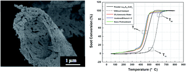
Porous biomorphic La0.9K0.1CoO3 catalysts were fabricated by the bio-templating method using tailored pinewood as the template and La0.9K0.1CoO3 sol as the precursor. The specimens were characterized by SEM, TEM, XRD, FT-IR, EDX, and N2-adsorption techniques. SEM and TEM results proved that the biomorphic cellular morphology of the initial pinewood structure was retained after heat treatment. The XRD and FT-IR results suggested a perovskite-type crystal structure for the microcellular catalysts. The generated catalysts possessed a relatively high surface area (147.97 m2 g−1) and porosity with a mean pore size of approximately 4–25 μm. The catalytic activity for the combustion of diesel soot was tested by a technique using a temperature-programmed reaction. The biomorphic La0.9K0.1CoO3 exhibited better catalytic activity for diesel soot combustion at low temperature than the La0.9K0.1CoO3 powders prepared from the same precursor sol due to enhanced contact efficiency between the soot and the catalysts, and the value of T10 was about 340 °C.


1 Introduction

Atmospheric pollution has drawn increasing attention due to its chronic threat to human health and ecological security.1,2 The wide application of diesel vehicles has undoubtedly deteriorated the situation due to the large amount of soot emissions, which has contributed a lot to particulate pollution. To efficiently reduce soot emissions from diesel vehicles many methods have been exerted, among which diesel particulate filters (DPF) are a promising method. An inevitable process in the application of DPF is regeneration, which has a great influence on the efficiency and lifetime of the DPF itself.3 The regeneration of DPF requires an incineration cleaning process of particulate matter, which is the primary ingredient of soot. Generally, the combustion temperature of soot is approximately 600 °C, whereas typical diesel engine exhaust temperatures fall within the 200–500 °C range. As a solution to this problem, oxidation catalysts have been introduced to lower the combustion temperature of soot and, consequently, to efficiently remove the emissions of particulate matter.4

Among the many oxidation catalysts, supported noble metal catalysts have been widely used for their excellent catalytic activity for catalytic soot combustion.5,6 However their widespread application is limited for a number of reasons such as high cost, deficiency in sulfur resistance and instability,7 while some substitute catalysts such as transition metal oxides,8,9 alkali metal oxides,10 perovskites11 and some cerium-based oxides12 are playing increasingly important roles on the grounds of their low cost and outstanding chemical and thermal stability. Among these, perovskite-type catalysts are regarded as potential substitutes for noble metal catalysts for soot removal due to their extraordinary structural characteristics and their capability to accommodate a wide range of substituting and doping elements to tailor their properties.13 In the past decades, perovskite-type oxides have been widely used in the field of catalytic soot removal from diesel exhausts.14–17 In particular manganese-based and cobalt-based perovskites have shown better catalytic activity for diesel soot combustion than other transition metal-based perovskites.17 It was reported that the La0.9K0.1CoO3 catalyst gave the highest catalytic activity for soot oxidation.18 However, the initial ignition temperature of soot over perovskite catalysts is relatively high.

As a complicated solid–solid–gas reaction, the contact between the catalysts and soot particles is a crucial issue that needs to be considered. The conventional catalysts normally have a low surface area and a smaller pore size owing to the high temperatures and long heat treatment time used in the preparation process, which limit the process of mass transfer of the reactant and restrict the amount of available active sites.19 Porous catalysts are introduced to solve this problem.

Templated synthesis is one of the promising methods to synthesize porous materials. Over the last decade, the manufacture of porous ceramics with controllable porosity by mimicking the cellular anatomy of bio-templates has attracted much interest. Organic structures available in nature are particularly used as biological templates for the preparation of porous materials with various microstructures, including fibrilla macroscopic structures,20 cellular structures,21 or complex micro/macropore structures,22 which have been applied to synthesize metal oxides,23–25 silicon carbides,26 zeolitic tissues27,28 and hierarchical ordered oxides29,30 to be further applied in many areas like filters, catalyst carriers or biomedical materials. However, reports about the fabrication of perovskite oxides from bio-templates have been rare.

Pinewood has been used as a bio-template to synthesize various materials such as hexagonal ferrites31 and magnesium oxides32 and the results were very attractive. Our paper demonstrates a simple and viable synthesis route to produce porous biomorphic La0.9K0.1CoO3 catalysts directly from pinewood impregnated with La0.9K0.1CoO3 sol. This kind of biomorphic porous Co-based catalyst exhibits high porosity, surface area and degree of order. This accounts for the good catalytic activity for diesel soot oxidation at low temperature, as the T10 of soot combustion is about 340 °C which is better than that of the other powder cobalt based perovskites reported previously.18,33 These results will be helpful to expand the potential application of porous materials in the area of soot removal from diesel exhausts.

2 Experimental Section

2.1 Synthesis of biomorphic La0.9K0.1CoO3

Pinewood pieces with dimensions of 30 × 25 × 10 mm3 were cut into transverse sections and dried at 110 °C for 24 h. They were first pretreated in a mixture of acetone/ethanol at 2[thin space (1/6-em)]:[thin space (1/6-em)]1 for 3 days to get rid of the redundant organic compounds for better infiltration later. Afterwards, they were washed with distilled water and dried at 60 °C for 3 days. As a comparison, the pinewood was also treated with 5% weak ammonia water for 6 hours, followed by the same treatment process as the first one. A sol corresponding to the final composition La0.9K0.1CoO3 was prepared by dissolving stoichiometric amounts of (0.033 mol) La(NO3)·6H2O (Tianjin Jiangtian Chemicals, Tianjin, China), (0.033 mol) Co(CH3COO)2·4H2O (Tianjin Jiangtian Chemicals, Tianjin, China), (0.0033 mol) CH3COOK (Tianjin Jiangtian Chemicals, Tianjin, China) and (0.067 mol) C6H12O6·H2O (Tianjin Jiangtian Chemicals, Tianjin, China) in 100 ml distilled water (31.1 wt%) for 10–15 min under stirring. Subsequently, the mixture was put into a water bath at 60 °C for 2 days to give the La0.9K0.1CoO3 sol.

The pinewood templates were immersed in a mixture of 20 ml La0.9K0.1CoO3 sol, 10 ml ethanol and 5 ml H2O, which served as the low viscosity precursor, for 3 days to make sure the precursor thoroughly infiltrated the templates. Then the immersed wood templates loading La0.9K0.1CoO3 sol were air-dried at 60 °C for 3 days, followed by heat treatment in the tube furnace under argon. The final process is listed as follows: first, the sample was heated to 120 °C at the rate of 3 °C min−1 followed by it remaining at 120 °C for 30 min. Afterwards, the temperature was increased to 400 °C with the heating rate of 2 °C min−1 and the samples were kept at 400 °C for 2 hours. Thirdly, at the rate of 10 °C min−1, the temperature was increased to 800 °C at which point the specimens were held at peak temperatures for 2 hours and naturally cooled to room temperature. Lastly, the obtained products were annealed at 800 °C in air flow for another 2 h to give porous La0.9K0.1CoO3 oxides.

2.2 Characterization of biomorphic La0.9K0.1CoO3

The crystal phases of the specimen were analyzed by an X-ray diffractometer (XRD; Rigaku, Tokyo, Japan), on which patterns were recorded for 2θ angles between 10° and 90° with increments of 0.02° and a counting time of 0.5 per s per step. The structures of the prepared catalysts were analyzed by Fourier transform infrared spectrometry (FT-IR; Nicolet 6700, US) with a 2 cm−1 resolution. The microstructures were observed by scanning electron microscopy (SEM; XL-30ESEM, Philips). The SEM was conducted using accelerating voltages from 0.5–30 kV, and 0.1 kV per step, and the specimens were cut into small pieces and stuck to the sample holder with a conducting resin and then coated with a gold layer to improve the images obtained. Energy dispersive X-ray spectroscopy (EDX, Genesis XM2, EDAX, USA) analysis was also performed to give the composition. The porosity was investigated by N2 adsorption measurements at 77 K using a NOVA 2000 gas sorption analyzer and the PSD (Pore size distribution) of samples were calculated using the Barrett–Joyner–Halenda (BJH) algorithm. Specific surface areas were calculated by the Brunauer–Emmett–Teller (BET) method.

2.3 The catalytic activity test

The catalytic activity of the catalysts towards soot combustion was tested by a TG-DSC (Mettler Toledo, USA). The specimens were placed in an alumina crucible, then were heated under an air flow (100 ml min−1) at a rate of 10 °C min−1 at a temperature range of 50 °C and 800 °C. The performance of the samples was evaluated using the values of T10, T50 and T90, which were defined as the temperatures at which 10%, 50% and 90% of carbon black was oxidized, respectively. The carbon black used in this research was purchased from Evonik Degussa and used in the test as a replacement for diesel soot due to its similar chemical components and specific surface area. The specimen was mixed with soot in a mass ratio of 20[thin space (1/6-em)]:[thin space (1/6-em)]1. As a comparison, the thermal analysis of the mixtures of soot with powder samples prepared from the La0.9K0.1CoO3-sol was also performed. Moreover, to ensure the same contact chance between air and carbon, inert SiO2 was added to carbon (20[thin space (1/6-em)]:[thin space (1/6-em)]1, w/w) for the non-catalyzed combustion experiments.

3 Results and discussion

3.1 Structural, morphological and textural properties

The biomorphic microstructures of the specimens heat-treated at 800 °C in argon flow are first investigated by scanning electron microscopy (SEM) and transmission electron microscopy (TEM), as shown in Fig. 1. The results reveal that a well-aligned porous framework has formed, which means that the specimens retained the pinewood's porous configuration faithfully, and the pretreatment has no obvious influence on the porous structure. The pore size of the specimen after heat treatment at 800 °C was approximately 4–25 μm while the thickness of the cell wall was approximately 1–2 μm. From the TEM results, the cell wall was composed of oxide nanoparticles, which can be further proved by XRD results shown in Fig. 3.
image file: c6ra12640e-f1.tif
Fig. 1 SEM and TEM micrographs of the specimens heat-treated at 800 °C in argon flow with three different pretreatments: (a) SEM image of the specimen after acetone/ethanol pretreatment, (b) SEM image of the specimen after ammonia water pretreatment, (c) SEM image of the specimen with no pretreatment, (d) TEM image of the specimen after acetone/ethanol pretreatment, (e) TEM image of the specimen after ammonia water pretreatment and (f) TEM image of the specimen with no pretreatment.

The fracture surfaces of the porous La0.9K0.1CoO3 with different pretreatments after annealing at 800 °C in air are shown in Fig. 2(c) and (d). For comparison, the SEM images of the pinewood and the pinewood loaded with the sol are also shown in Fig. 2(a) and (b), respectively. It is obvious that the mechanical properties of the pinewood templates decrease after annealing, and collapse has happened. This phenomenon can be attributed to the conversion of C element into CO2 during the annealing process in air, which remained in the process of heat treatment in argon flow. In other words, the departure of the support from the original C bio-template resulted in the decrease in strength of the material. Fortunately, the porous microstructures remained after annealing. The technological process can be specified as follows: the cell wall of the pinewood partially transformed into the C bio-template during the carbonization and released CO2 or some other organic gas, followed by the departure of C element through the process of annealing. Afterwards, the final structure formed, and the porous structure of the bio-template remained completely. The composition of the specimen after annealing was also investigated by energy dispersive X-ray spectroscopy (EDX). As shown in Fig. 2(e), the ratios of K, La and Co in the EDX measurements are about 1[thin space (1/6-em)]:[thin space (1/6-em)]9[thin space (1/6-em)]:[thin space (1/6-em)]13. Compared to the molar ratio, 1[thin space (1/6-em)]:[thin space (1/6-em)]9[thin space (1/6-em)]:[thin space (1/6-em)]10, the Co element exceeds this within a tolerable range. This is due to the presence of some impurity phases such as Co3O4 and the intrinsic error of EDX in the quantitative analysis. The peak without a label belongs to the Au element.


image file: c6ra12640e-f2.tif
Fig. 2 SEM micrographs of (a) the original pinewood, (b) the pinewood after impregnation, (c) porous La0.9K0.1CoO3 after annealing observed at a lower magnification, (d) porous La0.9K0.1CoO3 oxides at a fracture end observed at a higher magnification and (e) EDX analysis of the specimen after annealing.

In order to validate the crystal structure of the heat-treated specimen, the samples were subjected to X-ray diffraction (XRD) afterwards. Fig. 3 gives the powder XRD patterns of the heat-treated specimens formed in different atmospheres.


image file: c6ra12640e-f3.tif
Fig. 3 XRD patterns of the infiltrated specimens heat-treated at 800 °C in argon (a), and annealed at 800 °C in air (b).

According to these patterns, as for all the pinewood samples with a different pretreatment method, perovskite structures were formed after the heat treatment of the infiltrated specimens at 800 °C in flowing argon. However, other phases such as La2O2CO3 and Co3O4 co-exist, which was proved by the FT-IR results. This is due to the incomplete reaction of La0.9K0.1CoO3-sol at 800 °C in argon atmosphere and the lack of oxygen to combust the C element. The process of annealing can minimize the effects of these problems. After annealing in air atmosphere, the intensity of the diffraction lines associated with the perovskite phase becomes stronger. Although the small diffraction peaks from the Co3O4 phase, which makes up less than 6.57% (w/w) of the perovskite structure, remained in all specimens, the crystallinity is extraordinarily higher than that after the pyrolysis in argon. The XRD pattern of the biomorphic La0.9K0.1CoO3 catalyst annealed at 800 °C in flowing air is highly consistent with the JCPDS card: PDF 48-0123, and the peaks that appeared at 23.3°, 32.9°, 33.2°, 40.6°, 47.4°, and 58.8° are indexed to the (012), (100), (104), (202), (024), and (300) crystal face of the lanthanum cobalt phase, respectively. These results proved that the biomorphic oxides of La0.9K0.1CoO3 possessed ABO3 perovskite-type structures.

The FT-IR spectra of the infiltrated but un-heated specimens and the end biomorphic La0.9K0.1CoO3 catalyst are shown in Fig. 4. As for the infiltrated samples with different pretreatments, the absorption band at 3429 cm−1 in Fig. 4a is due to the stretching mode of O–H from hydroxyl and water; the peak absorption at 1628 cm−1 refers to the asymmetric stretching vibration of C[double bond, length as m-dash]O while the 1383 cm−1 band corresponds to the bending mode of C–H. The absorption band at 1059 cm−1 can be ascribed to the bending vibration of hydroxyl from a dextrose precursor. These spectra features indicate that the La0.9K0.1CoO3-sol has completely immersed in the pinewood template. The IR spectra of the biomorphic La0.9K0.1CoO3 catalyst are exhibited in Fig. 4b. It can be seen that a group of absorption bands at 665, 839 and 1066 cm−1 can be assigned to the vibrations of chemical bonds of LaCoO3.34,35 The IR spectrum of Co3O4 consists of an absorption peak at 578 cm−1, which corresponds to the XRD analysis. The appearance of the band at 1385 cm−1 represents the bending mode of C–H. Moreover, the strong absorption peak for C–H at 1479 cm−1 agrees well with the literature and the band at 3413 cm−1 refers to the stretching mode of OH in water. These spectra characteristics confirm that after annealing at 800 °C, the perovskite-type structures have come into being.


image file: c6ra12640e-f4.tif
Fig. 4 FT-IR spectra of the infiltrated specimens (a) and the biomorphic La0.9K0.1CoO3 catalyst after annealing at 800 °C in air (b).

From N2 adsorption measurements, the isotherm curves for the biomorphic products after annealing and the pore size distribution (PSD) are shown in Fig. 5. According to the curves at low relative pressure, the specimens with three different kinds of pretreatment all contain a large number of micropores. As for the curve of the middle relative pressure section, the gradual increase in the quantity indicates the abundant existence of mesopores or even macropores. We believe that the reason for the increase in the adsorption quantity is the occurrence of multilayer adsorption and the capillary condensation of mesopores and channels.


image file: c6ra12640e-f5.tif
Fig. 5 The results of the N2 adsorption measurements and the pore size distribution of the specimens with different pre-treatments after annealing: (a) acetone/ethanol = 2, (b) 5% ammonia water, (c) no pretreatment and (d) PSD results of the specimens.

The porosity and surface area of the original pinewood and samples sintered at 800 °C are shown in Table 1. The BET surface areas of the specimens after annealing are approximately 84 m2 g−1 and 147 m2 g−1 depending on whether the specimen was pre-treated, while their counterparts prior to the heat treatment are 0.148 m2 g−1 and approximately 1 m2 g−1, respectively. As compared in Table 1, the surface area increased a lot after pre-treatment while the average pore radius decreased. This may be due to the pores in the original pinewood being blocked by natural organic compounds such as cellulose and lignin, which were subsequently dissolved during the process of pre-treatment, resulting in numerous micropores and mesopores being unblocked and the average pore radius being lowered. Moreover, when the specimens were exposed to heat treatment and annealing, the residues of natural organic compounds were further pyrolysed, increasing both surface area and total pore volume. According to previous research, the BET surface area of traditional powdered La0.9K0.1CoO3 was 11.1 m2 g−1 using the citric acid-ligated method.18 It is obvious that the La0.9K0.1CoO3 derived from pinewood bio-templates shows a large enhancement and subsequently great potential for application in this area as a catalyst.

Table 1 Porosity of the specimensa
  Pretreatment type BET surface area (m2 g−1) Total pore volume (cm3 g−1) Average pore radius (nm)
a BET surface area, pore volume, and average pore diameter were calculated from nitrogen adsorption isotherms.
The pinewood prior to the heat treatment None 0.148 0.002 22.364
Acetone/ethanol = 2 1.217 0.001 1.559
5% ammonia water 0.931 0.002 1.578
The specimens after annealing None 84.09 0.050 1.193
Acetone/ethanol = 2 147.97 0.101 1.367
5% ammonia water 146.64 0.101 1.382


3.2 Catalytic performance for diesel soot combustion

In order to assess the activity of the biomorphic catalysts for diesel soot combustion, a soot and La0.9K0.1CoO3 mixture with a mass ratio of 1[thin space (1/6-em)]:[thin space (1/6-em)]20 was tested by TGA (Thermal Gravimetric Analysis). The combustion curves under tight contact are shown in Fig. 6 and the corresponding temperatures are listed in Table 2. Specimens with different pretreatments have similar characteristic temperatures, which suggests that the pretreatment has no obvious effect on the combustion temperature of soot. Compared with the mixture of soot and SiO2, T10, T50 and T90 of the biomorphic sample with the best performance decreased by 182 °C, 137 °C, 122 °C, respectively, suggesting that this perovskite has excellent catalytic activity for soot combustion. Moreover, upon comparing to the powder La0.9K0.1CoO3 (ref. 34) catalyst and some other cobalt-based perovskites,18 the T10 of the biomorphic La0.9K0.1CoO3 is much lower, which illustrates that this porous La0.9K0.1CoO3 possesses terrific catalytic activity at low temperature. The better catalytic activity at low temperature compared with the powder counterpart might come from the intrinsic advantages of the porous structure, which were derived from a pinewood bio-template in this case. It is well known that the contact condition between the catalysts and soot has much influence on the catalytic reactivity. The details of the related combustion temperatures of the different specimens are listed in the Table 2.
image file: c6ra12640e-f6.tif
Fig. 6 Thermal analysis results of different mixtures.
Table 2 Different combustion temperature of the specimens
Pretreatment type T10 (°C) T50 (°C) T90 (°C)
None pretreatment 342 450 493
Acetone/ethanol = 2 338 443 488
5% ammonia water 348 454 490
Powder La0.9K0.1CoO3 408 470 529
None catalyst 520 580 610


4 Conclusions

A biomorphic La0.9K0.1CoO3 perovskite-type oxide was prepared from a pinewood template by the bio-casting method and its catalytic properties towards diesel soot combustion were investigated. The porous oxides of La0.9K0.1CoO3 were proved by XRD to possess ABO3 perovskite-type structures while its pore size was approximately 4–25 μm. The result of TGA-DSC revealed that the T10 of this biomorphic catalyst towards soot combustion was lowered by approximately 59 °C, suggesting its relatively higher fraction of converted soot than powder La0.9K0.1CoO3 at the initial phase under low temperature. Further investigations on the simultaneous removal of NOx and diesel soot particulate over the obtained specimens are in progress.

Acknowledgements

This study was supported by the Natural Science Foundation of Tianjin (12JCQNJC05700).

References

  1. B. Brunekreef and S. T. Holgate, Lancet, 2002, 360, 1233–1242 CrossRef CAS.
  2. M. Kampa and E. Castanas, Environ. Pollut., 2008, 151, 362–367 CrossRef CAS PubMed.
  3. M. J. Kleeman, J. J. Schauer and G. R. Cass, Environ. Sci. Technol., 2000, 34, 1132–1142 CrossRef CAS.
  4. E. D. Banús, V. G. Milt, E. E. Miró and M. A. Ulla, Appl. Catal., B, 2013, 132–133, 479–486 CrossRef.
  5. J. Oi-Uchisawa, A. Obuchi, S. Wang, T. Nanba and A. Ohi, Appl. Catal., B, 2003, 43, 117–129 CrossRef CAS.
  6. M. V. Twigg, Appl. Catal., B, 2007, 70, 2–15 CrossRef CAS.
  7. R. Jiménez, R. Zamora, G. Pecchi, X. García and A. L. Gordon, Fuel Process. Technol., 2010, 91, 546–549 CrossRef.
  8. S. Waglöhner, D. Reichert, H. Bockhorn and S. Kureti, Chem. Ing. Tech., 2013, 85, 686–695 CrossRef.
  9. D. Reichert, T. Finke, N. Atanassova, H. Bockhorn and S. Kureti, Appl. Catal., B, 2008, 84, 803–812 CrossRef CAS.
  10. L. Castoldi, R. Matarrese, L. Lietti and P. Forzatti, Appl. Catal., B, 2009, 90, 278–285 CrossRef CAS.
  11. C. R. Gong, C. L. Song, Y. Q. Pei, G. Lv and G. L. Fan, Ind. Eng. Chem. Res., 2008, 47, 4374–4378 CrossRef CAS.
  12. G. Zhang, Z. Zhao, J. Xu, J. Zheng, J. Liu, G. Jiang, A. Duan and H. He, Appl. Catal., B, 2011, 107, 302–315 CrossRef CAS.
  13. H. Liu, P. Jin, Y.-M. Xue, C. Dong, X. Li, C.-C. Tang and X.-W. Du, Angew. Chem., 2015, 127, 7157–7160 CrossRef.
  14. J. Liu, Z. Zhao, C.-M. Xu and A.-J. Duan, Appl. Catal., B, 2008, 78, 61–72 CrossRef CAS.
  15. N. RUSSO, D. FINO, G. SARACCO and V. SPECCHIA, J. Catal., 2005, 229, 459–469 CrossRef CAS.
  16. A. Civera, M. Pavese, G. Saracco and V. Specchia, Catal. Today, 2003, 83, 199–211 CrossRef CAS.
  17. W. Y. Hernández, M. N. Tsampas, C. Zhao, A. Boreave, F. Bosselet and P. Vernoux, Catal. Today, 2015, 258, 525–534 CrossRef.
  18. W. Hong, L. Jian and Z. Zhen, et al., Catal. Today, 2012, 184(1), 288–300 CrossRef.
  19. R. Spinicci, M. Faticanti, P. Marini, S. De Rossi and P. Porta, J. Mol. Catal. A: Chem., 2003, 197, 147–155 CrossRef CAS.
  20. D. Fino, S. Bensaid and M. Piumetti, Appl. Catal., A, 2016, 509(5), 75–96 CrossRef CAS.
  21. J. Ma, Y. Ning, C. Gong, G. Xue and G. Fan, RSC Adv., 2015, 5, 53441–53447 RSC.
  22. Y. Shin, J. Liu, J. H. Chang, Z. Nie and G. J. Exarhos, Adv. Mater., 2001, 13, 728–732 CrossRef CAS.
  23. W. Zhang, D. Zhang, T. Fan, J. Ding, Q. Guo and H. Ogawa, Nanotechnology, 2006, 17, 840–844 CrossRef CAS.
  24. Z. Liu, T. Fan, W. Zhang and D. Zhang, Microporous Mesoporous Mater., 2005, 85, 82–88 CrossRef CAS.
  25. N. Adachi, M. Kuwahara, C. K. Sia and T. Ota, Materials, 2009, 2, 1923–1928 CrossRef CAS.
  26. Z. Liu, T. Fan, J. Gu, D. Zhang, X. Gong, Q. Guo and J. Xu, Mater. Trans., 2007, 48, 878–881 CrossRef CAS.
  27. T. Ota, M. Takahashi, T. Hibi, M. Ozawa, S. Suzuki and Y. Hikichi, J. Am. Ceram. Soc., 2005, 78, 3409–3411 CrossRef.
  28. A. Dong, Y. Wang, Y. Tang, N. Ren, Y. Zhang and Y. Yue, Adv. Mater., 2002, 14(12), 926–929 CrossRef CAS.
  29. A. Zampieri, H. Sieber, T. Selvam, G. T. P. Mabande, W. Schwieger, F. Scheffler, M. Scheffler and P. Greil, Adv. Mater., 2005, 17, 344–349 CrossRef CAS.
  30. X. Li, T. Fan, Z. Liu, J. Ding, Q. Guo and D. Zhang, J. Eur. Ceram. Soc., 2006, 26, 3657–3664 CrossRef CAS.
  31. Y. Pu, H. Zou and J. X. Wang, et al., Mater. Lett., 2011, 65(14), 2213–2215 CrossRef CAS.
  32. R. Q. Sun, X. Zhou and L. B. Sun, et al., Chem. J. Chin. Univ., 2007, 28(12), 2333–2337 CAS.
  33. N. Russo, S. Furfori, D. Fino, G. Saracco and V. Specchia, Appl. Catal., B, 2008, 83, 85–95 CrossRef CAS.
  34. H. Wang, Z. Zhao and P. Liang, et al., Catal. Lett., 2008, 124(1–2), 91–99 CrossRef CAS.
  35. N. A. Merino, B. P. Barbero and P. Grange, et al., J. Catal., 2005, 231(1), 232–244 CrossRef CAS.

Footnote

Electronic supplementary information (ESI) available. See DOI: 10.1039/c6ra12640e

This journal is © The Royal Society of Chemistry 2016
Click here to see how this site uses Cookies. View our privacy policy here.