Zhongping Yao*,
Yaqiong He,
Qixing Xia,
Han Wei and
Zhaohua Jiang
School of Chemistry and Chemical Engineering, Harbin Institute of Technology, Harbin 150001, PR China. E-mail: yaozhonging@hit.edu.cn; Tel: +86-451-86402805
First published on 23rd May 2016
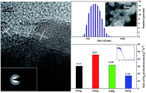
In this work, a series of Zn0.83Cd0.17S with high photocatalytic activity were hydrothermally synthesized and the effects of the hydrothermal temperature on the structural, chemical, morphological properties of the samples were investigated by X-ray diffraction (XRD), scanning electron microscopy (SEM), transmission electron microscopy (TEM), particle size analyzer and X-ray photoelectron spectroscopy (XPS), respectively. The photoabsorption properties were measured using a UV-vis diffused reflectance spectrophotometer and the photocatalytic activities of the samples for hydrogen production were evaluated under 300 W Xe lamp irradiation. The results show that the sub-micro sized spherical Zn0.83Cd0.17S particles are uniform and mainly composed of cubic zinc-blende phase. An increase of temperature improves the crystallinity of the samples and the ratio of Cd and Zn in the solid solution and consequently makes the absorption edges gradually shift monotonically to longer wavelengths. Also, the hydrothermal temperature influences the particle size and distribution of sulfide solid solution. The sample synthesized at 160 °C exhibits the best photocatalytic activity for H2 evolution with a hydrogen production rate of 45.97 mmol h−1 g−1 when the amount of the sample is 0.03 g in a 200 ml aqueous solution containing 0.35 M Na2S and 0.25 M Na2SO3.
A variety of methods have been established to synthesize ZnxCd1−xS solid solution recently, including solid state reactions,18 cation-exchange reactions,19 microwave syntheses,20 self-assembly approach,21 solvothermal method.22 Among the various fabrication methods, solvothermal synthesis was regarded as a promising route for its simplicity, low cost, high efficiency and good crystallization of the products.23,24 In the solvothermal process, different solvents have been reported to synthesize ZnxCd1−xS solid solution, such as dimethyl sulfoxide (DMSO),25 ethylene glycol,26 and ethanol.27 However, the above organic solvents are not better choices due to their toxicity and great harm to the environment. On the contrary, the hydrothermal synthesis with the water as the solvent has attracted more and more attention in these years.4
In our previous study, ZnxCd1−xS has been synthesized in the glycol solvent under 140 °C and among them, Zn0.83Cd0.17S showed the best photocatalytic hydrogen production activity with the rate of 4.02 mmol h−1 g−1.28,29 However, the samples presented the poor crystallization in composition and two absorption edges in UV-vis diffuse reflectance spectrum. In this paper, we selected water as solvent to synthesize a series of Zn0.83Cd0.17S photocatalysts. The effects of temperature on the structural, chemical, morphological and optical properties of the Zn0.83Cd0.17S solid solution were investigated in detail. Also, the photocatalytic activities of the samples for the hydrogen production from water splitting were evaluated, the results show that Zn0.83Cd0.17S photocatalysts synthesized at 160 °C in this work exhibits the best photocatalytic activity for H2 evolution with an initial rate of 45.97 mmol h−1 g−1, which is almost 11.4 times higher than that in the glycol in our previous work.30
:
1 were dissolved in 25 ml deionized water. After the complete dissolution, the mixture dripped to the thiourea aqueous solution (60 mmol thiourea dissolved in 25 ml deionized water) under continuous stirring and then the mixed solution was added into the Teflon-lined stainless steel autoclave. Before the hydrothermal reaction, the mixed solution was purged with N2 for 5 min in order to evacuate the air in the solution, and then the autoclave was sealed and maintained in an oven at different temperatures (120, 140, 160, 180 °C) for 12 h with an average heating rate of hydrothermal of 3.5 °C min−1 and cooled down to room temperature. After the hydrothermal reaction, the samples were centrifugally separated, rinsed with deionized water and washed with ethanol several times and dried at 70 °C for 12 h.
sin
θ = nλ, the decrease of 2θ means the decrease of sin
θ and accordingly the increase of interplanar crystal spacing (d). Due to the radii of Zn2+ (0.74 Å) is smaller than that of Cd2+ (0.97 Å), it is believed that Cd2+ insert into the ZnS lattice and formed the sulfide solid solution, which, meantime, leads to the increase of the d value and the lattice expansion of the materials. Besides, the increase of the d value also illustrates the increase of Cd content in the solid solution, which is bound to influence the photoabsorpion and photocatalytic property of the materials.
![]() | ||
| Fig. 1 XRD patterns of the samples synthesized at different temperatures for 12 h. (a) 120 °C (b) 140 °C (c) 160 °C and (d) 180 °C. | ||
Moreover, a small peak (★) appearing at 27.13° matches with JCPDS card number (89-2171) is indexed to ZnS, which means that the sample synthesized at 180 °C is composed of sulfide solid solution and zinc sulfide. This may be due to the quicker crystal nucleus formation rate of ZnS than that of sulfide solid solution under the higher temperature. Therefore, the sample synthesized at 180 °C is not homogeneous. Quite oppositely, the homogeneous sulfide solid solutions can be formed when the hydrothermal temperature is no more than 160 °C.
To further understand the properties of the samples, the elemental composition and valence states of the samples were probed by XPS. The spectra of the overall survey scan of the samples indicates the presence of Zn, Cd, and S, along with O and C (seen in ESI Fig. S2†). The presence of very small amounts of C and O2 in the spectrum is due to the carbon tape used for measurement and the gaseous molecules such as CO2 and H2O in the environment, respectively. Fig. 2 shows the high-resolution XPS spectra for Zn 2p, Cd 3d and S 2p of the samples synthesized at different temperatures. In the Zn 2p spectra, two peaks of Zn 2p3/2 and Zn 2p1/2 at approximately 1022.3 eV and 1045.4 eV (Fig. 2a) are attributed to the existence of Zn2+, which exactly matches the binding energy of Zn–S.33,34 The peaks of Cd 3d5/2 and Cd 3d3/2 (Fig. 2b) are centered at 405 eV and 411.8 eV, indicating the presence of the Cd–S bonding.35 The Zn 2p and Cd 3d peaks at binding energies are attributed to ZnxCd1−xS molecular environment, illustrating that the samples are the sulfide solid solution.22 The binding energies of S 2p3/2 and S 2p1/2 (Fig. 2c) are best fitted with two peaks at around 161.5 eV (monosulfide) and 162.5 eV (predominant disulfide anion).35,36 Moreover, the binding energies of Zn 2p and Cd 3d are decreased with the hydrothermal temperature increasing from 120 to 160 °C, and increased when the temperature is 180 °C. These slight changes are probably related to the increase of the content of Cd in the solid solution and the formation of the second phase ZnS in the materials. Meanwhile, a regular decrease of the binding energy in the XPS spectra of S 2p with the increase of temperature is observed.
![]() | ||
| Fig. 2 High-resolution XPS spectra of the samples synthesized at different temperatures: (a) Zn 2p, (b) Cd 3d and (c) S 2p. | ||
The morphology of the samples synthesized at different hydrothermal temperatures was investigated through SEM and TEM. ESI Fig. S3† and 3 are the SEM and TEM analysis of the samples synthesized at different temperatures, which shows that all the samples are uniform, spherical and sub-micro sized. TEM image in Fig. 3c presents that the synthesized sub-micrometer particles are about 180–200 nm in diameter and the HRTEM image in Fig. 3d shows clearly lattice fringes, suggesting a well-defined crystal structure. The fringes with lattice spacing of 0.322 and 0.198 nm correspond to the cubic (111) and (220) plane of the sulfide solid solution in XRD analysis. The associated selected electron diffraction (SAED; the inset in Fig. 3d) shows ring patterns, which is typical of polycrystalline material.
![]() | ||
| Fig. 3 SEM of the samples synthesized at (a) 140 °C, (b) 160 °C, TEM (c), HRTEM image and the associated SEAD pattern (d and its inset) of the sample synthesized at 140 °C for 12 h. | ||
| Number diameter (nm) | 120 °C | 140 °C | 160 °C | 180 °C |
|---|---|---|---|---|
| D10 | 105.707 | 220.194 | 105.709 | 43.821 |
| D50 | 164.182 | 255.002 | 164.183 | 58.771 |
| D90 | 255.004 | 824.992 | 255.002 | 122.42 |
| Span | 0.91 | 2.37 | 0.91 | 1.34 |
The width of particle size distributions was measured by span (span is determined by the equation: span = (D90 − D10)/D50) according to a British Standards. A smaller span value indicates a narrower particle size distribution and more uniform size. The span values of the samples synthesized at 120 °C and 160 °C are much lower than the other samples, which means that samples obtained at 120 °C and 160 °C are more homogeneous. Furthermore, the sample synthesized at 160 °C is a little more uniform than that at 120 °C and possesses an average pore diameter of 2–9 nm with the BET specific surface area of 47.36 m2 g−1 (seen in ESI Fig. S4†). Besides, there is a protuberance (◆) appeared in Fig. 4b, which is probably due to that the measured sample is not dispersed well during the preparation. In Fig. 4d, the formation of the tailing part marked (#) shows the severe agglomeration of the particles, which may be related to the following reasons: (1) the smallest size of the particles and (2) the effect of high temperature during the synthesis, which is consistent with the SEM images.
![]() | ||
| Fig. 5 The UV-vis spectrum for the samples synthesized at (a) 120 °C (b) 140 °C (c) 160 °C and (d) 180 °C for 12 h. | ||
Surprisingly, the samples synthesized at 180 °C presents a different absorption curve in shape from the other samples, whose absorption feature is similar with the results given in ref. 26 and 31. Based on the above references, it can be inferred that the sample synthesized at 180 °C is a composite of ZnS and solid solution rather than homogeneous solid solution which discussed in XRD. Generally, ZnS has a wider energy gap than the sulfide solid solution, which leads to the appearance of shape – different absorption curve.
The H2 evolution rate of the sample synthesized at 180 °C is lower than that synthesized at 160 °C, which is opposite from our expectation on the basis of the characterization as what mentioned before. The sample synthesized at 180 °C has a better crystallization and a large absorption edge than other samples. Meanwhile, the average diameter of the sample synthesized at 180 °C is less than other samples which means a higher specific surface area that is beneficial for the photoabsorption. All of these would indicate that the sample synthesized at 180 °C should possess a higher H2 evolution rate than that other samples. However, it is just the contrary, which means that other factors should be considered. Firstly, although the particles have smallest size, but its severe agglomeration reduces the specific surface area greatly. Among other samples, the sample synthesized at 160 °C has the second smallest size and good dispersity and homogeneity, which would lead to a larger specific surface area than that at 180 °C. Secondly, based on the discussion in 3.1, the sample synthesized at 180 °C may have both ZnS and solid solution. ZnS is only responding to ultraviolet light, its existence in the sample surely reduces the absorption and utilization of the visible light from xenon lamp. And meantime the sample synthesized at 160 °C presents as obvious red shift than those at 140 °C and 120 °C. Consequently, the sample synthesized at 160 °C presents the best photocatalytic activity in the hydrogen production.
On the other side, the amounts of the photocatalyst is another important factor to influence photocatalytic property during the photocatalysis process. Increase the amount of the catalyst, the incident rays from the light source can be sufficiently used for the photocatalysis with the improved phoatocatalytic activity. On the contrary, the increase of the catalyst is also bound to lead to the shadowing effects among catalyst particles, which is not conductive to photocatalytic reaction. Therefore, the effect of the amount of catalyst on the H2 evolution rate over the samples was investigated with the result shown in Fig. 6b. Clearly, the hydrogen production rate is first improved and then reduced with the increase of the catalyst amount. The highest H2 generation rate (45.97 mmol h−1 g−1) is obtained when the amount of the sample is 0.03 g.
(1) All the spherical samples are of different sizes and composed of cubic zinc-blende phase. Temperature influences the structure and composition of the samples and the particle size and distribution as well. Increasing the temperature, the crystallization of the samples is improved and the content of Cd in the solid solution gradually increases.
(2) All the samples exhibit intense absorption bands with steep edges corresponding to wavelength from 448 to 471 nm. The absorption edges of the samples gradually red-shift with the hydrothermal temperature due to the increase of the content of Cd in the solid solution. The absorption curve of the sample synthesized at 180 °C is different from the others in shape, which is related to the heterogeneous composition of the sample.
(3) The Zn0.83Cd0.17S synthesized at 160 °C exhibits the best photocatalytic activity for H2 evolution with an initial rate of 45.97 mmol h−1 g−1. The best amount of the sample catalyst is 0.03 g in a 200 ml aqueous solution containing 0.35 M Na2S and 0.25 M Na2SO3 under 300 W Xe lamp irradiation.
Footnote |
| † Electronic supplementary information (ESI) available. See DOI: 10.1039/c6ra12556e |
| This journal is © The Royal Society of Chemistry 2016 |