Shimo Yu,
Zhiming Han,
Xiuling Jiao,
Dairong Chen* and
Cheng Li*
National Engineering Research Center for Colloidal Materials, School of Chemistry and Chemical Engineering, Shandong University, Shanda'nan Road 27, 250100 Ji'nan, Shandong Province, China. E-mail: chengli@sdu.edu.cn; cdr@sdu.edu.cn
First published on 6th July 2016
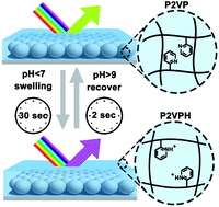
A new optical motif compatible with an ultrathin polymer gel film for chemical sensing was fabricated by infiltrating polymer gel into an inactive scaffold of monolayer colloidal crystals. The composite film, denoted as PG-MCC, features submicron thickness and presents thin film interference with only one reflectance peak in the visible region because of appropriate optical thickness. Consequently, the film exhibits distinct reflective color, which can be easily tuned by adjusting the fabrication parameters. In this study, PG-MCC was demonstrated as a pH sensor by using a weak polyelectrolyte poly-(2-vinyl pyridine) (P2VP). The pH-induced swelling and deswelling of P2VP gel led to a substantial thickness change and hence a reflectance peak shift of PG-MCC. Highly reversible, linear, reliable and very fast responses to pH variations were exhibited. Moreover, a colorimetric readout was readily achieved, making it promising for continuous monitoring of environmental analytes by the naked eye.
For optical elements, photonic materials that exhibit color as a result of refractive index periodicity in one, two, or three dimensions (1-, 2-, or 3D), have been proven promising in optical sensing in a non-destructive and label-free way.6 Until now, a number of photonic transduction modes have been applied to stimuli-responsive polymer gels.7–19 Asher et al. designed the motif of polymerized crystalline colloidal array (PCCA) by embedding well ordered crystalline colloidal arrays inside a hydrogel film.7–10 Recently, they have extended this 3D motif to 2D by attaching the hydrogel film to a monolayer of crystalline colloidal array.11–13 VPT of the polymer network in response to the analyte alters the particle spacing of the array and hence its diffraction. Based on a similar principle, inverse opal sensors of polymer gels have been fabricated by using opals (or colloidal crystals) as the hard template.14–19 However, their practical uses have been hampered, largely by their slow responses to solutes, such as pH, or molecular analytes. For the PCCA glucose sensor, it takes over 90 min to fully respond to the introduction of 1 mM analyte.8 For inverse opal pH sensor, it takes more than 20 min to reach equilibrium upon a change of 1 pH unit.15 The slow responses are directly associated with the slow VPTs of the polymer gels being used. Although several methods have been taken to speed the VPT process,9,20 they lack feasibility and generality. The most straightforward solution is to down size the gels, form bulk to ultrathin films, as the VPT rate is inversely proportional to the square of the dimension of the gel.21 Nevertheless, it remains a big challenge to utilize ultrathin polymer gel film for sensing because of the lack of compatible optical transducer.
Recently, Zhang et al. have fabricated ultrathin hydrogel films with thickness on submicrometer or micrometer scale by layer-by-layer assembly,22 which presented Fabry–Perot fringes with multiple peaks in their reflection spectra. Analyte-induced swelling of the hydrogel was reported by the shift of these fringes. While multiple spectra peaks generated by thin film interference can be used for sensing, a single reflectance peak in the visible region is more desirable because it produces distinct colors both in the absence and presence of an analyte. Instead of thin films, Serpe's group has fabricated microgel based etalons with various kinds of responsiveness.23,24 However, this method needs the pre-synthesis of microgels, which is inconvenient to extend to a wide range of materials. Recently, we have fabricated monolayer inverse opals of polymer gels, which provided both eigenmodes of photonic crystals and Fabry–Perot fringes for signal transduction.25 Due to its submicron thickness and interconnected macropores in the film, a full response to 7 pH unit change in less than 1 min was achieved. Unfortunately, the skin layer with a thickness of tens of nanometers on top of the macropores suffered from structural instability induced by intense swelling, which hampered the linearity of response.
To overcome these obstacles, here we propose a new optical motif for ultrathin polymer gel film sensors, which is polymer gel infiltrated monolayer colloidal crystals (PG-MCC). Monolayer colloidal crystals (MCC) composed of a monolayer array of polystyrene (PS) spherical particles are the inactive component that provides the solid scaffold for the polymer gels. By infiltration of the polymer solution using a controlled spin-coating method, an ultrathin film with a thickness on the submicrometer scale was formed. Due to an appropriate optical thickness for thin film interference, the film presents only one reflectance peak in the visible region, resulting in distinct color visible to naked eyes. Analyte-induced VPT of the polymer gel can lead to significant expansion of PG-MCC in thickness and hence shift of its reflection peak. Specifically, a typical pH-responsive polyelectrolyte gel poly-(2-vinyl pyridine) (P2VP) was chosen in this study. A pH sensor based on the optical motif of PG-MCC was fabricated and exhibited highly reversible and very fast response with improved linearity and reliability. More attractively, colorimetric readout is readily achieved with the presented sensor, making it promising for continuous monitoring analyte changes by naked eyes.
:
H2O2 = 7
:
3 v/v Caution!). After washed with copious water and dried, it was placed at the center of a glass Petri dish (60 mm in diameter). Then, water was added into the Petri dish with a pipette to level the upper surface of the glass but not cover it. Polystyrene (PS) spheres (380 nm in diameter, standard deviation < 10%) were synthesized by standard emulsion-free polymerization.26 The as-obtained suspension was diluted with an equal volume of ethanol and treated with ultrasonication. Then, 10 μL of this suspension was dropped onto the glass. Due to the hydrophilicity of the glass surface, the suspension spread automatically onto the water surface, and within a few seconds a film with iridescence was observed, indicating the formation of MCC at the air–water interface. To compact the film, a few drops of SDS solution (aq. 2 wt%) were added to vary the surface tension. Finally, the MCC was deposited onto the silicon wafer by inserting the wafer underneath it and subsequently picking it up from the wafer. The as-deposited MCC was kept at ambient conditions for drying.
Fig. 1 demonstrates the characteristic scanning electronic microscopy (SEM) images of the fabricated structures in each step. The as-assembled MCC shows a close packed structure composed of uniform PS particles of 380 nm in diameter (Fig. 1a). A cross-sectional view reveals the ordered array forms a monolayer on the Si wafer (Fig. 1b). After infiltration of the polymer precursor by spin-coating, the interstices among PS particles were filled and only imprints of the particles were observed from the upper side of the composite film (Fig. 1c). Its cross section shows clearly that the polymer precursor has well infiltrated into the MCC without impinging the PS particles, and a wavy profile in consistent with the pristine MCC was exhibited (Fig. 1d). By thermal annealing at 120 °C, a temperature above the glass transition temperature of PS (∼100 °C), the PS spheres turned glassy, and no imprints of particles were observed anymore, indicating the fusing of particles (Fig. 1e). In the mean time, the whole film became flattened, but retained the periodic structure inherited from the composite film (Fig. 1f).
The optical properties of fabricated structures were studied by reflectance spectra measurement (Fig. 2). Fig. 2a shows the spectra evolution from the MCC, to the as-spin-coated film, and to the PG-MCC. When the MCCs is supported on a substrate with a high refractive index (RI), like Si wafer, it act essentially like a dielectric film, showing peaks that are known as Fabry–Pérot fringes, which stems from interferences between beams reflected at the film–air and film–substrate interfaces.29 At normal incidence the wavelength of interference peak (λ) conforms to
| mλ = 2nd | (1) |
By spin-coating the polymer precursor onto MCC, the fringe exhibits only one peak in the visible region, and no dip associated with the photonic eigenmode was found because of the eliminated RI contrast upon the formation of composite film. After thermal annealing the peak shifted toward short wavelength, from 584 nm to 544 nm, due to a decrease in the film thickness.
The reflection spectra and hence the reflective color of the PG-MCC can be readily tuned by controlling the fabrication parameters. As the spin-coating speed increased from 1000 to 2000 rpm, the film thickness decreased due to a smaller amount of gel deposited per unit area. As a result, the reflection peak shifted toward short wavelength, from 544 nm to 490 nm, and the color exhibited by PG-MCC changed from light green to cyan (Fig. 2b), correspondingly, which is in accordance with formula (1). Using an increased precursor concentration of 4.7 wt% the fringe shifted towards longer wavelengths and presented two reflection peaks at 452 and 670 nm in the visible region, and hence exhibited a purple color (Fig. 2c). This is ascribed to the increase of film thickness by increasing the precursor concentration.
The sensitive dependence of interference peak on the structural parameters of PG-MCC renders it promising for optically encoded sensing. The swelling of the polyelectrolyte gel upon external stimuli will lead to changes in the optical thickness, which can be directly read out from the shift of the reflection peak. P2VP network is known for its fast and significant swelling under acidic conditions due to protonation of the pyridine group. Therefore, a pH sensor is demonstrated here using PG-MCC (Fig. 3). The original reflection peak was located at 539 nm. For pH values of 9.26, 8.12, and 7.07, the peak position remained nearly unchanged because P2VP gel only swell under acidic conditions. As the pH value decreases gradually from 6.05 to 2.44, the reflection peak of the PG-MCC shifted almost linearly towards longer wavelengths to 663 nm, and the specific position of reflection peak is 544 nm for pH 6.05, 560 nm for pH 5.09, 584 nm for pH 4.36, 642 nm for pH 3.06, and 663 nm for pH 2.44. Unlike our previously reported monolayer inverse opals of P2VP gel,25 the PG-MCC presented here overcomes the problem of structural instability at high acidity because of a much denser texture, and hence exhibited improved linearity for a wide pH range. Interestingly, the well-defined peak in the reflectance spectra and its large wavelength shift in the range of tens of nanometers can directly lead to visible color changes in response to environmental pH variations, which allows for simple detection of pH conditions by naked eyes (inset of Fig. 3b). The color of PG-MCC varied from light green, to orange, and to purple red in response to pH values of 7.07, 4.36, and 2.44, respectively, which was consistent with the reflectance spectra.
The swollen state of the PG-MCC caused by protonation of the pyridine group under acidic conditions can be captured by SEM measurement (Fig. 4). Compared to its initial morphology (Fig. 1f), the PG-MCC has exhibited significant volume expansion, and the film thickness is measured to increase from the original 340 nm to 350 nm for pH 4.36, and 365 nm for pH 3.06. However, it is worth noting that the conditions of high vacuum during SEM measurement could lead to the loss of swollen state to a large extent, thus the film thickness characterized by SEM would be much smaller than that of the in situ swollen state. Notably, unlike a homogeneous dense ultrathin P2VP gel film which folds upon swelling because of the different strain imposed by the supported Si wafer,31 the PG-MCC remains well attached to the underneath Si wafer without any folding or peeling off. This is probably because in PG-MCC the P2VP gel has a smaller contact area to the substrate than in the homogeneous dense film, and hence results in a higher flexibility that inhibits buckling deformation of the film.
![]() | ||
| Fig. 4 SEM images of the PG-MCC after equilibration in pH 4.36 (a) and pH 3.06 (b) buffer solutions. | ||
For practical uses, a fast, reversible, and reliable detection is highly desirable. Remarkably, the PG-MCC exhibits a quite fast optical response to pH variations (Fig. 5a). Within 10 s of soaking in a pH 3 buffer solution, the peak shift already reached 96% of that of the full response, and the equilibrium of PG-MCC in the solution was established at around 2 min. This fast response is comparable to our previously reported inverse opal monolayers of P2VP gels,25 and greatly surpasses the inverse opal hydrogel pH sensor reported by Braun et al.,15 and the 2D-PCCA pH sensor reported by Asher et al.,11 which need 20 and 30 min to reach full responses, respectively. The rapid kinetics of PG-MCC is ascribed to its ultrathin structure, which efficiently speeds the gel swelling.
To study the reversibility of PG-MCC for pH sensing, the wavelength shift of reflection peak between pH 9.26 and pH 3.06 was recorded for many cycles (Fig. 5b). As can be seen, the deviation of peak position is less than 1% for tens cycles of measurement. This result shows that the optical response of PG-MCC toward pH is highly stable, and reflects well the reversible swelling/deswelling of the polymer network.
Finally, we studied the possible optical hysteresis of the PG-MCC, because it is important for a sensor to have low hysteresis and hence reliable output regardless of historical inputs. The peak shifts of PG-MCC via two pathways of pH inputs were recorded in a continuous manner, one from high to low pH value, and the other in a reverse sequence. As can be seen from Fig. 5c, peak shifts resulted from the two pathways exhibited very small differences, indicating the existence of only a tiny hysteresis, which makes our sensor suitable for the continuous monitoring of environmental pH conditions.
| This journal is © The Royal Society of Chemistry 2016 |