DOI:
10.1039/C6RA12100D
(Paper)
RSC Adv., 2016,
6, 91349-91360
Dual functional polylactide–hydroxyapatite nanocomposites for bone regeneration with nano-silver being loaded via reductive polydopamine†
Received
10th May 2016
, Accepted 18th September 2016
First published on 19th September 2016
Abstract
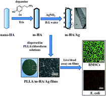
In bone tissue engineering, failure might take place in treating infected bone defects. Scaffolding materials with antibacterial function should be a potential solution for this problem. Herein, poly(L-lactide) (PLLA)–hydroxyapatite (HA) nanocomposites containing antibacterial silver nanoparticles (AgNPs) were prepared. In the preparation, surface coating of polydopamine (PDA) on HA nano-rods was a critical step to ensure both the homogeneous dispersion of HA nano-rods in the PLLA matrix and the successful loading of AgNPs onto HA nano-rods. With the total content of HA being fixed at 30 wt%, PLLA/HA nanocomposites containing different amounts of AgNPs (∼0.54 wt%, ∼0.27 wt% and ∼0.18 wt%) were prepared by mixing PDA-coated HA nano-rods (m-HA) with or without AgNPs at different ratios into PLLA. The resulting PLLA/HA/Ag nanocomposites demonstrated strong antibacterial activity against Escherichia coli at low content of AgNPs being incorporated, while they did not show significant cytotoxicity to bone mesenchymal stromal cells (BMSCs). In comparison with those cells cultured on pure PLLA film and tissue culture polystyrene, on the contrary, osteogenic differentiation of BMSCs was enhanced on PLLA/HA/Ag nanocomposite films benefiting from the HA component. The results suggested that PLLA/HA/Ag nanocomposites are anticipated to be good scaffolding materials for bone regeneration with dual functions of both antibacterial activity and osteocompatibility.
1. Introduction
Nanocomposites composed of poly(L-lactide) (PLLA)-based polyesters and nano-hydroxyapatite (nano-HA) have been intensively studied and used as substrates for bone regeneration, benefiting from their well-known biocompatibility, biodegradability and osteogenic bioactivity.1–3 In clinical applications, however, potential bacterial infection during or after surgery is threatening the safe use of implant materials,4 furthermore, influencing the efficiency of tissue recovery.5–7 In infected bone areas, the substrates for bone regeneration are required to have antibacterial performance for the purpose of achieving satisfactory bone regeneration.8–11 Besides, the occurrence of implant-associated infection is also of high probability in orthopaedic surgery.12 Therefore, the idea of introducing antibacterial components into bone repairing biomaterials has been widely verified in numerous published studies.13–16
In general, antibacterial substances such as chitosan,17,18 silver,19,20 zinc,21,22 and other materials23–26 had been incorporated into bone repairing biomaterials for the purpose. Among them, silver nanoparticles (AgNPs) were the most intensively studied due to their effective and broad-spectrum antibacterial activity.27 In addition to biological applications for pharmaceutical purposes,28 AgNPs were also widely evaluated as antibacterial components into substrates for bone tissue engineering.13–16 As for the risk concerns of using AgNPs in biomedical purposes, present studies demonstrated that both the cytotoxicity and the antibacterial activity of AgNPs were highly dose-dependent.29–31 And there was a possible therapeutical window by carefully weighing their antibacterial benefits and potential health risks.
AgNPs could be incorporated into a variety of organic and inorganic biomaterials targeting for bone regeneration.14,32–34 In preparing a kind of gelatin/nano-silver scaffold, Yazdimamaghani et al. directly took advantage of those –NH2 and –COOH moieties within gelatin to reduce silver ions into AgNPs, which distributed homogeneously throughout the lyophilized gelatin scaffold.32 Similarly, Saravanan et al. fabricated chitosan/nano-HA/nano-silver scaffolds by soaking porous chitosan/nano-HA scaffolds into aqueous solution containing silver nitrate, in which, AgNPs formed via the reduction function of those nitrogen and oxygen atoms in chitosan without using any external chemical reducing agent.14 Marsich et al. prepared a colloidal solution by reducing silver ions with ascorbic acid in the presence of a lactose modified-chitosan, and then this colloidal solution was adsorbed into porous alginate/HA scaffolds to get antibacterial nanocomposite bone grafts.33 In many other cases, while the preparation of Ag-doped apatite was attempted.15,34–36 For instance, Stanić et al. prepared Ag-doped nano-HA with different amounts of silver ions via neutralization method by fixing the atomic ratio of (Ca + Ag)/P at 1.67,34 and Geng et al. prepared Ag-doped nano-HA via a hydrothermal method.35 Logically, nanocomposites composed of widely used PLLA-based polyesters and Ag-doped nano-HA were envisioned excellent approaches to develop dual functional scaffolds with both osteocompatibility and antibacterial activity for bone regeneration.
In preparing nanocomposite scaffolds for bone regeneration, noticeably, the dispersibility of nano-HA in polymeric matrixes remains a knotty problem, which is also associated with Ag-doped nano-HA. In both literature and our previous work, surface modification with mussel-inspired dopamine has been proven able to significantly improve interfacial adhesion between inorganic fillers and polymeric matrixes, as well as ameliorate the dispersion of nano-scaled fillers in polymeric matrixes.37–41 The catechol group in dopamine was reported able to reduce silver ions into AgNPs and bind the nanoparticles firmly.42–45 For biomedical applications, besides, cell proliferation and differentiation were confirmed able to be positively promoted when dopamine modification was applied.46–48 Therefore, in this study, an idea was proposed and tested by preparing PLLA/HA/Ag nanocomposites via homogeneously incorporating AgNP-laden nano-HA (m-HA/Ag) into PLLA with the aid of dopamine surface modification on nano-HA (m-HA). Self-polymerization of dopamine would generate an active polydopamine (PDA) layer on nano-HA, and AgNPs were deposited onto m-HA by in situ nucleation and growth via the reducing and binding feature of PDA. The addition amount of HA into nanocomposites was fixed at an optimized value of 30 wt% according to our previous study.41 To alter the addition amounts of AgNPs into the nanocomposites, mixtures of m-HA/Ag and m-HA at different weight ratios were blended with PLLA. Evaluations on the cytotoxicity and the antibacterial activity of resulted PLLA/HA/Ag nanocomposites were performed by applying both bone mesenchymal stromal cell (BMSC) culture and Escherichia coli (E. coli) cloning. Meanwhile, the capacity of PLLA/HA/Ag nanocomposites in enhancing osteogenic differentiation of BMSCs was evaluated to assess the potential of these nanocomposites being used as dual functional substrates for bone regeneration.
2. Materials and methods
2.1. Materials
PLLA (Mw = 100000) was purchased from Purac (Netherlands). Dopamine hydrochloride, tris(hydroxymethyl)aminomethane (Tris) and silver nitrate (AgNO3) were purchased from Sigma and used directly. Solvents and other chemicals involved in this study were of analytically pure grade, which were obtained from Beijing Chemical Plant (China) and used as received.
2.2. Preparation of AgNP-laden nano-HAs
AgNP-laden nano-HAs were prepared in three steps. Referring to our previous work,41 nano-HAs were freshly made via a chemical precipitation of Ca(NO3)2·4H2O and (NH4)2HPO4 mixture solution (pH 10, Ca/P = 1.67) and the wet powders were used for following preparation to achieve good dispersion of nano-HAs in aqueous solution. The wet nano-HAs (0.2 g) were dispersed in 100 mL Tris–HCl buffer solution (10 mM, pH 8.5) containing 2 mg mL−1 dopamine hydrochloride. The system was continuously stirred at room temperature for 48 h, followed by centrifugation to collect m-HAs. After being washed with deionized water for three times, the wet m-HAs (1 g) were re-dispersed in 100 mL deionized water and AgNO3 (0.05 g) was added. The suspension was continuously stirred for another 4 h, followed by centrifugation, deionized water washing and freeze-drying to get the AgNP-laden nano-HAs (m-HA/Ag). For comparison, original nano-HAs and m-HAs were also freeze-dried.
2.3. Preparation of PLLA/HA nanocomposite films
Referring to our previous work,41 solution-casting method was applied to prepare PLLA/HA nanocomposite films. Briefly, 0.214 g HA powders (nano-HA, m-HA, m-HA/Ag, or mixtures of m-HA/Ag and m-HA) were dispersed in 5 mL chloroform under magnetic stirring for 24 h at room temperature, and the suspension was ultrasonically treated (500 W, 5 min) before use. Then the suspension was homogeneously mixed into 5 mL PLLA solution in chloroform (0.1 g mL−1), in which, the content of HA was fixed at 30 wt% in reference to the total weight of PLLA and HA. The mixture solution was cast onto glass plate, standing for 48 h at room temperature to allow for solvent evaporation, followed by vacuum-drying at 37 °C to constant weight to remove any residual solvent. To alter the addition amount of AgNPs into PLLA/HA nanocomposites, the mixtures of m-HA and m-HA/Ag were added at weight ratios of 1:1 and 2:1, in addition to the cases of only m-HA/Ag or m-HA being added. In other words, the resulted PLLA/HA/Ag nanocomposite films contained 30 wt%, 15 wt%, 10 wt%, and 0 wt% of m-HA/Ag, respectively, although all the nanocomposites contained 30 wt% of HA component. For comparison, nanocomposite films containing 30 wt% of original nano-HAs (PLLA/nano-HA) and pure PLLA films were also prepared.
2.4. Characterizations
Chemical compositions of prepared samples were determined by X-ray photoelectron spectroscopy (XPS), which was performed on a XPS spectrometer (ESCALAB 250, USA) with a monochromatized Al Kα X-ray source (1486.6 eV photons) under vacuum (10−8 Torr or lower) using an incidence angle of 45° at a power of 150 W. The amount of AgNPs depositing onto m-HA was determined by thermal gravity analysis (TGA) using a Q50 thermogravimetric analyzer (TA instruments, USA) in nitrogen atmosphere from room temperature to 800 °C at a heating rate of 20 °C min−1. The crystalline structures of synthetic nano-HA, m-HA and m-HA/Ag were evaluated by wide-angle X-ray diffraction (WAXD) from 20° to 80° using a Ultima III X-ray diffractometer (Rigaku, Japan) with a Cu Kα X-ray source (2500VB2+/PC, 40 kV, 200 mA). Dispersibility and morphology of various synthetic HA powders were observed under transmission electron microscope at an accelerating voltage of 200 kV (TEM, Hitachi 800) by dispersing them in chloroform for 24 h and dropping onto 800-mesh copper grids. Morphological observations of HA powders and PLLA/HA nanocomposite films were conducted with scanning electron microscope (SEM, S-4700, Hitachi) at an accelerating voltage of 20 kV after being sputter-coated with platinum (30 mA, 20 s) using a sputter-coater (Polaron E5600, USA). Silver element mapping was performed under the same parameters of SEM observation with an exposure time of 180 s.
2.5. Silver ion release
Release of silver ions from different nanocomposite films were measured by inductively coupled plasma optical emission spectrometer (ICP, ICPS-7500, Shimadzu, Japan). Patches of sample films (0.03 g) were placed into 10 mL of deionized water and incubated at 37 °C for 28 days with continuous agitation (50 rpm). The liquids were collected at day 0, 3, 7, 14 and 28, and submitted to ICP measurements to quantify the released silver ions. At the same time, fresh deionized water was replenished at each time point to continue the release experiment.
2.6. Antibacterial activity evaluation
The antibacterial activities of PLLA/HA/Ag nanocomposite films containing different amounts of AgNPs were evaluated using E. coli by live/dead assay. After E. coli was cultured in Brain Heart Infusion (BHI, Beijing Solarbio Science & Technology Co., Ltd) broth at 37 °C for 24 h, the bacterial density in the system was diluted to 107 colony-forming units (CFU) per milliliter, basing on the absorbance measured at 600 nm wavelength using a previously established standard curve. PLLA/HA/Ag nanocomposite films were cut into circular specimens (ϕ = 15 mm), and fitted into the 24-well tissue culture plate. To each well, 1 mL of the aforementioned E. coli suspension was added. The systems were incubated in an incubator thermoset at 37 °C for 24 h, and then the specimens were gently washed with phosphate buffered saline (PBS). After being stained with acridine orange/ethidium bromide (AO/EB), the fluorescent images were captured using a laser confocal scanning microscope (LCSM, TCS SP8, Leica) to judge the live/dead state of E. coli. For comparison, specimens of pure PLLA films and nanocomposite films containing original nano-HA or m-HA were performed the E. coli experiment similarly.
2.7. Biological evaluations
Rat BMSCs (purchased from Cell Culture Center, Peking Union Medical College, China) were cultured in Dulbecco's modified Eagle's medium (DMEM, Hyclone) supplemented with 10% fetal bovine serum (FBS, PAA, Germany), 100 IU per mL penicillin (Sigma), and 100 mg mL−1 streptomycin (Sigma). The culture was maintained with 5% CO2 at 37 °C and saturated humidity until 80% confluence prior to use.
2.7.1. Cytotoxicity assay. PLLA/HA nanocomposite films with or without AgNPs loading, together with pure PLLA film as reference, were sterilized with ultraviolet light exposure and 70% ethanol soaking for 2 h. To evaluate the potential cytotoxicity of loaded AgNPs against BMSCs, the materials were then soaked in DMEM for 24 h to get the extracts for BMSC culture according to ISO 10993-12: 200. BMSCs were seeded in 96-well plates at 1 × 10⁴ cells per well and incubated with extracts at 37 °C in a humidified atmosphere with 5% CO2 for 7 days, using DMEM as control. The medium was refreshed every other day. Cell proliferation was analyzed using Cell Counting Kit-8 (CCK-8, Beyotime, China). Briefly, at different culture time points (1, 3, 5 and 7 days), 20 μL of CCK-8 solution was added into each well and incubated at 37 °C in 5% CO2 for 4 h, and then OD values were measured using a microreader (Biorad 680, USA) at the wavelength of 490 nm.
2.7.2. Cell viability. Proliferation of BMSCs in the presence of PLLA/HA/Ag nanocomposite films was conducted in two ways. In the conventional way (contact mode), BMSCs were directly seeded onto the films and incubated. In the transwell chamber (0.4 μm, Costar) culture way (transwell mode), BMSCs and films did not contact directly. In both cases, circular specimens (ϕ = 9 mm) were used with 1 × 10⁴ BMSCs and 0.5 mL culture medium being added. The culture medium was refreshed every other day. Thus, the effects of released silver ions and the contact effect on cell viability could be identified, respectively. Both the cultures were conducted for 7 days, CCK-8 assay and AO/EB live/dead staining were applied at day 1, 3, 5 and 7 after cell seeding.
2.7.3. Cell morphology. Circular specimens (ϕ = 9 mm) were fitted into 48-well plates and 1 × 10⁴ BMSCs were seeded into each well. The systems were incubated for 3 days at 37 °C in a humidified atmosphere with 5% CO2. Subsequently, Hoechst 33528 and phalloidin staining were conducted, and cell morphology was observed using LCSM.
2.7.4. Osteogenic differentiation. To conduct osteogenic differentiation assay, 0.05 mmol L−1 vitamin C (Sigma), 10 mmol L−1 β-sodium glycerophosphate (Sigma), and 1 × 10⁻⁸ mol L−1 dexamethasone (Sigma) were added to the culture medium. Also, contact mode and transwell mode were applied for this assay. Circular specimens (ϕ = 9 mm) were used with 1 × 10⁵ BMSCs and 0.5 mL osteogenic inductive culture medium being added. The medium was refreshed every other day. After the systems were incubated for 3, 7, 14 and 21 days, in both culture manners, expressions of alkaline phosphatase (ALP) and collagen type I (Col-I), the two classic markers for osteogenic differentiation, were determined using enzyme-linked immunosorbent assay (ELISA) method.Briefly, the cells or the cell/matrix complexes were retrieved from the transwell chamber or the culture plate, and rinsed by PBS for three times. According to manufacturer's standard protocols, the ALP activity and Col-I synthesis were determined respectively using corresponding ELISA kit (R&D, Minneapolis, MN, USA). The ALP activities were normalized to the total protein content determined using the BCA assay kit (Thermo, USA).
2.8. Statistical analysis
All the biological quantitative data were represented as mean ± standard deviation for n = 3. Statistical analysis was made basing on t-test and difference between groups of *p ≤ 0.05 was considered significant and **p ≤ 0.01 was considered highly significant.
3. Results
3.1. Loading of AgNPs onto nano-HAs
Nano-HAs were prepared via chemical precipitation, and directly used for the following dopamine surface modification without drying. After being soaked in the dopamine aqueous solution for 24 h, the white nano-HAs visibly turned into dark-brown powders due to the self-polymerization of dopamine and the coating of resulted PDA. Subsequently, the resulted m-HAs were dispersed in an AgNO3 aqueous solution, in which, nucleation and growth of AgNPs would take place without extra reductive reagent due to the reductive feature of catechol group in dopamine. The prepared m-HA/Ag powders were systematical characterized with XPS, XRD, TGA, SEM and TEM.
XPS was applied to identify the surface chemical composition of m-HA/Ag powders. In comparison with the XPS spectrum of original nano-HA, as shown in Fig. 1A, the XPS spectrum of m-HA displayed the presence of an extra N1s signal (400 eV), apparently, which was originated from the dopamine structure.41 In the case of m-HA/Ag, the intensity of N1s signal could be seen decreasing in comparison with that of m-HA. Similarly, the intensities of Ca2p signal (347 eV) and P2p signal (133 eV) also decreased in the XPS spectrum of m-HA/Ag in comparison with those signals of m-HA. While at the same time, a group of new signals were detected around 365–377 eV in the XPS spectrum of m-HA/Ag, which should be assigned to Ag3d.49–51 From the enlarged profile of the Ag3d signal (Fig. 1B), it demonstrated two individual peaks at 368 eV and 374 eV with a spin–orbit separation of 6.0 eV, which were in accordance with the binding energies of Ag3d3/2 and Ag3d5/2, respectively. These data confirmed the presence of pure Ag0 species in m-HA/Ag.49
|
| Fig. 1 Characterizations of AgNP-laden nano-HA (m-HA/Ag) in comparison with original nano-HA and PDA modified m-HA: (A) XPS spectra; (B) enlarged XPS spectrum of Ag3d signal from (A); (C) XRD patterns; and (D) TGA curves. | |
XRD analysis was then conducted for all the nano-HA, m-HA and m-HA/Ag powders. As shown in Fig. 1C, the XRD patterns of the three kinds of HA powders were quite similar, except three extra diffraction peaks being identified at 38.12°, 64.45° and 77.38° in the XRD pattern of m-HA/Ag. The main XRD patterns of all the samples resembled the characteristic diffraction signals of crystalline HA, while the three peaks at 38.12°, 64.45° and 77.38° in the XRD pattern of m-HA/Ag were assigned to the (1 1 1), (2 2 0) and (3 1 1) crystal face of elemental silver, respectively.51,52 In combination XPS data with XRD results, thus, it was confirmed that the loading of elemental silver onto m-HA was successful. The absence of peak at 44.2° for the (2 0 0) crystal face of elemental silver was thought due to the low content of loaded AgNPs and the possible interference of HA signals.53,54
TGA data of nano-HA, m-HA and m-HA/Ag are shown in Fig. 1D. The original nano-HA displayed weight loss of ∼5.2 wt% upon being heated to 800 °C due to the elimination of moisture and other possible residuals. After the dopamine modification, the weight loss of m-HA increased to ∼15.7 wt% with the decomposition of PDA. While in the case of m-HA/Ag, the final weight loss at 800 °C was ∼13.9 wt%, which was higher than that of original nano-HA but lower than that of m-HA. The reason was that the elemental silver was stable in nitrogen atmosphere upon heating, which contributed no extra weight loss. Therefore, the loading amount of atomic silver on m-HA/Ag was able to be calculated ∼1.8 wt% by comparing the weight loss of m-HA and m-HA/Ag. The loading amount of Ag component could be seen not high, as the result, the Ag0 signals in both XPS and XRD spectra were weak accordingly as aforementioned. In preparing the m-HA/Ag listed in Fig. 1, the concentration of AgNO3 in aqueous solution was 0.05 g/100 mL, and the reaction time was 4 h. These parameters were optimized from repeating experiments by altering both the AgNO3 concentration and the reaction time. More information on preparation of m-HA/Ag was provided as Fig. S1.†
Morphology and dispersibility of the m-HA/Ag prepared under the optimized parameters were observed with SEM and TEM, in comparison with original nano-HA and m-HA. As those SEM images in Fig. 2 shown, the m-HA/Ag (Fig. 2C) displayed similar size and shape to both the original nano-HA (Fig. 2A) and the m-HA (Fig. 2B). The polymeric coating on HA nano-rods in both the cases of m-HA and m-HA/Ag could be easily identified as the surface PDA layer in comparison with the original nano-HA. From Fig. 2C, however, no visible AgNP could be detected. It was thought that the AgNP was too small to be seen at this scale. To prove the existence of silver component, thus, silver element mapping was conducted on m-HA/Ag. As shown in Fig. 2D, silver element was detected definitely and distributed homogeneously throughout the sample, indicating the successful loading of silver component onto HA nano-rods. From the TEM images shown in Fig. 3, the dopamine modification endowed both the m-HA (Fig. 3B) and the m-HA/Ag (Fig. 3C) excellent dispersibility in chloroform in comparison with those seriously aggregated nano-HA (Fig. 3A). Additionally, the excellent dispersibility of m-HA/Ag was further confirmed by dynamic light scattering (DLS) analysis (Fig. S2†). After being suspended in chloroform for 48 h, m-HA/Ag could achieve stable dispersion with narrow particle size distribution around 700 nm. The phenomenon was quite similar to the DLS data of m-HA as previously reported,41 indicating the loading of AgNPs onto m-HA not causing adverse effect on its dispersibility. From the enlarged TEM images shown in Fig. 3E and F, a surface layer of several nanometers in thickness was detected in both the cases of m-HA and m-HA/Ag. Besides, spherical nanoparticles of ∼6 nm in size were clearly detected binding to the surface layer on HA in the case of m-HA/Ag (Fig. 3F), showing excellent dispersion. With all the aforementioned facts, it was undoubted to say that these spherical nanoparticles in m-HA/Ag were AgNPs, which were reduced from silver ions by the PDA surface layer and subsequently bound to the PDA layer.
|
| Fig. 2 SEM observations on the morphology of original nano-HA (A), m-HA (B) and m-HA/Ag (C), together with the silver element mapping of m-HA/Ag (D). | |
|
| Fig. 3 TEM observations on the dispersibility in chloroform and the morphology of original nano-HA (A and D), m-HA (B and E) and m-HA/Ag (C and F). Image (D), (E) and (F) are magnifications of corresponding image (A), (B) and (C) respectively. | |
3.2. PLLA/HA nanocomposite films
To prepare PLLA/HA nanocomposite films, m-HA and m-HA/Ag powders were firstly dispersed in chloroform, which was a good solvent for PLLA. As shown in Fig. 3B and C, both m-HA and m-HA/Ag displayed excellent dispersion in the solvent, thus, it guaranteed the well blending of nano-scaled HA with PLLA in preparing various PLLA/HA nanocomposite films by solution-casting method. To control the loading amount of AgNPs into nanocomposites, the simplest way was to change the mixing ratio of m-HA to m-HA/Ag. By fixing the addition amount of total HA as 30 wt% according to our previous study,41 four kinds of PLLA/HA nanocomposite films were prepared by introducing m-HA, m-HA/Ag, or mixtures of m-HA and m-HA/Ag in weight ratios of 1:1 and 2:1, respectively. Their morphology was observed using SEM and the images are presented in Fig. 4.
|
| Fig. 4 Morphological observations on PLLA/HA nanocomposite films containing different proportions of m-HA and m-HA/Ag at the fixed HA content of 30 wt%: (A) m-HA; (B) m-HA/Ag; (C) m-HA:m-HA/Ag = 1:1; (D) m-HA:m-HA/Ag = 2:1. Those insets in image (B)–(D) are the silver element mappings of corresponding PLLA/HA/Ag nanocomposite films. | |
From the figures, all the nanocomposite films displayed similar morphology with visible HA nano-rods, which dispersed homogeneously throughout the films. Those AgNPs on the surface of m-HA/Ag did not show adverse effect on their dispersibility in PLLA matrix. Silver element mapping was then conducted on nanocomposite samples containing m-HA/Ag and are shown as insets in corresponding images. As shown in Fig. 4B–D, silver element was detected obviously in all the three PLLA/HA/Ag films, and the signal strength apparently decreased along with less m-HA/Ag being introduced. According to the TGA data, the practical loading contents of AgNPs in the three PLLA/HA/Ag nanocomposite films were calculated ∼0.54 wt%, ∼0.27 wt% and ∼0.18 wt%, respectively, as the mixing ratio of m-HA/Ag decreasing. Tensile properties of PLLA/HA nanocomposite films containing 30 wt% of m-HA or m-HA/Ag were evaluated and are provided as Fig. S3.† No significant difference in tensile strength and stress–strain behavior was detected between the two cases. The results confirmed that the introduction of AgNPs at low content almost had no adverse effect on mechanical properties of PLLA/HA nanocomposites.
3.3. Release of silver ion from PLLA/HA/Ag nanocomposite films
The silver ion release experiment was performed in deionized water, and the cumulative release of silver ions from PLLA/HA/Ag films containing different amounts of AgNPs are illustrated in Fig. 5. Within the experimental period, sustained release of silver ions was detected from all the three samples until 28 days. And the release would surely continue for longer time as indicated by the going up trend of silver ion release profiles. At the time of 28 days, the release percentages of silver ions were all estimated only ∼1.0% from all the three PLLA/HA/Ag nanocomposite films, while the released amount of silver ions demonstrated positive relationship to the loading content of AgNPs. The slight initial burst release detected for samples containing higher content of AgNPs was common in controlled drug release studies, which was related to the diffusion of water molecules into the composite and silver ions out of the composite.
|
| Fig. 5 Release profiles of silver ions from PLLA/HA/Ag nanocomposite films containing different proportions of m-HA and m-HA/Ag by soaking the films in deionized water at 37 °C for 28 days. | |
3.4. Antibacterial activity of PLLA/HA/Ag nanocomposite films
A popular Gram-negative E. coli was cultured on PLLA/HA/Ag nanocomposite films to identify their antibacterial activity and the dependence of antibacterial activity on the loading amount of AgNPs in the nanocomposite. E. coli cultured on pure PLLA film and PLLA/HA nanocomposite films containing only nano-HA or m-HA were used as comparisons. The live/dead staining results of E. coli cultured on all these films are presented in Fig. 6. The green and red staining represented the live and dead E. coli, respectively. For the three AgNP-free films, as shown in Fig. 6A–C, green stained E. coli had fully covered the pure PLLA film (Fig. 6A), the PLLA/nano-HA film (Fig. 6B) and the PLLA/m-HA film (Fig. 6C), almost showing no visible red spot. Besides, the strength of green fluorescence was the strongest on the PLLA/m-HA film. On the contrary, those E. coli cultured on PLLA/HA/Ag nanocomposite films were stained both green and red, which indicated both live and dead E. coli being detected. When the loading amount of AgNPs in the nanocomposite film was 0.54 wt%, only sparse green spots could be detected (Fig. 6D), suggesting the film having strong antibacterial activity. While in the cases of PLLA/HA/Ag nanocomposite films containing less AgNPs (0.27 wt% and 0.18 wt%), only a portion of E. coli was killed (Fig. 6E and F). These results clearly indicated that the antibacterial activity of PLLA/HA/Ag nanocomposites depended strongly on the loading amount of AgNPs.
|
| Fig. 6 Live/dead assay of E. coli cultured on pure PLLA film and various PLLA/HA nanocomposite films with the fixed HA content of 30 wt% by AO/EB staining (green: live; red: dead): (A) pure PLLA; (B) PLLA/nano-HA; (C) PLLA/m-HA; (D) PLLA/HA/Ag, m-HA/Ag; (E) PLLA/HA/Ag, m-HA:m-HA/Ag = 1:1; (F) PLLA/HA/Ag, m-HA:m-HA/Ag = 2:1. | |
3.5. Cytotoxicity of PLLA/HA/Ag nanocomposite films to BMSCs
The antibacterial activity of PLLA/HA/Ag nanocomposite films had been confirmed, however, their potential cytotoxicity against BMSCs was also required to be evaluated. Referring to ISO 10993-12: 200 standard, initially, extracts were made and used to perform the proliferation of BMSCs, using culture medium as control. As shown in Fig. 7, BMSCs proliferated continuously in all the extracts along with culture time. The average proliferation rates of BMSCs in all the extracts were found slower than that of BMSCs in the control case. This suggested that some soluble ingredients might have entered into the medium and showed some effects on cell proliferation. By statistical analysis, it was found the proliferation of BMSCs was comparable among the groups of control and extracts from pure PLLA, PLLA/nano-HA and PLLA/m-HA after 5–7 days of culture. For the three extracts from PLLA/HA/Ag nanocomposite films, however, BMSCs proliferated slightly slower than those cells in the three AgNP-free groups. In considering the release data of silver ion (Fig. 5), it was proposed that silver ions had been released into the extracts and caused some adverse effect on cell proliferation. Although the released amounts of silver ions were different for the three PLLA/HA/Ag nanocomposites, they did not show significant difference in influencing the proliferation of BMSCs.
|
| Fig. 7 Proliferation of BMSCs in extracts made from pure PLLA and various PLLA/HA nanocomposite films containing original nano-HA, m-HA, m-HA/Ag or mixtures of m-HA and m-HA/Ag, using DMEM as control. The total content of various HA powders was fixed at 30 wt% in all the nanocomposite films (*p < 0.05; **p < 0.01). | |
Subsequently, proliferation of BMSCs was evaluated in the presence of various films by using both the transwell mode and the contact mode. In the transwell culture mode, BMSCs did not contact the material while the soluble ingredients from the material could enter into the culture medium. As shown in Fig. 8A, BMSCs proliferated gradually in all the cases, being relatively slower in all the groups containing materials in comparison with the control group, but showing insignificant difference. It was coincident with those cell growth results in extracts (Fig. 7) that the materials could release some soluble ingredients to influence cell proliferation. In the contact culture mode, similar trend in the proliferation of BMSCs was detected on all the films in comparison with the control (Fig. 8B). The inhibition of PLLA/HA/Ag nanocomposite film containing 30 wt% m-HA/Ag on cell proliferation seemed more obvious than it in the previous case of transwell culture. This should be due to the contact effect of AgNPs on cell proliferation. In comparing Fig. 8A and B, noticeably, BMSCs proliferated faster in the contact culture mode than in the transwell culture mode, although the seeded cell densities being the same in both the cases. It not only revealed that the contact cytotoxicity of PLLA/HA/Ag nanocomposite films against BMSCs was insignificant. Moreover, it was suggested that the contact effect of the films could partly offset the adverse effect of release silver ions on cell proliferation.
|
| Fig. 8 Proliferation of BMSCs cultured in transwell mode and contact mode by applying materials including pure PLLA and various PLLA/HA nanocomposite films containing original nano-HA, m-HA, m-HA/Ag or mixtures of m-HA and m-HA/Ag, using TCPS as control. The total content of various HA powders was fixed at 30 wt% in all the nanocomposite films (*p < 0.05; **p < 0.01). | |
To further confirm this statement, after 1–7 days of culture, BMSCs growing on the films were evaluated by live/dead staining to judge their viability and proliferation visually. From Fig. 9, live cells could be seen being stained green on all substrates at all culture time points and red staining spots for dead cells could hardly be found. Besides, BMSCs proliferated continuously on all the substrates, reaching quite high cell densities after 7 days of culture. In addition, morphology of BMSCs cultured on PLLA/HA/Ag nanocomposite films was observed by fluorescent nucleus and cytoskeleton staining. As shown in Fig. 10, BMSCs attached well and spread into normal spindle-like shape on pure PLLA, PLLA/nano-HA, PLLA/m-HA and PLLA/HA/Ag nanocomposite films, showing no difference between groups. These results clearly confirmed BMSCs remained high cell viability on all the substrates including those on PLLA/HA/Ag nanocomposite films.
|
| Fig. 9 Live/dead assay of BMSCs cultured for 1–7 days on pure PLLA and various PLLA/HA nanocomposite films containing original nano-HA, m-HA, m-HA/Ag or mixtures of m-HA and m-HA/Ag by AO/EB staining (green: live; red: dead), using TCPS as control. The total content of various HA powders was fixed at 30 wt% in all the nanocomposite films. | |
|
| Fig. 10 Fluorescent staining of BMSCs cultured for 3 days on pure PLLA and various PLLA/HA nanocomposite films with the fixed HA content of 30 wt% by Hoechst 33528 and phalloidin to show the morphology of cell nuclei and cytoskeleton, respectively: (A) pure PLLA; (B) PLLA/nano-HA; (C) PLLA/m-HA; (D) PLLA/HA/Ag; (E) PLLA/HA/Ag, m-HA:m-HA/Ag = 1:1; (F) PLLA/HA/Ag, m-HA:m-HA/Ag = 2:1. | |
3.6. Effect of loaded AgNPs on osteogenic differentiation of BMSCs
Targeting for bone regeneration, the ability of PLLA/HA/Ag nanocomposite in enhancing osteogenic differentiation of BMSCs was paid a lot of attention. Similar to cell proliferation, the evaluation on osteogenic differentiation was also conducted in both the transwell mode and the contact mode.
In the transwell culture mode, the effect of sustained silver ion release from AgNP-laden nanocomposite on differentiation was investigated by analyzing ALP activity and Col-I content with culture time. As shown in Fig. 11A and C, the osteogenic differentiation of BMSCs was slightly promoted in the presence of materials in comparison with the control group. The PLLA/m-HA group demonstrated higher ability in enhancing the differentiation than the pure PLLA group and all the PLLA/HA/Ag groups. Among the three PLLA/HA/Ag groups, the osteogenic differentiation displayed negative dependence on the loaded amount of AgNPs. However, the difference between all the groups was insignificant.
|
| Fig. 11 ALP activity (A and B) and Col-I content (C and D) of BMSCs cultured in transwell mode and contact mode by applying materials including pure PLLA and various PLLA/HA nanocomposite films containing original nano-HA, m-HA, m-HA/Ag or mixtures of m-HA and m-HA/Ag, using TCPS as control. The total content of various HA powders was fixed at 30 wt% in all the nanocomposite films (*p < 0.05; **p < 0.01). | |
While in the contact culture mode, significant enhancements in osteogenic differentiation were identified on all PLLA/HA nanocomposite films in comparison with the control, especially on the PLLA/m-HA nanocomposite film (Fig. 11B and D). In comparison with PLLA/m-HA, the osteogenic differentiation of BMSCs cultured on the three PLLA/HA/Ag nanocomposite films were slightly inhibited, showing negative dependence on the loading amount of AgNPs. However, the inhibition was comparable in both the transwell culture mode and the contact culture mode. Moreover, the osteogenic differentiation of BMSCs was significantly promoted on the PLLA/HA/Ag nanocomposite films than on pure PLLA and control. Therefore, the PLLA/HA/Ag nanocomposite films did not demonstrate significant adverse effect on biological behaviors of BMSCs as they were against E. coli.
4. Discussions
Nanocomposites composed of PLLA and nano-HA are widely used substrates for bone regeneration.1–3 In concerning the potential infection associated with bone defect areas and implanted substrates, antibacterial scaffold materials for bone tissue engineering are demanded.8–11 AgNP is the most incorporated antibacterial component in various biomaterials,14–16,32–36 however, few details can be found in fabricating PLLA/HA/Ag nanocomposites. Besides, the balance between their antibacterial activity and osteocompatibility is also not quite clear currently.
Basing on our previous study,41 PLLA/HA nanocomposite films and porous scaffolds could be well fabricated with homogeneous dispersion of nano-HA in the PLLA matrix with the help of dopamine modification. The PDA-coated nano-HA demonstrated swelling behavior and had interaction with PLLA chains in chloroform, therefore, the PLLA/m-HA nanocomposites prepared from solution-casting displayed higher mechanical properties than those nanocomposites containing unmodified nano-HA. This finding provided a good way to further fabricate PLLA/HA/Ag nanocomposites.
The catechol group in dopamine was reported able to reduce silver ions into AgNPs and subsequently bind the AgNPs firmly.42–45 Accordingly, the surface PDA layer of m-HA was able to provide functional groups to reduce silver ions into AgNPs, and bind the elemental AgNPs onto the m-HAs due to its universal adhesive feature (Fig. 1–3). Both the dopamine modification and the reduction of silver ions were simple and mild reactions, which did not alter the crystalline structure of HA (Fig. 1C). Moreover, the loading of AgNPs on the surface of m-HAs did not bring significantly adverse effect on the dispersibility of m-HA/Ag in PLLA matrix (Fig. 4), as well as on the mechanical properties of resulted PLLA/HA/Ag nanocomposites (Fig. S2†). The amounts of AgNPs being incorporated into the PLLA/HA/Ag nanocomposites could be readily adjusted by adding m-HA and m-HA/Ag in different weight ratios. Thus, it endowed the flexibility in controlling the antibacterial activity of PLLA/HA/Ag nanocomposites.
AgNPs had been widely proven to be effective against a wide spectrum of Gram-positive and Gram-negative bacteria.27,55 It was proposed that the mechanism of antibacterial action of AgNPs were closely related to both elemental silver and silver ions:27,56 (1) AgNPs could interact with the bacterial membrane to increase permeability and disturb respiration, (2) the penetration of silver ions could disrupt adenosine triphosphate (ATP) production and DNA replication, (3) both AgNPs and silver ions could promote reactive oxygen species generation, which would attack membrane lipids and lead to destruction of membrane, mitochondria or DNA. Silver ions could have antibacterial activity even at quite low concentration (∼0.028 μg mL−1).57 From the release data of silver ions presented in Fig. 5, it was inferred that the PLLA/HA/Ag nanocomposites containing 0.54 wt% and 0.27 wt% of AgNPs might have good antibacterial performance. Confirmed by the culture of E. coli on various PLLA/HA nanocomposite films, as shown in Fig. 6, all the nanocomposites containing AgNPs demonstrated antibacterial activity, showing a clear dependence on the incorporated amount of AgNPs. Among them, the PLLA/HA/Ag nanocomposite containing 0.54 wt% of AgNPs seemed able to meet the anti-infection requirement for bone regeneration. Noticeably, those AgNP-free PLLA/HA nanocomposite films had no antibacterial activity, moreover, high viability and cloning of E. coli was even identified on the PLLA/m-HA nanocomposite film. This phenomenon was ascribed to the ability of PDA component in enhancing E. coli attachment and cloning, which was similar to those cases that PDA was able to promote cell attachment and proliferation.46–48
With these antibacterial PLLA/HA/Ag nanocomposites at hand, the potential cytotoxicity of AgNPs towards BMSCs was evaluated. Cytotoxicity of AgNPs towards mammalian cell had been reported in literature.27,29–31 For instance, Hsin et al. reported the critical concentration of silver ion in inducing apoptosis of fibroblasts was ∼50 μg mL−1.58 From Fig. 5, the silver ions released from PLLA/HA/Ag nanocomposites were below this value. It was suggested that the cytotoxicity relating to the released silver ions might be insignificant for these PLLA/HA/Ag nanocomposites. By culturing BMSCs both in the extracts of these materials (Fig. 7) and in the transwell containing the materials (Fig. 8A), continuous cell growth was detected in all the cases. Although the proliferation rate was slightly slower when the system contained more released silver ions from PLLA/HA/Ag nanocomposite, the results were comparable for all the AgNP-free and AgNP-laden PLLA/HA nanocomposites. Therefore, it was inferred that the adverse effect of silver ions on cell growth was limited at the present concentrations. By culturing BMSCs on the materials directly, as shown in Fig. 8B and 9, continuous cell growth was also detected in all the cases. Confirmed by the live/dead staining assay (Fig. 9), the contact cytotoxicity of PLLA/HA/Ag nanocomposite films against BMSCs was also insignificant without causing apparent cell apoptosis. And the cells attaching on PLLA/HA/Ag nanocomposite films displayed normal spindle-like shape with oval nucleus (Fig. 10). In considering the results of antibacterial activity and cytotoxicity together, the PLLA/HA/Ag nanocomposite containing 30 wt% of m-HA/Ag was identified to be both high antibacterial active and low cytotoxic, which would be surely welcomed for bone regeneration.
Targeting for bone regeneration, however, the effect of the incorporated AgNPs on the osteogenic differentiation of BMSCs was also an important issue. When BMSCs were cultured in the transwell with osteogenic inductive medium, it was found the soluble ingredients from all the PLLA/HA nanocomposites could promote the osteogenic differentiation (Fig. 11A and C). The kind of soluble ingredients should be calcium and phosphate ions from the dissolution of HA component. However, the enhancement was not so significant in comparison with the control, which was thought due to the low solubility of crystalline HA.59,60 Similar to the cell proliferation, the released silver ions only had minor adverse effect on the osteogenic differentiation of BMSCs because of their low concentration in the medium. When BMSCs were cultured directly on these AgNP-free and AgNP-laden PLLA/HA nanocomposite films, noticeably, the osteogenic differentiation was all much promoted in comparison with the control (Fig. 11B and D). Among all the materials, it was the PLLA/m-HA nanocomposite film displaying the highest ability in promoting osteogenic differentiation of BMSCs. Apparently, it was ascribed to the contribution of the well-dispersed m-HA in the nanocomposite. The incorporation of AgNPs had caused some decrease in the ALP expression and the Col-I synthesis in comparison to the PLLA/m-HA nanocomposite film. However, no significant adverse effect on osteogenic differentiation of BMSCs could be identified from the direct contact of cells on the PLLA/HA/Ag nanocomposite films.
With all these discussions, in a summary, the proliferation and osteogenic differentiation of BMSCs cultured on PLLA/HA/Ag nanocomposite films would not be significantly affected whether by the contact with AgNPs or by those silver ions being released into culture medium.
5. Conclusions
AgNP-laden nano-HAs were readily prepared by sequential dopamine modification and reduction of silver ions. They could be well dispersed in PLLA matrix to get PLLA/HA/Ag nanocomposites. Sustained release of silver ions from the nanocomposites was detected, however, the concentration of released silver ion was not high enough to cause significant cytotoxicity to BMSCs. Antibacterial activities of PLLA/HA/Ag nanocomposites were confirmed by culturing E. coli on the materials, however, BMSCs cultured directly on PLLA/HA/Ag nanocomposite films could proliferate and differentiate quite well. The present study revealed that bacteria were more sensitive to AgNPs than BMSCs. It confirmed the hypothesis that a possible therapeutical window existed in using AgNPs for biomedical purpose by balancing their antibacterial benefits and potential risks. Promisingly, the PLLA/HA/Ag nanocomposites developed in this study were expected to serve as dual-functional substrates for bone regeneration.
Acknowledgements
The authors acknowledged the financial supports from the National Basic Research Program of China (2012CB933904), National Natural Science Foundation of China (No. 51473016, 51373016, 51272181), and Beijing Municipal Commission of Education (ZDZH20141001001).
Notes and references
- M. Okamoto and B. John, Prog. Polym. Sci., 2013, 38, 1487–1503 CrossRef CAS.
- N. Naveena, J. Venugopal, R. Rajeswari, S. Sundarrajan, R. Sridhar, M. Shayanti, S. Narayanan and S. Ramakrishna, J. Mater. Chem., 2012, 22, 5239–5253 RSC.
- M. R. Rogel, H. Qiu and G. A. Ameer, J. Mater. Chem., 2008, 18, 4233–4241 RSC.
- J. W. Costerton, P. S. Stewart and E. Greenberg, Science, 1999, 284, 1318–1322 CrossRef CAS PubMed.
- D. S. Uppu, S. Samaddar, C. Ghosh, K. Paramanandham, B. R. Shome and J. Haldar, Biomaterials, 2016, 74, 131–143 CrossRef CAS PubMed.
- J. R. Fuchs, B. A. Nasseri and J. P. Vacanti, Ann. Thorac. Surg., 2001, 72, 577–591 CrossRef CAS PubMed.
- R. O. Darouiche, N. Engl. J. Med., 2004, 350, 1422–1429 CrossRef CAS PubMed.
- P. Zhou, Y. Xia, X. Cheng, P. Wang, Y. Xie and S. Xu, Biomaterials, 2014, 35, 10033–10045 CrossRef CAS PubMed.
- K. Feng, H. Sun, M. A. Bradley, E. J. Dupler, W. V. Giannobile and P. X. Ma, J. Controlled Release, 2010, 146, 363–369 CrossRef CAS PubMed.
- D. S. Morais, J. Coelho, M. P. Ferraz, P. S. Gomes, M. H. Fermandes, N. S. Hussain, J. D. Santos and M. A. Lopes, J. Mater. Chem. B, 2014, 2, 5872–5881 RSC.
- K. Hankenson, K. Gagne and M. Shaughnessy, Adv. Drug Delivery Rev., 2015, 94, 3–12 CrossRef CAS PubMed.
- W. Metsemakers, T. Moriarty, S. Nijs, H. Pape and R. Richards, Injury, 2016, 47, 595–604 CrossRef PubMed.
- N. Torres, S. Oh, M. Appleford, D. D. Dean, J. H. Jorgensen, J. L. Ong, C. M. Agrawal and G. Mani, Acta Biomater., 2010, 6, 3242–3255 CrossRef CAS PubMed.
- S. Saravanan, S. Nethala, S. Pattnaik, A. Tripathi, A. Moorthi and N. Selvamurugan, Int. J. Biol. Macromol., 2011, 49, 188–193 CrossRef CAS PubMed.
- P. N. Lim, L. Chang and E. S. Thian, Nanomedicine, 2015, 11, 1331–1344 CAS.
- C. Liu, K. W. Chan, J. Shen, H. M. Wong, K. W. K. Yeung and S. C. Tjong, RSC Adv., 2015, 5, 72288–72299 RSC.
- X. Zhao, P. Li, B. Guo and P. X. Ma, Acta Biomater., 2015, 26, 236–248 CrossRef CAS PubMed.
- A. Xu, L. Zhou, Y. Deng, X. Chen, X. Xiong, F. Deng and S. Wei, J. Mater. Chem. B, 2016, 4, 1878–1890 RSC.
- A. Ewald, D. Hösel, S. Patel, L. M. Grover, J. E. Barralet and U. Gbureck, Acta Biomater., 2011, 7, 4064–4070 CrossRef CAS PubMed.
- M. Li, Q. Liu, Z. Jia, X. Xu, Y. Shi, Y. Cheng and Y. Zheng, J. Mater. Chem. B, 2015, 3, 8796–8805 RSC.
- X. Shen, Y. Hu, G. Xu, W. Chen, K. Xu, Q. Ran, P. Ma, Y. Zhang, J. Li and K. Cai, ACS Appl. Mater. Interfaces, 2014, 6, 16426–16440 CAS.
- K. Huo, X. Zhang, H. Wang, L. Zhao, X. Liu and P. K. Chu, Biomaterials, 2013, 34, 3467–3478 CrossRef CAS PubMed.
- Y. Huang, X. Zhang, H. Mao, T. Li, R. Zhao, Y. Yan and X. Pang, RSC Adv., 2015, 5, 17076–17086 RSC.
- Y. Liu, L. Ren, K. Long, L. Wang and Y. Wang, Acta Biomater., 2014, 10, 289–299 CrossRef CAS PubMed.
- D.-W. Lee, Y.-P. Yun, K. Park and S. E. Kim, Bone, 2012, 50, 974–982 CrossRef CAS PubMed.
- S. Ni, X. Li, P. Yang, S. Ni, F. Hong and T. J. Webster, Mater. Sci. Eng., C, 2016, 58, 700–708 CrossRef CAS PubMed.
- C. Marambio-Jones and E. M. Hoek, J. Nanopart. Res., 2010, 12, 1531–1551 CrossRef CAS.
- M. Oćwieja, Z. Adamczyk, M. Morga and K. Kubiak, Adv. Colloid Interface Sci., 2015, 222, 530–563 CrossRef PubMed.
- V. Alt, T. Bechert, P. Steinrücke, M. Wagener, P. Seidel, E. Dingeldein, E. Domann and R. Schnettler, Biomaterials, 2004, 25, 4383–4391 CrossRef CAS PubMed.
- F. Martínez-Gutierrez, E. P. Thi, J. M. Silverman, C. C. de Oliveira, S. L. Svensson, A. V. Hoek, E. M. Sánchez, N. E. Reiner, E. C. Gaynor and E. L. Pryzdial, Nanomedicine, 2012, 8, 328–336 Search PubMed.
- L. Pauksch, S. Hartmann, M. Rohnke, G. Szalay, V. Alt, R. Schnettler and K. S. Lips, Acta Biomater., 2014, 10, 439–449 CrossRef CAS PubMed.
- M. Yazdimamaghani, D. Vashaee, S. Assefa, M. Shabrangharehdasht, A. T. Rad, M. A. Eastman, K. J. Walker, S. V. Madihally, G. A. Köhler and L. Tayebi, Mater. Sci. Eng., C, 2014, 39, 235–244 CrossRef CAS PubMed.
- E. Marsich, F. Bellomo, G. Turco, A. Travan, I. Donati and S. Paoletti, J. Mater. Sci.: Mater. Med., 2013, 24, 1799–1807 CrossRef CAS PubMed.
- V. Stanić, D. Janaćković, S. Dimitrijević, S. B. Tanasković, M. Mitrić, M. S. Pavlović, A. Krstić, D. Jovanović and S. Raičević, Appl. Surf. Sci., 2011, 257, 4510–4518 CrossRef.
- Z. Geng, Z. Cui, Z. Li, S. Zhu, Y. Liang, Y. Liu, X. Li, X. He, X. Yu and R. Wang, Mater. Sci. Eng., C, 2016, 58, 467–477 CrossRef CAS PubMed.
- U. Anjaneyulu, V. K. Swaroop and U. Vijayalakshmi, RSC Adv., 2016, 6, 10997–11007 RSC.
- L. Yang, S. L. Phua, J. K. H. Teo, C. L. Toh, S. K. Lau, J. Ma and X. Lu, ACS Appl. Mater. Interfaces, 2011, 3, 3026–3032 CAS.
- Y. Song, Y. Shen, H. Liu, Y. Lin, M. Li and C.-W. Nan, J. Mater. Chem., 2012, 22, 16491–16498 RSC.
- Y.-C. Chung and J.-Y. Huang, Thin Solid Films, 2014, 570, 376–382 CrossRef CAS.
- B. Luo, X. Wang, Q. Zhao and L. Li, Compos. Sci. Technol., 2015, 112, 1–7 CrossRef CAS.
- H. Sun, M. Ai, S. Zhu, X. Jia, Q. Cai and X. Yang, RSC Adv., 2015, 5, 95631–95642 RSC.
- M. Sureshkumar, D. Y. Siswanto and C.-K. Lee, J. Mater. Chem., 2010, 20, 6948–6955 RSC.
- T. S. Sileika, H.-D. Kim, P. Maniak and P. B. Messersmith, ACS Appl. Mater. Interfaces, 2011, 3, 4602–4610 CAS.
- L. Guo, Q. Liu, G. Li, J. Shi, J. Liu, T. Wang and G. Jiang, Nanoscale, 2012, 4, 5864–5867 RSC.
- H. Xu, X. Shi, H. Ma, Y. Lv, L. Zhang and Z. Mao, Appl. Surf. Sci., 2011, 257, 6799–6803 CrossRef CAS.
- C.-Y. Chien and W.-B. Tsai, ACS Appl. Mater. Interfaces, 2013, 5, 6975–6983 CAS.
- Y. Li, Y. Shi, S. Duan, D. Shan, Z. Wu, Q. Cai and X. Yang, J. Biomed. Mater. Res., Part A, 2014, 102, 3894–3902 CrossRef PubMed.
- S. H. Ku, J. Ryu, S. K. Hong, H. Lee and C. B. Park, Biomaterials, 2010, 31, 2535–2541 CrossRef CAS PubMed.
- Z. Deng, M. Chen and L. Wu, J. Phys. Chem. C, 2007, 111, 11692–11698 CAS.
- W. Wang, Y. Jiang, S. Wen, L. Liu and L. Zhang, J. Colloid Interface Sci., 2012, 368, 241–249 CrossRef CAS PubMed.
- C. Wu, G. Zhang, T. Xia, Z. Li, K. Zhao, Z. Deng, D. Guo and B. Peng, Mater. Sci. Eng., C, 2015, 55, 155–165 CrossRef CAS PubMed.
- L. Shen, L. Yu, M. Wang, X. Wang, M. Zhu and B. S. Hsiao, J. Nanosci. Nanotechnol., 2015, 15, 5004–5012 CrossRef CAS PubMed.
- A. A. Yanovska, A. S. Stanislavov, L. B. Sukhodub, V. N. Kuznetsov, V. Yu. Illiashenko, S. N. Danilchenko and L. F. Sukhodub, Mater. Sci. Eng., C, 2014, 36, 215–220 CrossRef CAS PubMed.
- J. Suwanprateeb, F. Thammarakcharoen, K. Wasoontararat, W. Chokevivat and P. Phanphiriya, Mater. Sci. Eng., C, 2012, 32, 2122–2128 CrossRef CAS.
- S. Ahmed, M. Ahmad, B. L. Swami and S. Ikram, J. Adv. Res., 2016, 7, 17–28 CrossRef CAS PubMed.
- P. Lim, L. Chang, B. Tay, V. Guneta, C. Choong, B. Ho and E. Thian, ACS Appl. Mater. Interfaces, 2014, 6, 17082–17092 CAS.
- S. Saidin, P. Chevallier, M. R. A. Kadir, H. Hermawan and D. Mantovani, Mater. Sci. Eng., C, 2013, 33, 4715–4724 CrossRef CAS PubMed.
- Y.-H. Hsin, C.-F. Chen, S. Huang, T.-S. Shih, P.-S. Lai and P. J. Chueh, Toxicol. Lett., 2008, 179, 130–139 CrossRef CAS PubMed.
- S. M. Latifi, M. Fathi, J. Varshosaz and N. Ghochaghi, Ceram. Int., 2016, 42, 6355–6359 CrossRef CAS.
- G. Balasundaram, M. Sato and T. J. Webster, Biomaterials, 2006, 27, 2798–2805 CrossRef CAS PubMed.
Footnotes |
† Electronic supplementary information (ESI) available. See DOI: 10.1039/c6ra12100d |
‡ Contributed equal to the study. |
|
This journal is © The Royal Society of Chemistry 2016 |