
Synthesis and structure of ruthenium-fullerides†
F. Lengab,
I. C. Gerberc,
P. Lecanted,
W. Bacsad,
J. Millere,
J. R. Gallaghere,
S. Moldovanf,
M. Girleanufg,
M. R. Axet*ab and
P. Serp*ab
aCNRS, LCC (Laboratoire de Chimie de Coordination), Composante ENSIACET, 4 allée Emile Monso, BP 44099, F-31030 Toulouse Cedex 4, France. E-mail: philippe.serp@ensiacet.fr; rosa.axet@lcc-toulouse.fr
bUniversité de Toulouse, UPS, INPT, F-31077 Toulouse Cedex 4, France
cUniversité de Toulouse, INSA, UPS, CNRS, LPCNO (IRSAMC), 135 avenue de Rangueil, F-31077 Toulouse, France
dCentre d'élaboration des Matériaux et d'études Structurales UPR CNRS 8011, 29 Rue Jeanne-Marvig, BP 4347, 31055 Toulouse, France
eArgonne National Laboratory, Chemical Sciences and Engineering Division, 9700 S. Cass Ave, Building 200, Argonne, IL 60429-4837, USA
fInstitut de Physique et Chimie des Matériaux de Strasbourg, UMR 7504 CNRS-UdS, 23 rue du Loess BP43, 67034 Strasbourg Cedex 2, France
gInstitut de Recherche Biomédicales des Armées, Unité Imagerie, Place du Médecin Général Inspecteur Valérie André, BP73, 91220 Brétigny-sur-Orge, France
First published on 14th July 2016
Abstract
We report a simple and original procedure for preparing Ru–C60 polymeric chains, which spontaneously self-assemble as polymeric spherical particles. The size of the particles can be controlled by the choice of the solvent used during the reaction. In addition, these Ru–C60 polymeric spheres can be surface decorated with Ru nanoparticles using the same mild reaction conditions by changing the Ru/C60 ratio. Several techniques (TEM in high resolution, scanning and electron tomography modes, IR, NMR, Raman, WAXS, EXAFS and XPS) together with theoretical calculations allowed us to have an insight into the structure of these Ru–C60 species.
1. Introduction
Extensive investigations of fullerene C60, both in its pristine form as well as in its doped or intercalated variants, have definitively confirmed a strong tendency toward polymerization of this molecule.1 The combination of C60 and metals offer exciting perspectives for the production of novel fullerene-based architectures with unprecedented properties for catalysis,2–4 batteries,5 sensors6 or nanoelectronic devices.7 Indeed, the spherical shape, high degree of symmetry, and coordinating geometries of C60 make it an ideal candidate for the construction of well-defined supramolecular architectures. The metal fullerides have been the subject of intensive investigations over the past 20 years, and mostly compounds of C60 with alkali and alkaline metals (A-C60) have been synthesized.8 Metals in these compounds usually occupy interstitial sites in the octahedral or tetrahedral environment of the C60 structure. The bonding in these fullerides is ionic and the C60 molecules rotate freely. Li4C60, one of the most characteristic representatives of lithium intercalated fullerides, features an unusual type of two-dimensional polymerization.9 In contrast, only a few reports claiming the existence of fullerides with transition metals (TM) are known;10 and despite the progress in study of A-C60 polymerization,11,12 the knowledge about this phenomenon in the TM–C60 systems is scarce. Transition metal fullerides can be prepared by chemical reactions in solution between a suitable TM complex and C60,13 through electrochemical reduction from solutions of C60 and selected TM complexes,14 or by co-evaporation of TM and C60 from separate sources under ultra-high vacuum (UHV) conditions.15 Up to now, TM fullerides of Pd,13 Pt,16 Co,17 Ru,18 Fe,19 Rh,20 and Ti15 have been reported. Although an understanding of the structure and bonding of the proposed compounds is highly desirable, the structure of these phases is still under discussion due to the poor crystallinity of the obtained samples. Depending on the amount of metal the suggested structure could be polymeric with a chain-like, two- or three-dimensional coordination. Moreover, it is believed that metal atoms serve as bridges connecting neighboring C60 molecules.The first and the most studied, TM fulleride, Pdx–C60, was reported in 1992 by Nagashima et al.13 This compounds, insoluble in most organic solvents, precipitated by mixing [Pd2(dba)3] (dba = dibenzylideneacetone) and C60 benzene solutions at room temperature. Two and three-dimensional amorphous polymeric structures were already proposed based on the results of electron probe microanalysis for Pdx–C60, but the exact nature of these polymers remains really unclear. Additionally, various TEM studies on the Pdx–C60 compound suggest the possible presence of Pd nanoparticles (NPs) in the material.21–23 The presence of metallic clusters was also evidenced for Rux–C60 compounds produced at higher temperatures from [Ru3(CO)12].18,24 However, using TEM, Lavrentiev et al. have observed the polymeric chains in the Cox–C60 mixture, which inner structure has been evaluated as (–C60–Co–C60–) order.25 It is worth mentioning that most of these structural studies have used a single characterization techniques such as Raman spectroscopy,19 XPS,15 or TEM.21 Some theoretical works, using Density Functional Theory (DFT), on exohedral fullerenes have been mainly devoted to the interaction between C60 and alkali atoms: Na, K,26–29 and in a lesser extent TM, mainly V,30 Ni,31 Au32,33 and Ta34,35 and more recently with Pd and Pt atoms.36 To our knowledge, no theoretical studies on the existence or the possible structures of –C60–TM–C60– linear chains have been reported.
Considering the possible applications of these supramolecular architectures, it is important to have a better knowledge of their structure for the establishment of structure/properties relationships. In this context, we decided to reexamine the structure of such compounds in the case of ruthenium with a large variety of complementary characterization techniques, including TEM in high resolution, scanning and electron tomography modes, IR, NMR, Raman, WAXS, EXAFS and XPS, as well as DFT calculations. The combination of all these techniques allowed us to propose that –C60–Ru–C60– polymers are formed and that Ru exhibits a η2(6)–η6 coordination mode. In addition, the control of the reaction conditions allows us to synthesize C60–Ru–C60– polymers, nanospherical Ru–C60 polymers, Ru Nps decorated nanospherical Ru–C60 polymers, and Ru nanoparticles supported on a C60 matrix.
2. Experimental section
2.1 General methods
All operations were carried out under argon atmosphere using standard Schlenk techniques or in an MBraun glovebox. Solvents were purified by standard methods or by an MBraun SPS-800 solvent purification system. [Ru(COD)(COT)] was purchased from Nanomeps Toulouse, fullerene C60 from Sigma-Aldrich, CO and H2 from Air Liquide. All these reactants were used as received.The ruthenium content was established by inductively coupled plasma optical emission spectroscopy (ICP-OES) in a Thermo Scientific ICAP 6300 instrument. Solid state NMR (MAS-NMR) with and without 1H–13C cross polarization (CP) were performed on a Bruker Avance 400WB instrument equipped with a 4 mm probe with the sample rotation frequency being set at 12 kHz unless otherwise indicated. Measurements were carried out in a 4 mm ZrO2 rotor. ATR-IR spectra were recorded on a Perkin-Elmer GX2000 spectrometer available in a glovebox, in the range 4000–400 cm−1. The Raman spectra have been recorded with a Explora (Horiba) spectrometer in backscattering geometry using an optical objective ×100 (NA 0.9). The wavelength of the incident laser has been 532 nm and the laser power was set to 1 mW.
2.2 TEM analyses
Some TEM and HRTEM analyses were performed by using a JEOL JEM 1011 CX-T electron microscope operating at 100 kV with a point resolution of 4.5 Å and a JEOL JEM 1400 electron microscope operating at 120 kV. The high resolution analyses were conduct using a JEOL JEM 2100F equipped with a Field Emission Gun (FEG) operating at 200 kV with a point resolution of 2.3 Å and a JEOL JEM-ARM200F Cold FEG operating at 200 kV with a point resolution of >1.9 Å. The approximation of the particles mean size was made through a manual analysis of enlarged micrographs by measuring at least 200 particles on a given grid. Other TEM micrographs were acquired with a JEOL 2100F S/TEM microscope equipped with a FEG operating at 200 kV, a spherical aberration probe corrector and a GATAN Tridiem energy filter. The resolutions attained are 2 Å and 1.1 Å under parallel TEM mode and scanning STEM modes, respectively. For STEM-HAADF analyses the spot size was of 0.13 nm, a current density of 140 pA, the camera focal length was 10 cm, corresponding to inner and outer detection angle of the annular detector of about 60 mrad and 160 mrad. For tomography experiments, the acquisitions of the tilt images series were performed using a high tilt sample holder, under angles spanning from +72 to −72°, with projections taken every 2° according to Saxton scheme. The irradiation damage was limited by using low electron doses. The images were first roughly aligned using a cross-correlation algorithm. A refinement of this initial alignment and a precise determination of the tilt axis direction were then obtained using the IMOD software where the centers of several Au nanoparticles from the analyzed group were used as fiducial markers.37 The volume reconstructions have been computed using an iterative approach consisting of a simultaneous algebraic reconstruction technique implemented using the TOMO3D software,38 the number of iterations not exceeding 40. Visualization and quantitative analysis of the final volumes were carried out using ImageJ software.2.3 DFT calculations
DFT calculations were carried out using the Vienna ab initio simulation package VASP.39–42 The code uses the full-potential projector augmented wave (PAW) framework.43,44 Exchange-correlation effects have been approximated using the PBE functional45 and applied in spin-polarized calculations. Besides to correctly describe weak intermolecular forces between Ru–C60 complexes and solvent molecules, we have also used the optB86b-vdW functional.46–49 We have checked that this scheme provide accurate geometries when van der Waals forces are the major bonding origin, as for instance in the C60 crystalline phase. See ESI.15† for the direct comparison between simulated and experimental WAXS spectra. If a slight compression factor of 1.5% is applied on the DFT structure, the agreement becomes almost perfect between peaks up to 0.8 nm. A kinetic-energy cutoff of 400 eV was found to be sufficient to achieve a total-energy convergence within several meV, considering a k-point sampling with a (1 × 1 × 5) grid for the polymeric state or gamma-point only calculations for isolated molecules and complexes, in conjunction with a Gaussian smearing with a width of 0.05 eV. During geometry optimization runs and cell relaxations, all the atoms were fully relaxed until forces on individual atoms were smaller than 0.01 eV Å−1. Calculation cells for isolated molecules and complexes were 25 × 26 × 27 Å, to avoid spurious interactions between periodic images, when the same lattice parameters on (Ox) and (Oy) were kept fixed for the polymeric phase. Figures of the different geometries were produced thanks to the 3D visualization program VESTA.502.4 WAXS, EXAFS, and XPS analyses
Wide angle X-ray scattering measurements were performed on a diffractometer dedicated to Pair Distribution Function (PDF) analysis: graphite-monochromatized molybdenum radiation (0.07169 nm), solid state detection and low background setup. Samples were sealed in Lindemann glass capillaries (diameter 1.5 mm) to avoid any oxidation after filling in a glove box. For all samples data were collected on an extended angular range (129 degrees in 2 theta) with counting times of typically 150 s for each of the 457 data points, thus allowing for PDF analysis. Classic corrections (polarization and absorption in cylindrical geometry) were applied before reduction and Fourier transform.X-ray absorption measurements were made on the bending magnet beam line of the Materials Research Collaborative Access Team (MRCAT) at the Advanced Photon Source, Argonne National Laboratory. The data were collected in step-scan, transmission mode. The 3 pre-edge regions, from −250 to −50 eV, −50 to −10 eV and −10 to 30 eV, were scanned in 10, 5 and 0.4 eV steps, respectively. The EXAFS was also scanned in 3 regions, to 6, from 6–10 and from 10–13 Å−1. The data acquisition time in each region was increased to give a high signal to noise in the k2-weighted chi. The Ru on C60 samples were handled and loaded in the absence of air and water in a glove box. The samples were placed in an environmental cell for data acquisition. The samples were additionally treated in 4% H2/He at 150 °C, cooled to room temperature and data taken without exposure to air. A ruthenium foil spectrum was acquired simultaneously with each measurement for energy calibration. Samples were pressed into a cylindrical holder capable of holding 6 samples with amounts chosen to give a ruthenium edge step of ca. 0.5–1.0. The spectra were obtained at room temperature without treatment and after heating in 4% H2/He at 150 °C for 1 h and cooled to RT. RuO2, Ru(NH3)6Cl3, Ru(NH3)6Cl2, Ru(IV), Ru(III) and Ru(II), respectively, reference compounds were obtained from Aldrich and used to determine the shift in the XANES energy due to change in oxidation state. Phase shifts, backscattering amplitudes were obtained from reference compounds: RuO2 (4 Ru–O at 1.99 Å and 2 Ru–O at 1.94 Å, or an average of 6 Ru–O at 1.97 Å) and Ru foil (12 Ru–Ru at 2.68 Å). The XANES edge energy was determined from the inflection point of the leading edge, i.e., the maximum in the first derivative. Standard procedures using WINXAS3.1 software were used to extract the EXAFS data. The coordination parameters were obtained by a least square fit in k- and r-space of the isolated multiple-shell, k2-weighted Fourier transform data. The samples were also analyzed by X-ray photoelectron spectroscopy (XPS) using a VG Escalab MKII spectrophotometer, which operated with a non monochromatized Mg K source (1253.6 eV).
2.5 Synthesis of Ru–C60 nanostructures
In a typical experiment51 the [Ru(COD)(COT)] (COD = 1,5 cyclooctadiene, COT = 1,3,5-cyclooctatriene) complex was introduced in a Fisher–Porter bottle, and a solution of fullerene C60 in the desired solvent was then introduced in the reactor. The resulting purple solution was stirred for 30 min at room temperature, after which the bottle was pressurized with 3 bar of H2. The solution, which turned black after few minutes of reaction, was kept under stirring overnight at room temperature. After this period of time, excess of H2 was eliminated and the volume of solvent was reduced under vacuum. Pentane was then added to the colloidal suspension to precipitate the Ru–C60 nanostructures. After filtration under argon with a cannula, the black solid powder was washed twice with pentane and filtrated again before drying under vacuum. For each ratio studied, the quantities of reactants are detailed hereafter:Ru–C60 1/1: 30.0 mg (0.10 mmol) of [Ru(COD)(COT)]; 68.5 mg (0.10 mmol) of fullerene C60 and 300 mL of CH2Cl2. Yield: 68.1 mg. Ru: 10.6%.
Ru–C60 2/1: 80 mg (0.25 mmol) of [Ru(COD)(COT)]; 91.3 mg (0.126 mmol) of fullerene C60 and 400 mL of CH2Cl2. Yield: 100 mg. Ru: 16.7%.
Ru–C60 5/1: 200 mg (0.63 mmol) of [Ru(COD)(COT)]; 91.3 mg (0.126 mmol) of fullerene C60 and 400 mL of CH2Cl2. Yield:129 mg. Ru: 35.6%.
Ru–C60 10/1: 400 mg (1.27 mmol) of [Ru(COD)(COT)]; 91.3 mg (0.126 mmol) of fullerene C60 and 400 mL of CH2Cl2. Yield: 188 mg. Ru: 48.7%.
Ru–C60 20/1: 100 mg (0.32 mmol) of [Ru(COD)(COT)]; 11.4 mg (0.016 mmol) of fullerene C60 and 50 mL of CH2Cl2. Yield: 36.9 mg. Ru: 50.4%.
Ru–C60 30/1: 150 mg (0.48 mmol) of [Ru(COD)(COT)]; 11.4 mg (0.016 mmol) of fullerene C60 and 50 mL of CH2Cl2. Yield: 48 mg. Ru: 54.7%.
Ru–C60 50/1: 250 mg (0.79 mmol) of [Ru(COD)(COT)]; 11.4 mg (0.016 mmol) of fullerene C60 and 50 mL of CH2Cl2. Yield: 80 mg. Ru: 61.9%.
3. Results and discussion
3.1 Synthesis and TEM characterization of the Ru–C60 nanostructures
Ru–C60 nanoarchitectures were synthesized by decomposing [Ru(COD)(COT)] under H2 (3 bar) in the presence of C60 at room temperature. The Ru/C60 ratio was fixed to 2/1 and the effect of the solvent the first to be studied. Several solvents (toluene, chlorobenzene, 1,2-dichlorobenzene, dichloromethane, chloroform and decalin) were used under the same reaction conditions except for CHCl3 and CH2Cl2, for which higher dilutions were used because of the low solubility of C60 in these solvents. TEM analyses of the Ru–C60 structures (Fig. 1) show a marked effect of the solvent on the size and shape on the synthesized materials. | ||
| Fig. 1 TEM micrographs of Ru–C60 structures with Ru/C60 = 2/1 synthesized in different solvents: decalin (scale bar 100 nm), 1,2-dichorobenzene (scale bar 200 nm), chlorobenzene (scale bar 500 nm), toluene (scale bar 500 nm), chloroform (scale bar 1000 nm) and dichloromethane (scale bar 500 nm). | ||
The syntheses carried out in decalin lead to structures with very irregular shapes, decorated with small Ru NPs (1.23 ± 0.22 nm). The TEM analyses of the product of the reaction in toluene showed smaller shapeless structures with mean diameters of ca. 175 nm. In this case, no NPs were detected from the TEM and HREM analyses (ESI.1†). Spherical particles were systematically obtained using chlorinated solvents or mixtures of toluene and CH2Cl2. TEM micrographs of the Ru–C60 structures synthesized in chlorobenzene, 1,2-dichlorobenzene, CHCl3, and CH2Cl2 reveal spherical particles with mean diameters of 285 ± 3 nm, 200 ± 3 nm, 229 ± 1.5 nm, and 40 ± 0.7 nm, respectively. Ru–C60 particles synthesized in CH2Cl2 displayed a significantly smaller mean diameter and narrower size distribution; furthermore they were decorated with small Ru NPs (1.15 ± 0.02 nm). STEM-EDX mappings of the structures synthesized in toluene, 1,2-dichlorobenzene and dichloromethane confirm that they are composed of Ru and C even if Ru NPs are not observed (Fig. 2 and ESI.1†).
 | ||
| Fig. 2 STEM-EDX mappings of Ru–C60 (2/1) structures synthesized in: (a) toluene (scale bar 300 nm); (b) 1,2-dichlorobenzene (scale bar 200 nm); and dichloromethane (scale bar 50 nm). | ||
It is known that polymeric spheres of uniform size can be produced using mechanical methods.52 In a first step, the polymers form long threads, which break into smaller droplets of uniform size due to Rayleigh instability. The size of the spheres is determined by the applied stress, and weakly depends on the viscosity ratio between the dispersed and the continuous phases.53,54 The polymer concentration has also an impact on the structure formed.55 If we assume that the Ru–C60 spheres are polymeric and present similar properties (i.e. viscosity) in all the synthesis carried out, we can correlate the self-assembly of the polymer in spheres with the solvent viscosity but also permittivity (see ESI.2†). In decalin, which has the higher viscosity and the lower permittivity (see Table 1), no shape control is achieved, while in CH2Cl2 (lower viscosity and high permittivity), small spherical particles are produced.
| Solvent | Viscosity (mPa s)56 | C60 solubility ([C60], mg mL−1)57 | Relative permittivity (εr)56 | TEM analyses |
|---|---|---|---|---|
| cis-Decalin | 3.042 | 4.6 | 2.22 | No shape control – structures decorated with Ru NP of 1.23 ± 0.22 nm |
| trans-Decalin | 1.948 | 2.18 | ||
| 1,2-Dichlorobenzene | 1.324 | 27 | 10.12 | Spheres of 200 ± 3 nm |
| Chlorobenzene | 0.753 | 7 | 5.70 | Spheres of 285 ± 3 nm |
| Toluene | 0.560 | 2.8 | 2.38 | No shape control of the structures of ca. 175 nm |
| Chloroform | 0.537 | 0.16 | 4.81 | Spheres of 229 ± 1.5 nm |
| Dichloromethane | 0.413 | 0.26 | 8.93 | Spheres of 39.6 ± 0.7 nm decorated with Ru NP 1.15 ± 0.02 nm |
The other solvents, showing intermediate viscosity, allow obtaining spheres as well, however, with larger mean diameters. The only exception is the material synthesized in toluene, which has, as decalin, a low permittivity. In order to unravel the growth mechanism of the spherical particles we monitored the reaction by ex situ TEM analyses. The synthesis was realized in CH2Cl2 using a Ru/C60 ratio of 2/1 at low temperature (−20 °C) because of the fast formation of the spheres at r.t. TEM micrographs of samples taken at different times are displayed in Fig. 3. As observed for the synthesis of polymeric spheres,54 in a first step the big droplets deform into long threads: at 5 min of reaction, mainly elongated objects (with sizes ranging from 300 to 1000 nm) are observed together with some spherical particles with diameters ca. 130 nm. At 10 min, the size of these objects ranged between 20 and 400 nm.
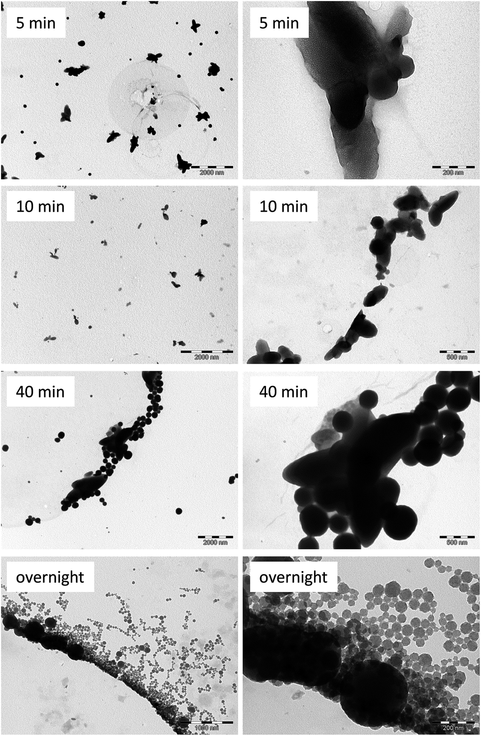 | ||
| Fig. 3 TEM micrographs of Ru–C60 structures synthesized in CH2Cl2 using a Ru/C60 ratio of 2/1 at −20 °C (scale bar: 5 min, 2000 and 200 nm; 10 min, 2000 and 500 nm; 40 min, 2000 and 500 nm; and overnight, 1000 and 200 nm). | ||
After 40 min of reaction, big spheres are observed (mean diameter of ca. 290 nm), which are clearly formed from the elongated objects. In the synthesis of polymeric particles a second step is observed, where the resulting droplets can again break up into daughter droplets. This second mechanism is much slower. We stirred the reaction overnight and a last sample was observed by TEM after 12 h. This sample shows spherical particles of 37.0 ± 0.3 nm mean diameter and big spherical particles of about 330 nm. These results indicate that a second step towards the formation of smaller spheres is indeed active. TEM images clearly show that the small spheres (≈40 nm) are formed from the large 300 nm spheres.
The STEM-EDX maps of the Ru–C60 structures synthesized at −20 °C in CH2Cl2 (see ESI.3†) confirm that the structures observed in the early stages of the reaction are constituted by Ru and C, as well as the spheres observed at 40 min of reaction. Nagashima et al. studied the synthesis of PdnC60 polymer from [Pd2(dba)3(CHCl3)] (dba = dibenzylideneacetone) in toluene using several Pd/C60 ratios.13 From microprobe analyses, they proposed a mechanism for the formation of C60Pdn, in which a one dimensional polymer C60Pdl is formed at the first stage. Then, insertion of additional Pd atoms between the polymer chains make bridges to form C60Pdn (n > 1). For C60Pdn (n > 3), an excess of Pd atoms would be deposited on the surface of C60Pd3. In a study on C60–Pd film electrodeposition from palladium acetate, Grądzka et al. have shown that in the presence of a large excess of palladium precursor, Pd NPs can be deposited on the polymer surface.58 In order to better understand the Ru system, we decided to explore the effect of the Ru/C60 ratio on the produced Ru–C60 structures. Using CH2Cl2 as solvent we fixed the concentration of C60 and we progressively increased the ruthenium content to obtain Ru/C60 ratios of 1, 2, 5, 10, 20, 30 and 50. The TEM images of the synthesized materials are shown in Fig. 4. The TEM and HREM analyses show that the Ru–C60 1/1 sample does not contain Ru NPs. Increasing the Ru content, Ru NPs are observed. Ru NPs display in all cases a small mean diameter, even at high Ru/C60 ratios, ranging from 1.10 to 1.35 nm (see Table 2 and ESI.4†). Interestingly, the Ru–C60 spheres do not change significantly in size for Ru/C60 ratios from 1/1 to 10/1 (≈40 nm, ESI.5†). At a 20/1 ratio, two size distributions are observed for the spheres: the major one centered at 39.8 ± 1.1 nm together with some bigger spheres with mean diameter of 78.8 ± 0.7 nm (see ESI.6 for size histograms†). The Ru–C60 30/1 sample also displayed spheres with a bimodal size distribution (56.4 ± 4.7 nm and 103.2 ± 1.0 nm). At 50/1 ratio the mean diameter of the spheres is 63.3 ± 0.8 nm (Fig. 4 and Table 2). The 30/1, and more particularly, the 50/1 samples are also characterized by the presence of aggregated Ru NPs. These aggregated Ru NPs are very similar to the ones obtained by decomposition of the [Ru(COD)(COT)] precursor in the absence of C60 (ESI.7†).
 | ||
| Fig. 4 TEM images of Ru–C60 structures synthesized in CH2Cl2 using a Ru/C60 ratio of 1/1 (scale bar 100 nm and 5 nm), 2/1 (scale bar 50 nm), 5/1 (scale bar 50 nm), 10/1 (scale bar 100 nm), 20/1 (scale bar 50 nm), 30/1 (scale bar 200 nm) and 50/1 (scale bar 200 nm). | ||
| Ru/C60 | Ru (%) | Nanoparticles mean sizea (nm) | Spheres mean sizea (nm) |
|---|---|---|---|
| a Manual measurement from enlarged micrographs of at least 200 objects. | |||
| 1/1 | 10.6 | Not observed | 39.1 ± 0.5 nm |
| 2/1 | 16.7 | 1.16 ± 0.02 nm | 31.6 ± 0.6 nm |
| 5/1 | 35.6 | 1.31 ± 0.03 nm | 42.6 ± 1.0 nm |
| 10/1 | 48.7 | 1.26 ± 0.03 nm | 32.4 ± 0.3 nm |
| 20/1 | 50.4 | 1.10 ± 0.01 nm | 39.8 ± 1.1 nm|85.2 ± 2.9 nm |
| 30/1 | 54.7 | 1.34 ± 0.01 nm | 56.4 ± 4.7 nm|103.2 ± 1.0 nm |
| 50/1 | 61.9 | 1.35 ± 0.02 nm | 63.3 ± 0.8 nm |
The extremely small size of the Ru species present in the Ru–C60 1/1 sample is a drawback for a classical TEM analysis. To overcome this inconvenient, a scanning TEM in high-angle annular dark field (STEM) approach, based on the Z-contrast dependence, can be useful to identify the small metallic species supported in/on the lighter matrix. For the 1/1 ratio of Ru–C60, the STEM-HAADF micrographs (Fig. 5a and b) show the presence of Ru atoms and/or clusters all over the surface of the spheres. The Ru clusters size does not exceed 0.6 nm. In the case of a representative sphere for the 30/1 ratio (Fig. 5c and d) the HAADF micrographs show the presence of small Ru NPs with a higher concentration of Ru on the surface of the sphere. The distribution of the Ru signal, acquired in STEM-EDX along a line scan, confirms the presence of Ru NPs on the outer sphere surface within the 30/1 sample (ESI.8†), whereas a more uniform distribution of Ru is observed for the 1/1 sample (ESI.8†). The above TEM or STEM analyses on the 2D projections of the objects do not provide clear information on the possible presence of Ru species (clusters or small NPs) in the interior of the spheres. Electron tomography analysis allowed the investigation within the volume of theses spheres.
 | ||
| Fig. 5 (a) STEM-HAADF micrographs of Ru/C60 1/1 (scale bar 20 nm), (b) (scale bar 10 nm), (c) STEM-HAADF micrographs of Ru/C60 30/1 (scale bar 200 nm), and (d) (scale bar 50 nm) nanostructures. In (b), the yellow and red arrows point to the Ru few atom clusters and larger clusters, respectively. | ||
Fig. 6 shows the results obtained for the 30/1 Ru–C60 ratio. From the slices views of 3D reconstructed volumes (Fig. 6b and c), the presence of small Ru NPs is obvious on the surface of the sphere, creating a Ru NP shell with a thickness around 7 nm, which correspond to a multilayered NP structure. This is in agreement with the Ru signal distribution observed in Fig. 5c and d, as the heavier elements appear most contrasted in STEM-HAADF, and also with the distribution of the Ru signal, acquired in STEM-EDX (ESI.8†). It is thus clear that no crystallized Ru NPs are present inside the spheres, but this analysis does not exclude the presence of atomic Ru inside the polymeric matrix. The analysis was also performed for the Ru–C60 sample with a 1/1 ratio, but the very small size of the clusters prevents their localization (see ESI.9†), the size of the clusters being below the resolution limit for the electron tomography analysis. The resolution attained in electron tomography is in the nanometer range, thus it is practically impossible to evidence the presence of any metal atoms and/or few-atom clusters within the spheres. It is therefore reasonable to propose that, in CH2Cl2, the decomposition of the [Ru(COD)(COT)] precursor leads to the formation of polymeric spheres containing Ru atoms or small clusters and fullerenes at low Ru–C60 ratio (≤1), and that further increase of the Ru–C60 ratio lead to Ru atoms, clusters or NPs deposition on the surface of these polymeric spheres.
 | ||
| Fig. 6 3D analysis of Ru/C60 30/1 by electron tomography: (a) bright field TEM images at 0° tilt from the tilt series; (b) and (c) representative cross-sectional slices along one direction in the plane of 3D reconstructed volume showing the location of shell of Ru 2 nm size NPs on the surface of the sphere. The thickness of the shell is 7 nm (scale bar 50 nm). | ||
The addition of extra Ru atoms in the polymeric spheres might be prevent by diffusional limitations. We checked independently that the decomposition of an excess of [Ru(COD)(COT)] on the Ru–C60 1/1 sample, to reach a 10/1 ratio, leads to NPs deposition outside the Ru–C60 1/1 spheres (ESI.10†).
Cold FEG confirmed de absence of clusters on the Ru–C60 matrix of 1/1 sample (Fig. 7).
 | ||
| Fig. 7 STEM-HAADF micrographs of Ru/C60 1/1. | ||
3.2 DFT calculations on the Ru–C60 nanoarchitectures
There are five possible sites on C60 surface where Ru atoms may be adsorbed: (i) an atop site (η1) on which the Ru atom is coordinated to a single carbon atom, (ii) a bridge site between two hexagonal rings (η2(6)), (iii) a bridge site between pentagonal and hexagonal rings (η2(5)) on which a Ru atom sits on the C–C bond and forms two Ru–C bonds, (iv) a hollow site above the center of a pentagonal ring of C atoms (η5), and (v) a hollow site above the center of a hexagonal ring of C atoms (η6).59 For a single Ru atom interacting with one C60, the most stable configuration is η2(6), with an adsorption energy of −48 kcal mol−1, followed by η2(5), η6, η5, with energies of −39, −38, and −35 kcal mol−1 respectively, when the atop site is instable. The better stability of the η2(6) site is not surprising, as the double bond shared by two hexagons corresponds to a maximum of the electronic density of the fullerene. Experimentally, this coordination mode has been observed for Pt complexes and suspected for Ru ones.60 These adsorption energies are significantly larger than the ones obtained for Ni, Au or Ta atoms for instance, with typical values smaller than −25 kcal mol−1,31,33,34 but remain in very good agreement with values obtained for Rh, Ir, Pd and Pt atoms.36 Pt atoms bind more strongly than Ru ones on C60 since, using the same computational settings; the adsorption energy of a Pt atom is already −66 kcal mol−1. This value is also in good agreement with previously reported values.36,61,62 For the η2(6) and η2(5) sites, the smallest Ru–C bond lengths are 2.06 Å. Then, the Ru–C bond length increases with the increase of adsorption energy for the different sites. For the η6 site, two Ru–C bond-lengths are 2.25, two others Ru–C distances are 2.34 when the last ones are 2.42 Å. It means that a single Ru prefers to lie in a displaced hollow site than a highly symmetric site. Finally, the Ru–C distances in the less stable site, i.e. in the η5 position, are 2.21 Å. Interestingly the most stable Ru–C60 complex is magnetic with a moment of 2 μB.If now, we take into account the real source of Ru in the calculation, the [Ru(COD)(COT)] precursor, we have to consider that it may coordinate even when it is partially decomposed. The [Ru(COD)] species adsorbs, in a η2(6) site, with two short Ru–C bond-lengths of 2.11 Å and adsorption energy of −58 kcal mol−1. The η1 site has almost the same energy but with a smaller Ru–C distance, i.e. 2.04 Å. On the contrary, the [Ru(COT)] species has a preference for the η1 site, with an adsorption energy of −38 kcal mol−1, the η2(6) site is 4 kcal mol−1 higher. However when using the following energy balances: [Ru(COD)(COT)] + C60 → [Ru(COT)]@C60 + COD or [Ru(COD)(COT)] + C60 → [Ru(COD)]@C60 + COT, meaning that we take into account the precursor dissociation energy, the energy differences become largely positive: +21 and +28 kcal mol−1, respectively. As a result the precursor has to be completely decomposed by the action of H2 in order to allow for the creation of Ru–C60 bond, and no partially decomposed Ru complexes can be stabilized on C60 surfaces.
In interaction with 2 fullerenes, a single Ru atom will preferably coordinate in a η2–η2 position, bridging two η2(6) positions, with 4 Ru–C bond-lengths of 2.07 Å, as shown in Fig. 8a. This complex adopts then a dumbbell like structure, as for a Ni31,63 or Pt atoms.36,61,62 The corresponding stabilization energy of this nonmagnetic complex is large, −44 kcal mol−1, when adding a C60 to an existing Ru–C60 complex. However, this binding energy for a Ru complex is still lower than the value of −65 kcal mol−1, obtained at a semi-empirical level, on a C60–Pt–C60 complex.62 It suggests again that a Ru atom has a little less affinity for C60 than a Pt one. Interestingly, another stable structure that exhibits a η2(6)–η6 coordination mode, as it can be seen from Fig. 8b, lies only 12 kcal mol−1 higher in energy. The corresponding Ru–C distances are ranging from 2.07 to 2.85 Å for this site that connects 8C to the Ru atom. This relatively small energy difference between the two coordination modes can be reduced by 5 extra kcal mol−1 due to the presence of adsorbed hydrogen atoms on the C60, as proposed experimentally, cf. Section 2.3, in the vicinity of the Ru atom (ESI.11†). Indeed a significant change of the Ru coordination is observed upon H2 adsorption since the most stable structure possesses a η2(6)–η4 character. More details on the H2 adsorption thermodynamics are provided in ESI.† In the case of the dumbbell like structure (Fig. 8a) it seems obvious that a third C60 can be easily coordinated to the central Ru atom. This reaction is still thermodynamically favorable but the energy gain is only −11 kcal mol−1, due to the coordination mode of the third fullerene that is η1. This stable Ru(C60)3 configuration might be viewed as a potential linker between ideal linear polymeric chains, as described in the following. The two C60–Ru–C60 complexes (Fig. 8a and b) can thus be viewed as elementary bricks for hypothetical 1D chains made of –C60–Ru–C60– with a 1Ru/1C60 ratio. Fig. 8d shows a first model made of a unit cell that contains only 1Ru and 1C60. The corresponding optimized lattice constant is around 10.4 Å. In this particular state, the four smallest Ru–C distances are lying in 2.04 to 2.11 Å range and two others are at 2.42 and 2.45 Å, when the next nearest C atoms are 2.66 Å away. As a consequence the Ru atom has a η2(6)–η4 coordination mode in this case. To allow for more flexibility in the coordination mode, we have also used a model made of 2Ru atoms and 2C60 in the calculation cell. As a result of rotating slightly one of the C60 molecule on a vertical axe, a η2(6)–η6 coordination mode is stabilized, as shown in Fig. 8e.
 | ||
| Fig. 8 (a) Side views of C60–Ru–C60 complex in the dumbbell like structure, (b) in the η2(6)–η6 coordination mode, and (c) of the most stable Ru–(C60)3 complex. (d) Ideal 1D chains and their corresponding lattice parameters for 1Ru/1C60 ratio for the η2(6)–η4 in state, and (e) for the η2(6)–η6 state. (f) Partially hydrogenated ideal polymeric chains with one Ru atom hydrogenated, and (g) with one C60 hydrogenated. Ru atoms are in grey, carbon atoms in brown and H in white. | ||
In this structure a Ru atom does not have a perfect hollow position with 8 different Ru–C bond-lengths: 2.04, 2.11, 2.14, 2.18, 2.34, 2.44, 2.53 and 2.57 Å. Almost the same values are yielded for the second metallic center (Fig. 9). This coordination mode is in good agreement with EXAFS results as shown below.
 | ||
| Fig. 9 Side view of an ideal 1D chain for 1Ru/1C60 ratio in the η2(6)–η6 state. Ru atoms are in grey, carbon atoms in brown. | ||
If we now try to complete the coordination sphere of one Ru atom by approaching a H2 molecule, it spontaneously dissociates and push the Ru to change its coordination to be η2(6)–η3 with two additional Ru–H distances are 1.59 Å, see Fig. 8f. When these two hydrides are transferred to one of the C60, the η2(6)–η6 coordination modes are recovered but with a slight elongation of the largest Ru–C distances, that are now between 2.60 and 2.70 Å (Fig. 8g). In these cases, the Ru atoms provide large electronic density to the neighboring C60, with a charge transfer of around 0.6e−. This value is not surprising since C60 is a well-known electron acceptor and it is in reasonable agreement with Raman spectroscopy results, see below.
From the different microscopy techniques, it seems obvious that outside the spheres that contain the polymeric phase, Ru NPs are formed. To propose an answer at the molecular level of this statement, we have addressed two issues: are the thermodynamics in favor of Ru NPs formation? Could it be the solvent that protects metallic atoms and avoid Ru–Ru formation bonds?
Starting from an ideal polymeric 1D chain, and thus adding a second metal atom is indeed energetically favorable; as well as adding 2 other ones, as it can be seen in Table 3. An interesting feature is that the lattice parameter values are increased to accommodate the creation of new Ru–C bonds, and thus could be experimentally detected. Moreover the distance between the two Ru1–Ru2 in Fig. 10a is very unusual for metallic bond with a value of 2.28 Å. With 4Ru atoms, see Fig. 10b, the Ru4–Ru2 bond-length is 2.26 Å, the Ru1–Ru2 is slightly elongated (+0.09 Å) when the last one is 2.35 Å. From Table 3, where energy gains per Ru atom are compiled in various binding situations, i.e. in single complex with different ratio Ru/C60, in some ideal 1D polymeric chains or even in small cluster models and finally in the bulk, we can provide some interesting insights of the reaction media behavior. Indeed, when comparing the stabilization energies of a Ru atom involved in a 1D chain, and one in small clusters or even worse in the bulk, it is clear that a Ru atom prefers to bind to other Ru atoms.
| Systems | Energy gain (kcal mol−1) |
|---|---|
| C60–Ru | −48 |
| (C60)2–Ru | −92 |
| (C60)3–Ru | −103 |
| …–C60–Ru–C60–… | −86 |
| …–C60–Ru2–C60–… | −94 |
| …–C60–Ru3–C60–… | −106 |
| …–C60–Ru4–C60–… | −76 |
| Ru13 | −96 |
| Ru55 | −123 |
| Ru bulk | −153 |
 | ||
| Fig. 10 (a) Side views and lattice parameters corresponding to a 2 Ru/C60 and (b) 4 Ru/C60 ratio. Ru atoms are in grey, carbon atoms in brown. | ||
If one considers Ru13 cluster formation energy (−96 kcal mol−1) the two processes are thermodynamically favorable. At this step, one can assume that the polymeric phase, with a ratio close to 1Ru/1C60 without excluding small clusters of Run (n ≤ 3) linking fullerenes is the kinetic product of the reaction and then larger Ru NPs are created, producing the thermodynamic products of the reaction. To support this idea we have also calculated the binding energy of Ru13–(C60)x complexes with x up to 6. The corresponding values are slightly lower than the other values given in Table 3: the energy gain per C60 is maximal for x = 1 and x = 2, with −87 and −88 kcal mol−1 respectively, and decreases when x increases: −77, −74, −61 kcal mol−1. See ESI.12† for molecular models of the stable Ru13–C60, Ru13–(C60)2, and Ru13–(C60)6 complexes. It means that when fullerenes are in excess, they have also the possibility to strongly bind metallic NPs.
However, in the present theoretical picture of the system, it is not clear why the polymeric phase is stabilized and has a net preference for the 1Ru/1C60 ratio. To propose a reasonable explanation, at the atomic scale, we have further investigated the effects of two different solvent molecules, i.e. toluene and dichloromethane, on the stabilization of the polymeric phase. Since the solvent molecules interact only weakly with the metallic center, see ESI.13† for a more detailed discussion, the main reason is only a steric effect that avoid the agglomeration of Ru atoms.
3.3 Infrared, NMR and Raman characterization
The attenuated total reflectance infrared (ATR-IR) spectra were recorded for Ru–C60 samples in the solid state (ESI.12†). Characteristic peaks of C60 are observed at 524, 576, 1182 and 1422 cm−1, together with other vibrations in the ranges 2800–3000 cm−1 (C–H stretching) and 400–1600 cm−1 (C–H bending, C–C stretching, C–H out of plane). Some of the peaks have been attributed to C60H18 (ref. 64) and C60H36 (ref. 65) species, the rest of the peaks are probably due to the presence of a mixture of hydrofullerenes with different number of hydrogen atoms.66,67 The presence of fullerene in the samples was also confirmed by MAS 13C{1H} NMR, with a signal at 143.2 ppm for Ru–C60 1/1, 2/1, 5/1 and 10/1 samples (see ESI.13†). The 13C{1H} MAS NMR also show a low intensity broad signal at 40 ppm. The CP-MAS 13C NMR (CP = 1H–13C cross polarization) allowed to clearly observe the broad signal at 40 ppm, which increased the intensity with increasing the Ru content. We attributed this broad signal to the presence of a mixture of hydrogenated fullerenes,64,68–72 as observed by ATR-IR. This was also confirmed by 1H MAS NMR with the presence of a large peak at 4.5 ppm. However, the low intensity of these peaks indicates that fullerene hydrogenation is only a side reaction. If usually high temperatures and pressures are necessary for fullerene hydrogenation,73,74 the hydrogenation of C60 on Rh/Al2O3 catalyst at ambient temperature and pressure has already been reported.75Since carbon monoxide is a sensitive probe for studying the surface composition of metal NPs, the CO adsorption on the Ru–C60 nanostructures was investigated by ATR-IR. Carbon monoxide was reacted with solid samples of Ru–C60 in a Fisher–Porter bottle under mild conditions (1.5 bar of CO, r.t., 24 h). Then, ATR-IR spectra were recorded with a spectrometer available in a glove box (ESI.12b†). After CO exposure Ru–C60 1/1, 2/1, 5/1 and 10/1 samples display three new peaks between 1900 and 2130 cm−1, which are typical of adsorbed terminal CO species. In the Ru–C60 1/1 nanostructure, the peaks appear at 1998, 2053 and 2120 cm−1. The [Ru(CO)3(alkene)2] complexes typically show three adsorption bands in the CO stretching region, in particular [Ru(CO)3(C2H4)2] complex display three bands at 2081, 2005 and 1995 cm−1.76–78 Considering that C60 acts as an electron-deficient olefin, the bands should be shifted to highest stretching frequencies in a [Ru(CO)3(η2–C60)2] species compared to [Ru(CO)3(C2H4)2], fitting with the spectrum obtained for the Ru–C60 1/1 nanostructure, i.e. CO molecules are likely to coordinate to the Ru atoms in a polymeric structure to form species such as [Ru(CO)3(μ-η2,η2-C60)]∞. However, species containing more or less CO ligands, as well as a mixture of species, cannot be discarded, as the signals are relatively broad. Ru–C60 2/1, 5/1 and 10/1 samples show the same pattern, although the signal at 1998 cm−1 becomes broader when increasing the Ru content. We assigned this broad signal, which increases in intensity with the number of Ru NPs present on the sample, to terminal CO adsorbed on the Ru NPs surface as it usually appears in the region of 1970–2000 cm−1.79,80 For higher Ru–C60 ratios, the ATR-IR signal intensity was too low for interpretation.
C60 is a well-known electron acceptor and Raman spectroscopy can give valuable information on charge transfer. Fig. 11a shows Raman spectra excited at 532 nm of C60 and Ru–C60 1/1, 2/1, 5/1 and 10/1 samples, in the spectral range of the tangential pitch mode Ag(2). It has been shown that the energy of the Ag(2) mode (1468 cm−1 for pure C60) is sensitive to charge transfer when evaporating C60 on metal surfaces,81 or for transition metal fullerides.19 While the charge transfer depends on the work function of the metal, other factors such as screened metal–adsorbate interactions and the effect of covalent bond formation may also influence the observed spectral shift, but are believed to be less important. It is commonly accepted that this mode is downshifted by approximately 6 cm−1 transferred to C60 in ionic fulleride compounds. For C60 alkali metals the downshift is directly proportional to the number of metal atoms since each atom donates one electron. The relationship between shift and composition is more complicated for the transition metal fullerides since these compounds may exhibit a large proportion of covalent bonding between metal and C60. The Ag(2) mode downshift observed for Pd–C60 and Pt–C60 fullerides is 15 cm−1, and it suggests that these fullerides have a structure with the metal atoms connected by a η2-bonding to two neighboring C60 molecules. From the work function of Ru (4.71 eV) one would expect a similar spectral shift as for Cu (4.70 eV) of −23 cm−1. We observe here a spectral shift as large as −10.1 cm−1 for the Ru–C60 10/1 sample, and a significant broadening with increasing amount of C60 caused by strong electron–phonon interaction. For the Ru–C60 1/1 sample, the spectral shift is of −6 cm−1. The spectrum of C60 without Ru is shown at the bottom of Fig. 11a for comparison. The fact that the spectra with Ru do not show a superimposed spectrum of C60, demonstrates that the C60 is strongly interacting with Ru. The Ag(2) mode downshift difference between the Ru–C60 and Pd–C60 and Pt–C60 fullerides might be due to a different coordination mode: a η2-bonding to two neighboring C60 for Pd–C60 and Pt–C60, and η2 and η6 for the Ru–C60 fulleride. The charge transfer to the C60 molecules is therefore not only dependent on the metal but also on the type of covalent bonds. The charge transfer was also evidenced by XPS, by comparing the binding energy of Ru3p3/2 (462.2 eV) with that of metallic ruthenium (461.2 eV). Fig. 11b shows how the spectral shift progressively saturates at higher molar concentration of C60. No sizeable spectral shifts are observed for the Hg(7) and Hg(8) modes, which is consistent with only smaller shifts observed for C60 on Cu (<7 cm−1). The smaller observed spectral shift observed for the Ru–C60 samples compared to metal surfaces, indicates that the work function of the Ru species is larger than for bulk Ru.
 | ||
| Fig. 11 (a) Raman spectra excited at 532 nm of C60 and Ru–C60 1/1, 2/1, 5/1 and 10/1 samples in the spectral range of the tangential pitch mode Ag(2); and (b) Raman spectral position of the Ag(2) band as a function of molar concentration of C60. | ||
3.4 WAXS and EXAFS characterization
Samples sealed in Lindemann glass capillaries were measured by WAXS using a diffractometer dedicated to pair-distribution function (PDF) studies (low background, Mo radiation). For the samples with high Ru/C60 ratio, the obtained diffractograms are very close and fully consistent with metallic Ru in the hcp system (Fig. 12a) with no significant contribution of C60. After corrections and Fourier transforms, the related PDF functions are as expected also very close, and consistent with metallic Ru NPs with low structural disorder and sizes (from coherence length) reaching 2.5 nm. From the shape of the envelope characterized by a rapid initial decrease and a secondary maximum for a larger value (ca. 1.5 nm) before the final decrease, size dispersion is likely, with a large proportion of NPs much smaller than the 2.5 nm value. This is in agreement with TEM measurements. Evolution is much different for smaller Ru/C60 ratios: for 10/1, 5/1 and 2/1, we first observe (Fig. 12b) a gradual decrease of the peaks characteristic of the Ru hcp structure, then for the 1/1 ratio the hcp crystalline signature totally vanishes and the diffractogram is closer to the one obtained for pure C60, excepted for the sharp peaks at low angle observed only for the highly ordered pure C60 sample. The main interest of PDF analysis is the capability to go beyond crystalline order and to analyze characteristic distances in the material, related to crystalline periodicity or not. In the present case, we observe on the PDF a decrease of the metallic Ru order, however mostly marked for the longer distances for the 10/1, 5/1 and 2/1 ratios, actually indicating Ru NPs with a more single size distribution and an average diameter close to 1.5 nm for the 2/1 ratio. For the 1/1 ratio (ESI.14†), we observe a drastic change for the distances in the 0.2–0.3 nm range: for 2/1, we can still observe the Ru–Ru bonding distance (0.265 nm in bulk Ru), it however fully vanishes for 1/1, which in this range of distances is indeed very close to pure C60. Distribution of distances in this range may also quite well accommodate bonds between Ru atoms and light elements, especially C (the 0.21–0.23 nm range for the Ru–C bonding distance is quite common). | ||
| Fig. 12 (a) Left – diffractograms for high Ru/C60 ratio together with Ru hcp reference data, right – related PDF. (b) Left – diffractograms for lower Ru/C60 ratio with Ru hcp reference data, right – related PDF. | ||
In addition, ab initio calculations, as well as the results of our DFT calculations, show a significant contraction of the bond lengths for Run clusters (n < 13): Ru–Ru bonding distance between 0.21 and 0.24 nm. However, since non-bonding C–C distances from C60 obviously also pile up there, such agreement with PDF is however not a clear evidence of Ru–C or Ru–Ru bonds. All these elements clearly indicate that for the 1/1 ratio, there is no evidence of Ru NPs, although we cannot completely discarded some metallic bonding for Ru atoms in small clusters in this sample. Order is dominated by the C60 structure for short distances, but discrepancies for distances above 0.7 nm strongly indicate more extended ordering, however difficult to safely characterize. Short distances are also in good agreement with eventual Ru–C bonding. For the 2/1 ratio, small Ru NPs (ca. 1.5 nm) can be observed. For the 5/1 ratio and above, these small NPs can still be observed but associated to an increasing proportion of larger NPs (2.5 nm from coherence length), which suggests increasing coalescence from small NPs.
 | ||
| Fig. 13 (a) Ru K-edge XANES from 22.09 to 22.17 keV (red: Ru–C60 1/1 N2 at RT, and blue: Ru–C60 1/1H2 at 150 °C); (b) magnitude of the Fourier transform of k2-weighted Ru EXAFS (k2: Δk = 2.8–11.3 Å−1) (blue: Ru–C60 2/1, H2 at 150 °C, red: Ru–C60 1/1, H2 at 150 °C); and (c) Ru K-edge XANES from 22.09 to 22.17 keV (blue: Ru–C60 1/1, H2 at 150 °C red: Ru–C60 2/1, H2 at 150 °C). | ||
| Sample | XANES energy, keV | Scatter | N | R, Å | Δσ2 (×103) | Eo, eV |
|---|---|---|---|---|---|---|
| Ru foil, ref | 22.1170 | Ru–Ru | 12 | 2.68 | 0.0 | 8.5 |
| RuO2 | 22.1285 | Ru–O | 5.8 | 1.97 | 3.0 | 0.5 |
| Ru(NH3)6Cl3 | 22.1268 | |||||
| Ru(NH3)6Cl2 | 22.1248 | |||||
| Ru–C60 1/1, N2 at RT | 22.1244 | Ru–C | 8.3 | 2.21 | 3.0 | 7.7 |
| Ru–C60 1/1, H2 at 150 °C | 22.1244 | Ru–C | 8.3 | 2.21 | 3.0 | 8.7 |
| Ru–C60 2/1, H2 at 150 °C | 22.1241 | Ru–C | 5.1 | 2.21 | 3.0 | 9.4 |
| Ru–Ru | 2.2 | 2.63 | 4.0 | 1.5 |
The XANES energy of Ru–C60 2/1 is the same as Ru–C60 1/1, e.g., 22.1244 keV; however, the shape of the XANES is slightly different, indicating some small difference in structure. The magnitude of the FT of Ru–C60 2/1 shows that there are fewer light scatters and a new higher R peak at about 2.5 Å (phase uncorrected distance). Fits of the EXAFS spectra indicate there are fewer Ru–C, 5.1 Ru–C at 2.24 Å, compared to sample Ru–C60 1/1. In addition, the scatter at longer R is due to Ru–Ru scatter with a coordination number of 2.2 at 2.63 Å, typical of metallic Ru NPs. Since non-metallic Ru–C has 8 bonds, a coordination number of 5.1 indicates that approximately 61% of the Ru is bonded to C in the Ru–C60 2/1 sample. The remaining Ru is, therefore, metallic, i.e., 39%. For the metallic fraction the true coordination number is the measured coordination number divided by the fraction of metallic Ru, or 2.2/0.39, or 5.6. For fcc and hcp metals a coordination number of 5.6 is consistent with a particle size of about 1.5 nm.82 In summary, sample Ru–C60 1/1 has 8 Ru–C bonds, which are stable to reduction in H2 at 150 °C; while in sample Ru–C60 2/1 approximately one-third of the Ru is reduced to 1.5 nm metallic Ru NPs. In the latter, the remaining two-thirds Ru–C are identical to those in Ru–C60 1/1 sample.
4. Conclusions
In summary, the decomposition reaction of [Ru(COD)(COT)] in the presence of C60 has been investigated, and the product(s) of the reaction characterized. The choice of the solvent affects the course of the reaction. Spherical particles are selectively produced in solvent with low viscosity and high permittivity, such as dichloromethane. The particle size depends on the nature of the solvent. According to the Ru/C60 ratio, these spherical particles can be surface decorated with metallic Ru NPs (1.5–2.5 nm), which are stabilized by C60. The structure of the spherical particles has been studied. TEM, EXAFS, WAXS, and DFT calculations point to a polymeric structure, in which each Ru atoms is coordinated to two C60, with a η2(6)–η6 coordination mode. Solvent molecules contribute to stabilize this fulleride. This polymeric phase is the kinetic product of the reaction. Then, larger Ru NPs are created on their surface, producing the thermodynamic products of the reaction. During the decomposition reaction under hydrogen, partial hydrogenation of the fullerene occurs, catalysed by the ruthenium. Significant charge transfer from ruthenium to fullerene has been evidenced by Raman spectrometry and XPS for all the prepared materials, which is an important factor to take into account, particularly if we consider the possible reactivity of this fulleride. We believe that these results should open the possibility to draw structure/properties relationships.Acknowledgements
This work was supported by the Centre National de la Recherche Scientifique (CNRS), which we gratefully acknowledge. The authors acknowledge financial support from the program of China Scholarships Council (CSC) for F. L. grant. I. C. Gerber also acknowledges the Calcul en Midi-Pyrénées initiative-CALMIP (Project p0812) for allocations of computer time. Part of this work was also performed using HPC resources from GENCI-IDRIS and CINES (Project x2015096649).References
- F. Giacalone, N. Martín and F. Wudl, in Fullerene Polymers, Wiley-VCH Verlag GmbH & Co. KGaA, 2009, pp. 1–14 Search PubMed.
- B. Coq, J. Marc Planeix and V. Brotons, Appl. Catal., A, 1998, 173, 175–183 CrossRef CAS.
- N. F. Goldshleger, Fullerene Sci. Technol., 2001, 9, 255–280 CrossRef CAS.
- B. Machado and P. Serp, in Nanostructured Carbon Materials for Catalysis, The Royal Society of Chemistry, 2015, pp. 312–411 Search PubMed.
- R. Loutfy and S. Katagiri, in Perspectives of Fullerene Nanotechnology, ed. E. Ōsawa, Springer, Netherlands, 2002, ch. 32, pp. 357–367 Search PubMed.
- B. S. Sherigara, W. Kutner and F. D'Souza, Electroanalysis, 2003, 15, 753–772 CrossRef CAS.
- C. Brosseau, Surf. Coat. Technol., 2011, 206, 753–758 CrossRef CAS.
- M. J. Rosseinsky, Chem. Mater., 1998, 10, 2665–2685 CrossRef CAS.
- M. Riccò, T. Shiroka, M. Belli, D. Pontiroli, M. Pagliari, G. Ruani, D. Palles, S. Margadonna and M. Tomaselli, Phys. Rev. B: Condens. Matter Mater. Phys., 2005, 72, 155437 CrossRef.
- V. N. Ivanova, J. Struct. Chem., 2000, 41, 135–148 CrossRef CAS.
- M. J. Rosseinsky, J. Mater. Chem., 1995, 5, 1497–1513 RSC.
- T. Yildirim, O. Zhou and J. E. Fischer, in The Physics of Fullerene-Based and Fullerene-Related Materials, ed. W. Andreoni, Springer, Netherlands, 2000, vol. 23, ch. 2, pp. 23–66 Search PubMed.
- H. Nagashima, A. Nakaoka, Y. Saito, M. Kato, T. Kawanishi and K. Itoh, J. Chem. Soc., Chem. Commun., 1992, 377–379 RSC.
- K. Winkler, K. Noworyta, W. Kutner and A. L. Balch, J. Electrochem. Soc., 2000, 147, 2597–2603 CrossRef CAS.
- L. Norin, U. Jansson, C. Dyer, P. Jacobsson and S. McGinnis, Chem. Mater., 1998, 10, 1184–1190 CrossRef CAS.
- G. H. M. Dias, Quim. Nova, 1995, 18, 592–596 CAS.
- V. Lavrentiev, H. Abe, S. Yamamoto, H. Naramotoand and K. Narumi, Mol. Cryst. Liq. Cryst., 2002, 386, 139–143 CrossRef CAS.
- M. Wohlers, B. Herzog, T. Belz, A. Bauer, T. Braun, T. Rühle and R. Schlögl, Synth. Met., 1996, 77, 55–58 CrossRef CAS.
- A. V. Talyzin and U. Jansson, Thin Solid Films, 2003, 429, 96–101 CrossRef CAS.
- A. S. Gurav, Z. Duan, L. Wang, M. J. Hampden-Smith and T. T. Kodas, Chem. Mater., 1993, 5, 214–216 CrossRef CAS.
- J. M. Cowley, M. Q. Liu, B. L. Ramakrishna, T. S. Peace, A. K. Wertsching and M. R. Pena, Carbon, 1994, 32, 746–748 CrossRef CAS.
- A. V. Talyzin, A. Dzwilewski and M. Pudelko, Carbon, 2007, 45, 2564–2569 CrossRef CAS.
- K. Winkler, K. Noworyta, A. de Bettencourt-Dias, J. W. Sobczak, C.-T. Wu, L.-C. Chen, W. Kutner and A. L. Balch, J. Mater. Chem., 2003, 13, 518–525 RSC.
- T. Braun, M. Wohlers, T. Belz, G. Nowitzke, G. Wortmann, Y. Uchida, N. Pfänder and R. Schlögl, Catal. Lett., 1997, 43, 167–173 CrossRef CAS.
- V. Lavrentiev, H. Abe, H. Naramoto, S. Sakai and K. Narumi, Chem. Phys. Lett., 2006, 424, 101–104 CrossRef CAS.
- D. Östling and A. Rosén, Chem. Phys. Lett., 1993, 202, 389–393 CrossRef.
- J. Roques, F. Calvo, F. Spiegelman and C. Mijoule, Phys. Rev. B: Condens. Matter Mater. Phys., 2003, 68, 205412 CrossRef.
- J. Pitarch-Ruiz, S. Evangelisti and D. Maynau, J. Mol. Struct.: THEOCHEM, 2004, 681, 203–207 CrossRef CAS.
- L. M. Ramaniah and M. Boero, J. Chem. Phys., 2010, 133, 134701 CrossRef PubMed.
- A. N. Andriotis, M. Menon and G. E. Froudakis, Phys. Rev. B: Condens. Matter Mater. Phys., 2000, 62, 9867–9871 CrossRef CAS.
- A. N. Andriotis and M. Menon, Phys. Rev. B: Condens. Matter Mater. Phys., 1999, 60, 4521–4524 CrossRef CAS.
- M. K. Shukla, M. Dubey and J. Leszczynski, ACS Nano, 2008, 2, 227–234 CrossRef CAS PubMed.
- Q. Zeng, X. Chu, M. Yang and D.-Y. Wu, Chem. Phys., 2012, 395, 82–86 CrossRef CAS.
- L. M. Ramaniah, M. Boero and M. Laghate, Phys. Rev. B: Condens. Matter Mater. Phys., 2004, 70, 035411 CrossRef.
- M. Matsubara, C. Massobrio, L. M. Ramaniah, E. Ruiz and M. Boero, Phys. Rev. B: Condens. Matter Mater. Phys., 2010, 81, 195433 CrossRef.
- O. Loboda, Quantum-chemical studies on Porphyrins, Fullerenes and Carbon Nanostructures, Carbon Nanostructures, Springer-Verlag Berlin, Heidelberg, 2013 Search PubMed.
- J. R. Kremer, D. N. Mastronarde and J. R. McIntosh, J. Struct. Biol., 1996, 116, 71–76 CrossRef CAS PubMed.
- C. MessaoudiI, T. Boudier, C. O. S. Sorzano and S. Marco, BMC Bioinf., 2007, 8, 288 CrossRef PubMed.
- G. Kresse and J. Furthmüller, Comput. Mater. Sci., 1996, 6, 15–50 CrossRef CAS.
- G. Kresse and J. Hafner, Phys. Rev. B: Condens. Matter Mater. Phys., 1993, 47, 558–561 CrossRef CAS.
- G. Kresse and J. Furthmüller, Phys. Rev. B: Condens. Matter Mater. Phys., 1996, 54, 11169–11186 CrossRef CAS.
- G. Kresse and J. Hafner, Phys. Rev. B: Condens. Matter Mater. Phys., 1994, 49, 14251–14269 CrossRef CAS.
- P. E. Blöchl, Phys. Rev. B: Condens. Matter Mater. Phys., 1994, 50, 17953–17979 CrossRef.
- G. Kresse and D. Joubert, Phys. Rev. B: Condens. Matter Mater. Phys., 1999, 59, 1758–1775 CrossRef CAS.
- J. P. Perdew, K. Burke and M. Ernzerhof, Phys. Rev. Lett., 1996, 77, 3865–3868 CrossRef CAS PubMed.
- M. Dion, H. Rydberg, E. Schröder, D. C. Langreth and B. I. Lundqvist, Phys. Rev. Lett., 2004, 92, 246401 CrossRef CAS PubMed.
- G. Román-Pérez and J. M. Soler, Phys. Rev. Lett., 2009, 103, 096102 CrossRef PubMed.
- J. Klimeš, D. R. Bowler and A. Michaelides, Phys. Rev. B: Condens. Matter Mater. Phys., 2011, 83, 195131 CrossRef.
- K. Jiří, R. B. David and M. Angelos, J. Phys.: Condens. Matter, 2010, 22, 022201 CrossRef PubMed.
- K. Momma and F. Izumi, J. Appl. Crystallogr., 2011, 44, 1272–1276 CrossRef CAS.
- C. Amiens, B. Chaudret, D. Ciuculescu-Pradines, V. Colliere, K. Fajerwerg, P. Fau, M. Kahn, A. Maisonnat, K. Soulantica and K. Philippot, New J. Chem., 2013, 37, 3374–3401 RSC.
- C. Vauthier and K. Bouchemal, Pharm. Res., 2009, 26, 1025–1058 CrossRef CAS PubMed.
- C. Mabille, F. Leal-Calderon, J. Bibette and V. Schmitt, Europhys. Lett., 2003, 61, 708–714 CrossRef CAS.
- V. Schmitt, F. Leal-Calderon and J. Bibette, Top. Curr. Chem., 2003, 227, 195–215 CrossRef CAS.
- S. K. Smoukov, T. Tian, N. Vitchuli, S. Gangwal, P. Geisen, M. Wright, E. Shim, M. Marquez, J. Fowler and O. D. Velev, Adv. Mater., 2015, 27, 2642–2647 CrossRef CAS PubMed.
- CRC Handbook of Chemistry and Physics, ed. W. M. Haynes, CRC Press, 95th edn, 2014 Search PubMed.
- R. S. Ruoff, D. S. Tse, R. Malhotra and D. C. Lorents, J. Phys. Chem., 1993, 97, 3379–3383 CrossRef CAS.
- E. Grądzka, M. Wysocka-Żołopa and K. Winkler, J. Phys. Chem. C, 2014, 118, 14061–14072 Search PubMed.
- M. R. Axet, O. Dechy-Cabaret, J. Durand, M. Gouygou and P. Serp, Coord. Chem. Rev., 2016, 308, 236–345 CrossRef CAS.
- P. J. Fagan, J. C. Calabrese and B. Malone, Science, 1991, 252, 1160–1161 CrossRef CAS.
- H. Nagashima, Y. Kato, H. Yamaguchi, E. Kimura, T. Kawanishi, M. Kato, Y. Saito, M. Haga and K. Itoh, Chem. Lett., 1994, 23, 1207–1210 CrossRef.
- A. A. Voityuk, J. Phys. Chem. A, 2009, 113, 11801–11808 CrossRef CAS PubMed.
- Y. Huang and B. S. Freiser, J. Am. Chem. Soc., 1991, 113, 8186–8187 CrossRef CAS.
- A. D. Darwish, A. G. Avent, R. Taylor and D. R. M. Walton, J. Chem. Soc., Perkin Trans. 2, 1996, 2051–2054 RSC.
- R. Bini, J. Ebenhoch, M. Fanti, P. W. Fowler, S. Leach, G. Orlandi, C. Ruchardt, J. P. B. Sandall and F. Zerbetto, Chem. Phys., 1998, 232, 75–94 CrossRef CAS.
- A. V. Talyzin, Y. O. Tsybin, T. M. Schaub, P. Mauron, Y. M. Shulga, A. Zuettel, B. Sundqvist and A. G. Marshall, J. Phys. Chem. B, 2005, 109, 12742–12747 CrossRef CAS PubMed.
- A. V. Talyzin, Y. O. Tsybin, J. M. Purcell, T. M. Schaub, Y. M. Shulga, D. Noreus, T. Sato, A. Dzwilewski, B. Sundqvist and A. G. Marshall, J. Phys. Chem. A, 2006, 110, 8528–8534 CrossRef CAS PubMed.
- R. Ghafouri and M. Anafcheh, Fullerenes, Nanotubes, Carbon Nanostruct., 2015, 23, 40–48 CrossRef CAS.
- S. M. C. Vieira, W. Ahmed, P. R. Birkett, C. A. Rego, S. Kotsiris and T. Drewello, Fullerenes, Nanotubes, Carbon Nanostruct., 2004, 12, 139–145 CrossRef CAS.
- T. Wagberg, D. Johnels, A. Peera, M. Hedenstroem, Y. M. Schulga, Y. O. Tsybin, J. M. Purcell, A. G. Marshall, D. Noreus, T. Sato and A. V. Talyzin, Org. Lett., 2005, 7, 5557–5560 CrossRef CAS PubMed.
- R. E. Haufler, J. Conceicao, L. P. F. Chibante, Y. Chai, N. E. Byrne, S. Flanagan, M. M. Haley, S. C. O'Brien and C. Pan, et al., J. Phys. Chem., 1990, 94, 8634–8636 CrossRef CAS.
- A. S. Lobach, Y. M. Shul'ga, O. S. Roshchupkina, A. I. Rebrov, A. A. Perov, Y. G. Morozov, V. N. Spector and A. A. Ovchinnikov, Fullerene Sci. Technol., 1998, 6, 375–391 CrossRef CAS.
- S. M. Luzan, Y. O. Tsybin and A. V. Talyzin, J. Phys. Chem. C, 2011, 115, 11484–11492 CAS.
- A. V. Talyzin, Y. M. Shulga and A. Jacob, Appl. Phys. A: Mater. Sci. Process., 2004, 78, 1005–1010 CrossRef CAS.
- L. Becker, T. P. Evans and J. L. Bada, J. Org. Chem., 1993, 58, 7630–7631 CrossRef CAS PubMed.
- Y. M. Wuu, J. G. Bentsen, C. G. Brinkley and M. S. Wrighton, Inorg. Chem., 1987, 26, 530–540 CrossRef CAS.
- Y. M. Wuu, C. Zou and M. S. Wrighton, Inorg. Chem., 1988, 27, 3039–3044 CrossRef CAS.
- S. L. Ingham and S. W. Magennis, J. Organomet. Chem., 1999, 574, 302–310 CrossRef CAS.
- P. Lara, T. Ayvali, M.-J. Casanove, P. Lecante, A. Mayoral, P.-F. Fazzini, K. Philippot and B. Chaudret, Dalton Trans., 2013, 42, 372–382 RSC.
- X. Qi, M. R. Axet, K. Philippot, P. Lecante and P. Serp, Dalton Trans., 2014, 43, 9283–9295 RSC.
- S. J. Chase, W. S. Bacsa, M. G. Mitch, L. J. Pilione and J. S. Lannin, Phys. Rev. B: Condens. Matter Mater. Phys., 1992, 46, 7873–7877 CrossRef CAS.
- J. T. Miller, A. J. Kropf, Y. Zha, J. R. Regalbuto, L. Delannoy, C. Louis, E. Bus and J. A. van Bokhoven, J. Catal., 2006, 240, 222–234 CrossRef CAS.
Footnote |
| † Electronic supplementary information (ESI) available. See DOI: 10.1039/c6ra12023g |
| This journal is © The Royal Society of Chemistry 2016 |