Microbial assisted industrially important multiple enzymes from fish processing waste: purification, characterization and application for the simultaneous hydrolysis of lipid and protein molecules

S. Hepziba Suganthi and K. Ramani*
Biomolecules and Biocatalysis Laboratory, Department of Biotechnology, SRM University, Kattankulathur, Kancheepuram District-603203, Tamil Nadu, India. E-mail: ramani.k@ktr.srmuniv.ac.in; microramana@yahoo.co.in; Fax: +91-44-27417770; Tel: +91-44-27417770

Received 7th May 2016 , Accepted 13th September 2016

First published on 15th September 2016


Abstract

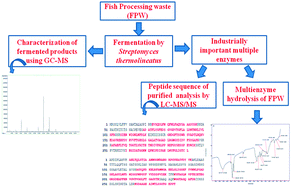
Fish processing waste (FPW) was evaluated as the substrate for the concomitant production of industrially important alkaline lipase and protease by Streptomyces thermolineatus for the hydrolysis of lipid and protein rich FPW. The FPW contributed to the effective growth of the organism and also aided the enzyme production. Media optimization was performed using response surface methodology for maximum enzyme production (lipase 402 U ml−1; protease 896 U ml−1). The enzymes were purified with ammonium sulphate precipitation, dialysis, and gel filtration chromatography and achieved a specific activity of lipase and protease of 903 and 2539 U mg−1 respectively, and purity of 8.6 and 10.8 fold respectively. The purified enzymes were stable over a wide range of temperatures (30–70 °C), pH (6.5–9.5), organic solvents and surfactants, with higher affinities for their substrates. Hydrolysis studies showed that the purified lipase and protease hydrolysed 76 and 86% of lipid and protein respectively. In conclusion, these enzymes have great potential for industrial applications especially treating waste containing multiple substrates.


1. Introduction

The food processing industries, slaughter houses, oil processing and refineries, dairy industries, etc. generate considerable amounts of lipid and protein-rich solid waste that are used in the production of low value-added products.1 The fish processing waste (FPW) produced during fish processing is rich in lipids and proteins. The FPW is usually discharged into the marine environment, dewatered and landfilled/composted or sent to a fishmeal plant which contributes to environmental problems and also increases cost in terms of transportation and processing.2 In order to minimise the problems related to disposing of the FPW, these wastes are used as ingredients in the preparation of low value-added products like animal feed.3

The fish processing industries face major problems in handling these wastes and their disposal. The presence of lipid and protein increases the COD, BOD and organic matter.4 Despite of the biodegradability of lipid and protein molecules, their degradation is difficult because of the varied structure, solubility and the substrate specificity among them.5 Hence, there is a need of specific enzymes which are produced from specific substrates/wastes (lipid or protein) to degrade the respective wastes effectively.

The present study dealt with the microbial utilization of FPW and production of high value-added enzymes such as lipase and protease for industrial applications. The usage of combination of enzymes like lipase and protease for the treatment of lipid and protein rich waste is gaining interest nowadays as it is specific and environmentally safe. There are several industrially important strains one being Streptomyces species which produces many secondary metabolites and enzymes with different substrate specificities.6,7 The variety of secondary metabolites and enzymes produced by the Streptomyces sp. are of extracellular nature which is considered as “generally regarded as safe” (GRAS) according to food and drug administration.8 Various Streptomyces sp. are reported to produce extracellular protease9–11 and lipase.12–14 But there is no report on the concomitant production of lipase and protease by Streptomyces sp. to the best of our knowledge. Also, there is no literature on the production of lipase or protease from Streptomyces thermolineatus. The Streptomyces sp. used in our study produced both lipase and protease by utilizing the FPW as the substrate.

The production of industrially important enzymes like lipase and protease using a single fermentation medium would greatly reduce the production and purification cost. Very few reports are available on the concomitant production and purification of lipase and protease which would be applicable for the processes requiring both lipase and protease.15 It is also mandatory that the produced lipase must be proteolytic resistant in order to perform its function in presence of protease.

The culture conditions like the physicochemical parameters, nutritional requirements and components present in the media not only interfere with the extracellular enzyme production but also vary for different organisms and different substrates and hence careful efforts must be taken to maximise the enzyme production. In the modern biotechnological era, the statistical approach, response surface methodology (RSM) is an effective tool, used for multiple variable optimization utilizing the basic principles of statistics, randomization, replication and duplication.16 RSM enables the identification of optimum conditions, significant factors involved and their interactions and also to quantify the relationship of the responses and important input factors in limited number of experiments.17

In the present investigation, the FPW was evaluated for its usability as substrate for the concomitant production of enzymes in a single fermentation system. In order to hydrolyse the FPW and to concomitantly produce lipase and protease, Streptomyces sp. was chosen as it is an industrially important organism that produces enzymes with different substrate specificity, high activity and a wide temperature and pH stability. The organism S. thermolineatus was used for the biodegradation of FPW containing lipid and protein and simultaneously produces lipase and protease. The culture conditions were optimized using RSM based on full factorial central composite design (CCD). The lipase and protease produced were characterized and used for the hydrolysis of FPW.

2. Materials and methods

Sample collection and preparation

The FPW obtained from a local fish market in refrigerated conditions was cooked until boiling and the hard parts were removed. The cooked FPW was blended in blender and the resulting slurry was stored at −20 °C until use. The sample was prepared in bulk quantity and stored in deep freezer (at −20 °C) as a stock and used for our entire study. Hence, the stability was maintained throughout the same. The lipid portion of FPW was extracted using chloroform and the lipid content was estimated by the procedure followed by Joseph et al.18 The residual sample containing protein was subjected to acetone precipitation at 4 °C, dissolved in distilled water and its protein content was checked using the method followed by Bradford.19

Screening and isolation of lipolytic and proteolytic organisms

The organism that produces alkaline lipase and protease, Streptomyces thermolineatus was isolated from oil contaminated soil of Chennai petroleum corporation limited (CPCL), Chennai using starch casein broth containing (g l−1) starch, 10; casein powder, 1.0; KNO3, 2.0; NaCl, 2.0; K2HPO4, 0.02; and CaCO3, 0.01. The organisms that showed the zone of clearance on both tributyrin agar and skim milk agar plates were acclimatized to degrade fish processing waste. Tributyrin in tributryin agar is a triglyceride, composed of butyric acid and glycerol. The organism producing lipase cleaves the tributyrin and forms halos around the colony forming clear zones. Skim milk agar is used to demonstrate the proteolysis by cleaving casein. The tributyrin and skim milk agar are the selective media for the isolation of lipase and protease producing strains. Out of 31 isolates, the Streptomyces thermolineatus showed a better degradation of lipid18 and protein19 in FPW as well as produced maximum amount of lipase and protease and hence, it was chosen for the further study. The quantitative activity of lipase was determined by titrimetry method using olive oil emulsion as the substrate as described in Ramani et al., 2013.5 Five ml of olive oil emulsion, 2 ml of 0.03% Triton X-100, 1 ml of 3 M NaCl, 2 ml of 0.075% CaCl2 and 4 ml of phosphate buffer (pH 8.0) was added to 1 ml of the enzyme solution. The enzyme–substrate mixture was incubated at 45 °C for 15 min and the reaction was terminated by adding ethanol[thin space (1/6-em)]:[thin space (1/6-em)]acetone (1[thin space (1/6-em)]:[thin space (1/6-em)]1, v/v) to the mixture. The liberated fatty acids were titrated against 0.02 N NaOH, using phenolphthalein as an indicator with the appearance of pale pink color is the end point. The quantity of fatty acids liberated is calculated based on the equivalents of NaOH used to reach the titration end point. The blank experiment was performed following the same procedure without the enzyme addition. One unit of lipase activity was defined as the amount of enzyme that released 1 μM of fatty acid per min under assay conditions.

The quantitative assay of protease was determined using casein as the substrate with slight modification in the method followed by Anson, 1938.20 Five hundred microlitre of enzyme solution was added to 0.5 ml of 1% (w/v) substrate solution (casein) with 50 mM citrate phosphate buffer, (pH 8) and incubated for 30 min at 45 °C and the reaction was stopped by adding 1 ml of 10% trichloro acetic acid (TCA). The TCA added to enzyme–casein mixture without incubation served as the blank. Both the blank and test solutions were centrifuged at 10[thin space (1/6-em)]000 rpm for 10 min. To 0.4 ml of supernatant, 1.0 ml of 50 mM Na2CO3 and 0.2 ml of Folin–Ciocalteau reagent were added, and the reaction mixture was incubated at room temperature for 30 min and the absorbance was measured at 660 nm. One unit (U) of proteolytic enzyme activity was defined as the amount of enzyme that liberated 1 μg tyrosine per ml per minute from casein under specified assay conditions and the amount of tyrosine liberated was determined from the tyrosine standard curve.

The organism was maintained on starch casein agar slants at 4 °C and sub-cultured every 15 days. The selected organism was then characterized by morphological analysis, staining methods, biochemical tests and 16S rRNA sequencing. The phylogenetic tree was constructed to identify the closely related species.

Optimization of culture conditions by response surface methodology (RSM)

The lipase and protease production by biodegrading FPW were screened by varying the different time points (0–144 h), pH (2.0–10), temperature (10–70 °C), ammonium sulphate concentration (0.5, 1.0, 1.5, 2.0 and 2.5 g l−1 of the mineral salt medium), inoculum concentration (2.5–12.5% (v/v)) and different concentrations of metal ions (g l−1) [K2HPO4 (0.5–2.5), MgSO4 (0.1–0.5), NaCl (0.5–2.5), CaCl2 (0.1–0.5), FeSO4 (0.05–0.25)]. The significant factors were then optimized by RSM.

Response surface methodology (RSM) is a mathematical modelling tool that was employed to study the significant factors such as temperature, pH, incubation time and nitrogen source and also their interaction among them on the lipase and protease production. Central composite design (CCD) in Design Expert software, version 8.0.7.1 (Stat Ease Inc. Minneapolis, USA, trial version) was employed to design the experiment and to analyse the interaction of significant factors on multiple enzyme production. The above mentioned independent variables were studied at three different levels and a series of experiments (n = 30) were carried out. The model equation used for the analysis was given below:

image file: c6ra11867d-t1.tif
where Y is the predicted response, k is the number of factors, α0 is the design factor of interest, αi and αij are coefficients. The significance of the model was analyzed statistically using F-test of ANOVA and the coefficient of determination to measure the goodness of fit. The R2 value determines the accuracy and quality of the above polynomial model. The model was validated by performing the experiment for three times using the optimized conditions obtained from RSM.

Enzymes production

The organism Streptomyces thermolineatus was inoculated in the mineral salt medium containing composition (g l−1) K2HPO4 (1.0); CaCl2 (0.4); MgSO4 (0.2); NaCl (0.5); FeSO4·7H2O (0.2); fructose (0.5); (NH4)2 SO4 (2.0) and FPW (150) at pH 8 and incubated for 96 h and 120 h for protease and lipase extraction respectively. The cell free culture supernatant obtained by centrifugation at 6500 rpm for 20 min at 4 °C was assayed for lipase and protease activity. All experiments were done in triplicates.

Characterization of substrates and fermented products

CHN analysis. The composition of carbon, hydrogen and nitrogen content of the FPW was analyzed using Perkin-Elmer Series II 2400 CHNS/O Elemental Analyser. Required quantity of (2–5 mg) of FPW was combusted at 1500 °C under argon atmosphere and the elements were detected by the detector.

Determination of fatty acid composition by gas chromatography and mass spectrophotometry (GC-MS)

The composition of fatty acid present in initial and fermented FPW was determined by preparing methyl esters according to the methodology followed in Ichihara and Fukubayashi, 2010.21 The fatty acid methyl esters (FAME) were then identified using gas chromatography coupled to mass spectroscopy (GC-MS) model (Agilent technologies, USA, 7890B GC system connected to 5977A MSD) which is equipped with HP_5MS 5% phenyl methyl silox column with dimensions 30 m × 250 μm × 0.25 μm. Helium was used as carrier gas with a flow rate of 1 ml min−1, in a split ratio of 100[thin space (1/6-em)]:[thin space (1/6-em)]1. The analysis was carried out using 60 °C for 2 min, 10 °C min−1 to 200 °C, 5 °C min−1 to 240 °C and held at 240 °C for 8 min. The temperature of the injector and detector were set at 250 and 260 °C respectively. The obtained MS spectra were compared with the reference spectra present in the NIST.Lib.

Determination of amino acid composition by HPLC

The amino acid composition present in initial and fermented FPW was analyzed by HPLC. The samples were hydrolyzed using 6 N HCL at 100 °C for 24 h and neutralized to pH 7 with 10 N NaOH. The samples were then analyzed using Agilent 1100 HPLC amino acid analyzer.5

Enzymes purification

For enzymes purification, the cell free supernatant obtained was subjected to gradient ammonium sulphate precipitation from 20–80% (w/v) with the increase of 20% (w/v) each time. The ammonium sulphate was added to the supernatant to a saturation of 20% (w/v) and incubated for 2 h and centrifuged at 12[thin space (1/6-em)]000 rpm for 15 min at 4 °C and same procedure was repeated for 40, 60 and 80% (w/v) saturation. The precipitate collected was resuspended in 50 mM phosphate buffer (pH 8.0) and it was dialysed against the same buffer at 4 °C. The dialysate was loaded onto a pre-equilibriated Sephadex G-100 column, set at a flow rate of 0.5 ml min−1 using phosphate buffer (pH 8.0) and the fractions were collected. The quantitative activity of lipase and protease were determined after ammonium sulphate precipitation, dialysis and gel filtration column chromatography. The fractions containing the lipase and protease were pooled separately, lyophilised and subjected to SDS-PAGE for molecular weight determination and homogeneity. The amino acid composition of purified lipase and protease were analyzed by HPLC as described earlier.5

Determination of optimum reaction conditions

Effect of pH and temperature on the activity and stability of purified enzymes. The optimum pH for the purified lipase and protease were studied at different pH ranging from 3 to 9 in the following buffers: 100 mM acetate buffer (pH 3.0–5.0), 100 mM phosphate buffer (6.0–8.0), 100 mM tris buffer (pH 9.0–10.0) using olive oil and casein as substrate for lipase and protease respectively under standard assay conditions. The stability of the purified lipase and protease were studied by incubating the purified enzymes in different buffers of varying pH ranging from 3.0 to 10.0 for 1 h at 37 °C and the activity of lipase and protease were measured under standard assay conditions.

The optimum temperature for the purified lipase and protease were determined by incubating the enzyme at varying temperatures ranging from 30 to 90 °C at optimized pH 8. The thermal stability was studied by incubating lipase and protease at various temperatures (30 to 90 °C) for 1 h and the activity of lipase and protease were measured under standard assay conditions.

Effect of solvents, metal ions, detergents, reducing agents and inhibitors. The effect of polar solvents (methanol, ethanol, isopropanol, acetone and acetonitrile) and non polar solvents (pentane, toluene, hexane, benzene and octane) on the purified lipase and protease were carried out by incubating the purified enzymes with different solvents for 1 h at 45 °C. The stimulatory or inhibitory effects of metal ions on the enzymes were studied by incubating 1 mM concentration of CuSO4, MgCl2, ZnCl2, KCl, CaCl2, FeSO4 and MnCl2 with the buffered (pH 8.0) enzymes for 1 h at 45 °C and the activity of the lipase and protease were recorded.

The detergents like SDS, Triton X-100, Tween 20 and Tween 80 of concentration 0.1% were incubated with the buffered (pH 8.0) enzyme for 1 h at 45 °C. The buffered (pH 8.0) lipase and protease were also incubated with the reducing agents such as β-mercaptoethanol and dithiothritol and inhibitor like PMSF for 1 h at 45 °C. After the incubation time, the activity of the lipase and protease were determined.

Amino acid sequence analysis with liquid chromatography and mass spectrophotometry/mass spectrophotometry (LC-MS/MS)

The amino acid sequence of the purified lipase and protease was determined by LC-MS/MS LC (Shimadzu UFLC, Japan) MS/MS (Bruker, Germany, impact HD MS/MS TOF). Samples of purified lipase and protease were prepared according to the protocol described by Shevchenko et al.22 with slight modification. Briefly, 5 μg of purified lipase and protease were run in SDS-PAGE and the protein bands were excised into cubes (1 × 1 mm) using a clean scalpel. The excised bands were washed with milliQ water. After washing, 500 μl of 50 mM ammonium bicarbonate[thin space (1/6-em)]:[thin space (1/6-em)]acetonitrile (1[thin space (1/6-em)]:[thin space (1/6-em)]1, v/v) was added and incubated for 15 min and repeated the step twice. Then, 500 μl of acetonitrile was added and incubated at room temperature for 15 min and repeated the step twice. The gel pieces were saturated with 50 μl of 12 ng of trypsin in ammonium bicarbonate buffer and incubated overnight for enzymatic digestion of proteins. Added 50 μl of extraction buffer containing 5% formic acid in acetonitrile to each tube and incubated for 15 min at 37 °C under shaking. The supernatant was collected and dried in vacuum centrifuge and stored at −20 °C until use. For further LC-MS/MS analysis, 20 μl of 0.1% (v/v) trifluoro acetic acid was added, vortexed and centrifuged. The digested peptides were analysed by LC-MS/MS with the column zorbax eclipse plus C18 (4.6 × 100 mm × 3.5 μm). Two microlitre of the sample was injected to the column through autosampler (SIL20ACHT) with the flow rate of 0.4 ml min−1. The column was equilibriated with water and 0.1% formic acid and a linear gradient was performed for 30 min to reach 70% acetonitrile and it remained the same for another 5 min and brought back to 2% acetonitrile with the total run time of 45 min. The obtained mass spectra were analysed by data analysis software and data were compared with Swissprot database through Mascot search engine.

Fourier transform-infrared spectroscopy (FT-IR) of purified enzymes

The functional groups present in the purified lipase and protease were identified using a Perkin Elmer FT-IR spectrophotometer and the spectrum was analyzed in the spectral range of 400–4000 cm−1.

Substrate specificity of purified lipase and protease

The substrate specificity of purified lipase was determined according to the procedure followed by Gururaj et al.27 with the following substrates: p-nitrophenyl alkanoate esters of varying carbon chain length like p-NP acetate (C2), p-NP butyrate (C4), p-NP decanoate (C10), p-NP myristate (C14), and p-NP palmitate (C16). The substrate specificity of purified protease was tested using casein, bovine serum albumin, gelatin and azocasein at pH 8.0 and temperature 45 °C.

Determination of kinetic parameters

The enzyme kinetic parameters like maximum velocity (Vmax) and Michaelis–Menten constant (Km) for the purified lipase and protease were studied using the different concentration of olive oil emulsion and casein (1–10%) at pH 8.0 and temperature 45 °C. The Lineweaver–Burk equation plot (eqn (1)) was used to determine the kinetic parameters.
 
image file: c6ra11867d-t2.tif(1)
where [S] is the substrate concentration (mM) and V is the initial reaction rate of the enzyme (mM min−1).

Kinetic studies on the hydrolysis of FPW using the purified enzymes

In order to determine the efficiency of the purified lipase and protease on the hydrolysis of FPW, a series of batch experiments were performed by varying the incubation time (1–24 h), pH (3.0–9.0) and temperature (30–65 °C). The experiments were performed in a 100 ml conical flask by adding 1 g of FPW into 15 ml of 50 mM phosphate buffer (pH 8) containing 1000 U of purified lipase and protease each and incubated at 45 °C for the hydrolysis of FPW. The residual lipid and protein content in the sample were then analyzed. The % hydrolysis of FPW was calculated by
image file: c6ra11867d-t3.tif

3. Results and discussion

Isolation and identification of lipolytic and proteolytic microorganism

The oil contaminated soil samples are rich in organisms that are capable of producing lipase and protease. Amongst 31 organisms isolated, one organism showed clear zones on both tributyrin and skim milk agar suggesting that the organism was capable of producing both lipase and protease and it also indicated that the produced enzymes are extracellular. Upon staining, the organism was found to be rod shaped and Gram positive. The organism grows well at 45 °C and pH 8.0. The biochemical characterization (data not shown) and 16S rRNA sequencing followed by the BLAST revealed that the isolate was Streptomyces thermolineatus (Fig. 1). The NCBI Gene bank assigned an accession number for the submitted sequence and is KT757685. The blast result showed that it is closely related to S. thermolineatus (accession no. NR0112442.1).
image file: c6ra11867d-f1.tif
Fig. 1 Phylogenetic tree showing the relationship of S. thermolineatus with other Streptomyces sp.

Optimization of culture conditions using response surface methodology (RSM)

The growth rate of S. thermolineatus was satisfactory using FPW as substrate indicating that the FPW catered the needs of the organism for its growth. The production of protease and lipase began after 24 and 48 h of incubation respectively when the organism was in the exponential phase and the enzyme production reached their maximum during the stationary phase.23 In order to examine the factors that influence the growth and the enzyme production, RSM was used. After the preliminary experiments, RSM using the full factorial CCD method was employed to study the interactions of four significant factors (incubation time, temperature, pH and nitrogen source) that had great impact on lipase and protease production. Table 1 shows the high and low levels with the coded levels for the various significant factors. The software generated 30 different experiments using different combinations of four factors such as temperature, pH, time and nitrogen concentration. After the experiments were conducted, the results were fed to the software, analyzed the results and were depicted in Tables 2 and 3 for lipase and protease respectively. The regression analysis shown on Table 4 depicts the effects of the significant factors on the lipase and protease activity and were predicted by the second order polynomial function as
Lipase activity (U ml−1) = +389.6 − 9.5 × A − 2.17 × B − 9.0 × C − 2.3 × D − 3.00 × A × B + 4.00 × A × C + 6.25 × A × D + 5.0 × B × C + 4.75 × B × D + 2.75 × C × D − 28.46 × A2 − 51.46 × B2 − 67.21 × C2 − 50.96 × D2

Protease activity (U ml−1) = +868.67 − 5.50 × A − 17.83 × B − 6.50 × C + 3.33 × D + 9.25 × A × B − 30.00 × A × C + 21.2 × A × D − 10.75 × B × C + 6.50 × B × D − 1.75 × C × D − 77.29 × A2 − 62.04 × B2 − 74.29 × C2 − 58.79 × D2
where A, B, C and D correspond to incubation time, pH, temperature and nitrogen source respectively.
Table 1 High and low levels of significant factors with coded levels
Factor Name Lipase Protease
Low level High level Low level High level
A Incubation time (h) 96 144 72 120
B pH 7 9 7 9
C Temperature (°C) 40 50 40 50
D Ammonium sulphate concentration (g l−1) 1 3 1 3


Table 2 Experimental design for RSM with four independent variables showing the observed and predicted values of lipase production
Run A (incubation time, h) B (pH) C (temperature °C) D (nitrogen source g l−1) Mean observed response (U ml−1) Predicted response (U ml−1)
1 144 9 50 1 170 165.5
2 72 8 45 2 300 294
3 144 9 40 3 190 192.8
4 120 8 45 2 390 389.6
5 96 7 50 3 160 191.6
6 120 6 45 2 196 188.1
7 120 8 45 2 390 389.6
8 144 7 40 1 190 200.8
9 96 9 50 1 186 195
10 144 9 50 3 172 188.3
11 144 7 50 1 168 175.3
12 120 8 45 0 200 195.5
13 96 7 40 1 240 234.3
14 120 8 45 2 386 389.6
15 144 7 40 3 192 193.6
16 120 8 45 2 402 389.6
17 96 9 40 3 200 203.3
18 96 9 50 3 206 192.8
19 144 9 40 1 172 171
20 96 7 40 3 200 202.1
21 96 7 50 1 188 192.8
22 144 7 50 3 188 179.1
23 168 8 45 2 260 256.8
24 120 8 55 2 110 102.8
25 120 8 45 2 386 389.6
26 120 8 35 2 140 138.3
27 120 8 45 2 384 389.6
28 120 8 45 4 180 181.1
29 120 10 45 2 180 199.5
30 96 9 40 1 210 216.5


Table 3 Experimental design for RSM with four independent variables showing the observed and predicted values of protease production
Run A (incubation time, h) B (pH) C (temperature °C) D (nitrogen source g l−1) Mean observed response (U ml−1) Predicted response (U ml−1)
1 96 6 45 2 656 656.1
2 72 9 40 3 576 552.2
3 72 9 50 1 616 600.5
4 96 8 45 2 816 868.6
5 72 7 50 1 680 689.2
6 96 8 55 2 540 558.5
7 120 7 50 1 544 557.2
8 144 8 45 2 540 548.5
9 72 9 50 3 580 574.2
10 96 10 45 2 554 584.8
11 96 8 35 2 572 584.5
12 96 8 45 2 896 868.6
13 96 8 45 2 864 868.6
14 96 8 45 2 880 868.6
15 96 8 45 2 868 868.6
16 120 7 40 3 640 644.9
17 96 8 45 4 616 640.1
18 120 9 50 3 560 564.2
19 96 8 45 2 888 868.6
20 72 9 40 1 580 571.5
21 48 8 45 2 548 570.5
22 120 7 40 1 620 605.2
23 120 9 50 1 538 505.5
24 120 9 40 3 578 596.5
25 96 8 45 0 620 626.8
26 72 7 50 3 666 636.9
27 120 9 40 3 692 662.1
28 72 7 40 1 632 617.2
29 120 7 50 3 602 589.9
30 72 7 40 3 560 571.9


Table 4 ANOVA for the second-order polynomial model for the lipase and protease production
Source Degree of freedom Mean square F value P value prob > F
Lipase
Model 14 15[thin space (1/6-em)]582.12 153.38 <0.0001 (significant)
Residual 15 101.59    
Lack of fit 10 133.63 3.56 0.0867 (non-significant)
Pure error 5 37.5    
Total 29      
R2       0.9931
[thin space (1/6-em)]
Protease
Model 14 27[thin space (1/6-em)]578.59 31.15 <0.0001 (significant)
Residual 15 885.31    
Lack of fit 10 923.43 1.14 0.4697 (non-significant)
Pure error 5 809.07    
Total 29      
R2       0.9667


Analysis of variance (ANOVA-partial sum of squares-type III)

The significance of the model for the second order polynomial model was determined by the F and p-values shown in Table 4. The p-value is less than 0.0001 for both lipase and protease and the F-value is 153.3 and 31.15 for lipase and protease respectively. The p values denote the significance of each coefficient which is mandate to infer the interaction between the significant factors. Ordinarily, smaller the p value, greater is the significance of their coefficient.24 The F-value depicts the model adjusted well to the experimental data. The F-value of lack of fit for lipase and protease are 3.56 and 1.14 respectively denotes the pure error is not significant which is the indicative of the goodness of model devised. The Prob > F less than 0.0500 indicate the model is significant. Typically, as the coefficient of variation (CV) value gets lowered, the reliability of the experiment becomes more eminent. CV is a statistical measure that describes the amount of variability relative to the mean. CV value of lipase and protease are 4.35 and 4.55 respectively suggesting a better precision and reliability of experiments.25 The regression coefficient R2 was used to determine the model precision and was calculated to be 0.9931 and 0.9667 for lipase and protease respectively. The regression coefficient shows that the model could explain 99.3 and 96.7% of variability in the responses indicating that the devised model is more accurate and consistent.

Localization of the optimum conditions

The effects of the individual, independent factors and the interaction among them can be studied by the 3D response surface plots that are graphical representations of the regression equation. The optimum level of each factor can also be evaluated from 3D response surface plot based on model regression equation. The 3D response surface plots showed that the lipase and protease were produced in maximum levels at an optimum condition of temperature 45 °C, pH 8, ammonium sulphate concentration 2 g l−1 and time 96 h and 120 h for protease and lipase respectively. Fig. 2a–f showed the effects of interaction of two significant factors while keeping the other factor at zero level. The optimum point for each component can be obtained from the coordinates of the central point within the highest contour levels from each of the Fig. 2a–f.26
image file: c6ra11867d-f2.tif
Fig. 2 Response surface curve for lipase activity (U ml−1) by S. thermolineatus as a function of (a) time (h) and pH, (b) time (h) and temperature (°C) and (c) time and ammonium sulphate (g l−1) and protease activity (U ml−1) as a function of (d) time (h) and pH, (e) time (h) and temperature (°C) and (f) time and ammonium sulphate (g l−1).

The activity of lipase and protease were 402 and 897 U ml−1 at the optimized condition of time 120 h for lipase enzyme production, 96 h for protease enzyme production, temperature 45 °C, pH 8.0, ammonium sulphate concentration 2 g l−1, and 1% substrate concentration suggesting that the optimum culture condition is required for the maximum enzyme production.5 The presence of protease did not affected the production or the activity of lipase suggesting that the produced lipase is proteolytic resistant lipase.

Product characterization

The FPW before and after the fermentation was characterized by using C, H and N content, GC-MS and amino acid composition analysis. The total lipid and protein concentration in FPW were 13.19 and 68.8 mg g−1 respectively. The maximum lipid and protein conversion was observed at 120 and 96 hours and pH 8.0, temperature 45 °C. After fermentation at the optimized conditions, the lipid and protein levels were greatly reduced to 2.96 and 16.44 mg g−1 respectively suggesting the effective fermentation of S. thermolineatus. The percentage (w/w) of carbon, hydrogen and nitrogen present in the FPW before and after fermentation were 52.39, 9.53 and 8.82 and 32.2, 5.11 and 7.19 respectively. The CHN result showed there is a considerable decrease in the percentage of carbon, hydrogen and nitrogen after fermentation. This result clearly indicates the conversion of complex biomolecules present in the FPW into simpler units. The GC-MS chromatogram revealed the fatty acid profile with retention time before and after fermentation (Fig. 3a and b). The predominant fatty acids that are present before fermentation are palmitic acid (C17H34O2 – 42%), stearic acid (C19H38O2 – 21%), oleic acid (C19H36O2 – 18%), tetradecanoate (C15H30O2 – 5.1%), palmitoleic acid (C17H32O2 – 4.09%) and arachidic acid (C21H42O2 – 1.12%) with the retention time of 18.082, 20.778, 20.565, 15.730, 17.844 and 23.499 min respectively. The peaks of palmitoleic acid and arachidic acid present before fermentation were absent after fermentation. Also, the peak area of palmitic acid, stearic acid, oleic acid and myristic acid with the retention time of 18.082, 20.653, 20.315 and 15.730 min respectively, were greatly reduced after fermentation confirming the lipid degradation. The composition of amino acid in the initial and fermented FPW was shown in the Table 5. The increase in the amount of free amino acid in the fermented FPW indicates the proteolytic ability of the organism. The S. thermolineatus produces protease and breaks down the complex proteins into peptides and amino acids and utilizes them for their growth and survival. The amino acids such as serine, glutamine, glycine, valine, methionine, leucine, tryptophan are greatly increased after fermentation in the medium.
image file: c6ra11867d-f3.tif
Fig. 3 GC-MS chromatogram of fatty acid (a) initial FPW and (b) fermented FPW using S. thermolineatus.
Table 5 Amino acid composition of initial and fermented FPW
S. no. Amino acid Initial (%) Fermented (%)
1 Aspartic acid 0.563 0.613
2 Glutamic acid 0.113 0.205
3 Asparagine 0.209 0.135
4 Serine 0.436 0.831
5 Glutamine 0.346 0.593
6 Glycine 0.536 0.893
7 Alanine 0.916 1.035
8 Tyrosine 0.332 0.356
9 Histidine 0.593 0.693
10 Valine 0.395 1.103
11 Methionine 0.693 0.955
12 Isoleucine 0.963 1.084
13 Phenylalanine 0.783 0.983
14 Leucine 0.291 0.883
15 Lysine 0.195 0.382
16 Proline 0.559 0.778
17 Trytophan 0.193 0.465


Purification of enzymes

The extracellular lipase and protease present in 120 h cell free culture supernatant was purified by ammonium sulphate precipitation and gel filtration chromatography. Table 6 depicts the summary of the purification steps showing the specific activity of purified lipase and protease as 904 and 2540 U mg−1 with a yield of 21 and 27% respectively. The purification fold of 8.64 and 10.8 were achieved for lipase and protease respectively. The molecular mass of the purified lipase and protease were determined to be 34 and 23 kDa respectively on SDS-PAGE (Fig. 4). Mander et al.13 reported that the lipase produced from Streptomyces sp. was 37.5 kDa whereas the molecular weight of the protease was comparatively lower than protease isolated from Streptomyces sp. MAB18.23
Table 6 Summary of purification steps of lipase and protease from S. thermolineatus
Purification steps Total activity (U) Total protein (mg) Specific activity (mg) Yield (%) Purification fold
Lipase
Crude lipase 402[thin space (1/6-em)]000 3844 105 100 1
Ammonium sulphate precipitation 232[thin space (1/6-em)]000 1865 124 58 1.18
Dialysis 168[thin space (1/6-em)]000 438 384 42 3.67
Sephadex G-100 86[thin space (1/6-em)]000 95 904 21 8.64
[thin space (1/6-em)]
Protease
Crude protease 895[thin space (1/6-em)]833 3844 233 100 1
Ammonium sulphate precipitation 580[thin space (1/6-em)]833 1865 311 65 1.3
Dialysis 465[thin space (1/6-em)]833 438 1064 52 4.56
Sephadex G-100 241[thin space (1/6-em)]667 95 2540 27 10.8


image file: c6ra11867d-f4.tif
Fig. 4 SDS-PAGE showing the molecular weight of purified lipase and protease. Lane M molecular weight marker (14.3 to 97.4), lane 1 purified protease (23 kDa), lane 2 purified lipase (34 kDa).

Determination of amino acid sequence of purified enzymes by LC-MS/MS analysis

The amino sequence of the purified lipase and protease were determined by LC-MS/MS analysis. The obtained sequence of lipase and protease from Mascot search were compared with Swissprot database for homology search. The purified lipase and protease of S. thermolineatus showed 74 and 60% homology with lipase of Thermomyces langinosus and protease of Streptomyces griseus respectively. To date, there is no sequence data available for the lipase and protease of S. thermolineatus. The possible amino acid sequence of the purified lipase and protease were shown in Fig. 5 and 6 respectively. The molecular mass and isoelectric point (pI) of purified lipase were determined as 32 kDa and 5.36 respectively whereas for protease the molecular mass and pI were found as 29.81 kDa and 5.60 respectively. The amino acid sequencing analysis suggested that the isolated lipase and protease exhibits the characteristic of lipase and protease enzyme family.27 The LC-MS/MS analysis also suggested that the protease produced from S. thermolineatus is an aminopeptidase that cleaves the amino acid from N-terminal side of a protein.28 This result suggests the exopeptidase activity of the protease isolated from S. thermolineatus.
image file: c6ra11867d-f5.tif
Fig. 5 LC-MS/MS analysis and peptide sequence matches of lipase. (a) MS/MS fragmentation of VVFTGHSLGGALATVAGADLR found in LIP_THELA in SwissProt, lipase OS = Thermomyces lanuginosus (b) observed ions of each type are shown in bold and (c) possible sequence of purified lipase. Matched peptides are shown in bold.

image file: c6ra11867d-f6.tif
Fig. 6 LC-MS/MS analysis and peptide sequence matches of protease. (a) MS/MS fragmentation of AHLTQLSTIAANNGGNR found in APX_STRGG in SwissProt, aminopeptidase S OS = Streptomyces griseus subsp. griseus (b) observed ions of each type are shown in bold and (c) possible sequence of protease. Matched peptides are shown in bold.

Characterization of purified enzymes

Effect of pH and temperature on the purified enzymes activity and stability. The maximum activity of purified alkaline lipase and protease from S. thermolineatus were best at pH 8.0 suggesting the alkaline nature of the enzymes which is preferred for industrial use especially in detergent industry.29,30 Fig. 7a shows the relative activity and stability of purified lipase and protease at different pH. The purified lipase showed above 86% activity in the pH range of 7–9 and it retained very good activity after 1 h confirming its stability at this pH range. At pH 9.5 and 6.5, the residual lipase activity was declined to 31.8 and 56.2% respectively, due to the denaturation of lipase. This result is in agreement with that of lipase produced from Streptomyces coelicolor A(3)2 showing pH stability in the range (6.0–9.0) reported by Cote and Shareck.31 The lipases produced by Streptomyces bambergiensis OC25-4[thin space (1/6-em)]14 and Streptomyces sp.12 were also found to be active at an optimum pH of 8.0. Though the protease showed its optimum activity at pH 8.0, it exhibited good stability retaining more than 80% of its initial activity in pH range between 6.5 and 9.0. At pH 6 and 9.5, the enzyme retained the residual activity of 75 and 64.8% respectively as reported by other workers.9,32,33 Ghorbel et al.8 also reported that the protease produced from Streptomyces flavogriseus HS1 was highly active between pH 6.0 and 8.0, with optimum pH at 7.0. Our data suggested that the purified alkaline lipase and protease from S. thermolineatus were stable over a broad range of pH and this property is highly desirable for industrial applications.
image file: c6ra11867d-f7.tif
Fig. 7 Relative activity and stability of purified lipase and protease at different (a) pH and (b) temperatures.

The activity of the purified lipase and protease were determined by varying temperatures from 30 to 90 °C and is depicted in Fig. 7b. The maximum activity of lipase and protease were observed at 45 °C and 68 and 77% of residual lipase and protease activity respectively were observed at 70 °C. The activity of both enzymes declined beyond 70 °C. The loss of activity above 70 °C could be due to the denaturation of enzyme structure by heat.34 Fig. 7b also showed that the lipase and protease produced by S. thermolineatus are highly stable (100%) at 45 °C and they can withstand temperatures from 30–60 °C retaining more than 85% of their initial activities whereas at 70 °C, purified lipase and protease retained 74 and 47% activity respectively, after 1 h of incubation. These results suggest that the produced lipase and protease can withstand high temperatures which are highly preferable for the industrial purpose. The enzymes that withstand high temperatures are good candidates for biotechnological applications especially in synthetic reactions.35

Effect of organic solvents and metal ions. The use of enzymes for many industrial purposes has gained attention in last few decades. The lipases are employed for the bioconversion in presence of organic solvents and the proteases are used in the peptide and ester synthesis under non-aqueous conditions. So there is a constant search for novel solvent tolerable enzymes to use for industrial applications.13 The stability of purified lipase and protease were tested on various organic solvents such as DMSO, methanol, acetonitrile, ethanol, acetone, isopropanol, benzene, toluene, hexane and octane with varying log[thin space (1/6-em)]P values from −1.378 to 4.783 at 10% solvent concentration (Table 7). The log[thin space (1/6-em)]P value is a logarithm of the partition coefficient between water and n-octanol and it provides a measure of differential solubility of the solvent.13 The purified lipase was more stable in the presence of non-polar solvents (log[thin space (1/6-em)]P value > 2.13–4.783) like benzene, hexane, toluene and octane showing 114–138% of residual activities. The purified lipase was moderately stable in the presence of polar solvents (log[thin space (1/6-em)]P < −1.378) like DMSO, methanol, ethanol, acetone and isopropanol with the residual activities of 52–93%. The lipase was more stable in non polar solvents than polar solvents. The polar solvents clear off the water molecule from the enzyme's active site attributing to lesser activity whereas the non polar solvents being immiscible with enzyme render it work at the interphase thereby enhancing the enzyme's activity.13
Table 7 Effect of organic solvents and metal ions on purified lipase and protease activity
Organic solvents (10%) log[thin space (1/6-em)]P value Relative lipase activity (%) Relative protease activity (%) Metal ions (1 mM) Relative lipase activity (%) Relative protease activity (%)
Control   100 100 Control 100 100
DMSO −1.378 97 93 Cu2+ 71 87
Methanol −0.764 77 80 Mg2+ 113 96
Acetonitrile −0.394 15 37 Zn2+ 53 69
Ethanol −0.235 60 74 Mn2+ 35 92
Acetone −0.208 43 52 Fe2+ 93 95
Isopropanol 0.074 49 60 K+ 98 94
Benzene 2.13 119 24 Ca2+ 96 123
Toulene 2.46 114 30 EDTA 33 63
Hexane 3.764 138 86      
Octane 4.783 115 94      


The purified protease was quiet stable in both polar and non-polar solvents of which DMSO showed maximum relative activity of 93%. DMSO enhanced the protease activity of Streptomyces sp.11 Benzene and toluene inhibited the protease activity retaining 24 and 30% respectively of its initial activity. This is in contrast with the results reported by Sangeetha et al.15 and Doddapaneni et al.36 Acetonitrile inhibited both lipase and protease retaining the residual activity of 15 and 37% respectively after 1 h of incubation whereas acetone moderately inhibited both enzymes with the residual activity of 43 and 52% for lipase and protease respectively. Acetonitrile found to strongly inhibit the lipase of Streptomyces sp. CS268[thin space (1/6-em)]13 and Pseudomonas otitidis.5 The above results showed that the purified lipase and protease from S. thermolineatus are stable in both polar and non-polar solvents. This property is highly applicable in various industrial applications which are processed with the polar and non-polar solvents particularly in pharmaceutical and food processing industries for flavour synthesis.37

Many enzymes require metal ions for their activity and certain enzymes use metal ions as a cofactor. In this study, the effect of various metal ions such as Cu2+, Mg2+, Zn2+, Mn2+, Fe2+, K+, Ca2+ and EDTA at a concentration of 1 mM was studied on the activity of purified lipase and protease and the results were tabulated in Table 7. The activity of lipase was enhanced by Mg2+ to 113% whereas Cu2+, Zn2+, Mn2+ and EDTA inhibited the lipase and it retained 71, 53, 35 and 33% respectively of its original activity. Similar results has been reported by Ayaz et al.12 in which it was observed that lipase from Streptomyces sp. OC119-7 showed stimulatory effect by Mg2+ and inhibitory effect by Zn2+ and Mn2+. The Fe2+, K+ and Ca2+ had least effect on the lipase activity. The calcium ion stimulated the activity of protease by 123%. Apart from Ca2+, other metal ions had least effect on purified protease except for Zn2+ and EDTA that inhibited the protease activity by 31 and 37% respectively. The Ca2+ enhances the activity of protease produced from Lactobacillus brevis, Anthrobacter sp., Corynebacterium sp. and Bacillus subtilis.38,39 The inhibitory effect of EDTA was observed on both purified lipase and protease suggesting that the enzymes from S. thermolineatus are metal dependent.15

Effect of detergents, inhibitors and reducing agents. In the last few decades, the usage of enzymes in the detergent industries is common and hence the need for enzymes compatible with detergent formulation is a common requisite. The effect of surfactants such as SDS, Triton X-100, Tween 20 and Tween 80 on the activity of purified lipase and protease were tabulated in Table 8. The activity of lipase and protease were unaffected by the non-ionic detergent Triton X-100. Tween 20 inhibited lipase activity by 50% whereas SDS and Tween 80 moderately inhibited it retaining the residual lipase activities of 71 and 73% respectively. Mander et al.13 reported that lipase produced from Streptomyces sp. CS268 was inhibited by Tween 20, Tween 80 and SDS but the Triton X-100 enhanced the lipase activity. The protease remains unaffected by the surfactant tested except for Tween 20 which moderately inhibited the activity by 21%. Tween 20, Tween 80 and Triton X-100 had little influence on the protease from Streptomyces flavogriseus HS1 whereas SDS strongly inhibited it retaining only 19% of protease activity.8 The above results demonstrated that the presence of surfactant did not have much impact on the produced lipase and protease activity suggesting their applicability in industries.
Table 8 Effect of detergents, inhibitors and reducing agents on purified lipase and protease activitya
Detergents (0.1%) Relative lipase activity (%) Relative protease activity (%) Inhibitors & reducing agents (1 mM) Relative lipase activity (%) Relative protease activity (%)
a The enzyme activity of the control was taken as 100%. SDS-sodium dodecyl sulphate, DTT – dithiothreitol.
Control 100 100 Control 100 100
SDS 71 99 PMSF 96 29
Triton X-100 97 96 β-Mercaptoethanol 75 80
Tween-20 50 79 DTT 82 88
Tween-80 73 92      


The purified lipase and protease were incubated with 1 mM PMSF, beta mercaptoethanol and dithiothritol for 1 h at 45 °C and their effect on enzyme activity has been tabulated in Table 8. The lipase remains unaffected by PMSF whereas residual activity of protease was greatly reduced to 29% suggesting that protease produced by S. thermolineatus is a serine protease.40 The reducing agent like β-mercaptoethanol and dithiothritol showed moderate inhibition on the activity of both lipase and protease suggesting the need for the disulphide bridges for the enzyme's activity.5 The disulphide bridges in the enzymes helps in the stabilization of the enzyme structure thereby establishing the function of the enzymes.

Amino acid composition of purified lipase and protease

The amino acid composition of purified lipase and protease from S. thermolineatus were determined using HPLC (Table 9). It was found that the purified lipase contained 30.81% of polar amino acid and 69.18% of non-polar amino acid whereas the protease had 42.7 and 57.2% of polar and non-polar amino acids respectively. The ratio of polar/non-polar amino acid of lipase and protease was found as 0.44 and 0.74 respectively. The presence of high percentage of non-polar amino acids present in the lipase attributes to the hydrophobicity of the lipase.
Table 9 Amino acid composition of purified lipase and protease
Amino acids in lipase Mol (%) Amino acids in protease Mol (%)
Polar amino acids
Aspartic acid 4.14 Aspartic acid 4.15
Glutamic acid 11.14 Glutamic acid 2.24
Serine Traces Serine Traces
Glutamine 4.08 Glutamine 1.84
Threonine 2.41 Threonine 2.48
Arginine 6.75 Arginine 3.86
Lysine 2.74 Lysine 2.36
Tyrosine 9.08 Tyrosine 10.01
Histidine 2.36 Histidine 3.83
[thin space (1/6-em)]
Non-polar amino acids
Glycine 2.49 Glycine 2.04
Valine 2.47 Valine 6.04
methionine 6.75 Methionine 10.83
Isoleucine 11.39 Isoleucine 16.58
Leucine 10.18 Leucine 11.8
Alanine 7.62 Alanine 4.03
Proline 1.52 Proline 10.7
Cysteine 11 Cysteine 2.36
Phenylalanine 2.79 Phenylalanine 2.36
Trptophan 1.01 Trptophan 2.36


Functional groups of purified lipase and protease

The FT-IR spectra reveal the major functional groups present in the purified lipase and protease and is shown in the Fig. 8a and b. The spectral region from 1300 to 1800 cm−1 represents the peaks corresponding to the peptide group vibration. The peaks at 1427.92 cm−1, 1643.661 cm−1 and 1449.97 cm−1, 1648.5 cm−1 in the spectra of purified lipase and protease respectively, can be attributed to the C–N stretching vibrations of amide and C[double bond, length as m-dash]O stretching vibrations of amide I of enzymes.41 The broad band at 3426.98 cm−1 and 3429.46 cm−1 in the spectra of lipase and protease respectively, could be attributed to secondary amine and peptide bond.42
image file: c6ra11867d-f8.tif
Fig. 8 FT-IR spectra of purified (a) lipase and (b) protease from S. thermolineatus.

Substrate specificity of purified lipase and protease

The purified lipase showed more specificity towards long and intermediate carbon length of p-nitrophenyl esters than the short carbon chain fatty acid (Table 10). The specific activity towards the C16 substrate was about 4.5 fold higher than that of C4 carbon chain length. The specificity of purified protease towards various substrates like casein, bovine serum albumin, gelatin and azocasein was evaluated. The protease exhibited highest specificity for casein (610 U ml−1), followed by BSA (530.83 U ml−1), azocasein (436.67 U ml−1) and gelatin (304.16 U ml−1) (Table 10). This suggests that the protease showed specificity for a wide range of substrates. The specificity towards a range of substrate shows that the protease produced from S. thermolineatus could be used for the hydrolysis of large polypeptides and complex proteins.43
Table 10 Substrate specificity of purified lipase and protease
Substrate Enzyme activity (U ml−1)
Substrate specificity of lipase (U ml−1)
p-Nitrophenyl acetate (C2) 50
p-Nitrophenyl butyrate (C4) 66.99
p-Nitrophenyl decanoate (C10) 118.91
p-Nitrophenyl myristate (C14) 124.34
p-Nitrophenyl palmitate (C16) 223.11
[thin space (1/6-em)]
Substrate specificity of protease (U ml−1)
BSA 530.833
Casein 610
Azocasein 436.67
Gelatin 304.166


Determination of enzyme kinetic parameters

The Michaelis Menten constant (Km) and maximum velocity (Vmax) of the purified lipase and protease using olive oil and casein respectively were determined by Lineweaver–Burk plot (Fig. 9a and b). The Km and Vmax of purified lipase were 2.25 mM and 149.54 mM min−1 respectively, which was 30 times higher than the Vmax of lipase from Streptomyces sp. CS326.44 The Km and Vmax of purified protease were 5.6 mM and 32.38 mM min−1 respectively. The Km represents the enzyme's affinity towards the substrate while Vmax denotes the catalytic efficiency. The lower the Km greater is the affinity of the enzyme towards the substrate which in turn show better hydrolysis of substrate. This hydrolytic property can be used to hydrolyse the lipids and proteins present in FPW.5,45
image file: c6ra11867d-f9.tif
Fig. 9 Lineweaver–Burk plot of (a) purified lipase using olive oil and (b) purified protease using casein as substrate.

Kinetic studies on the hydrolysis of FPW

The microorganism produces enzymes to utilize the available substrate present in the surrounding environment. The lipase produced by the organism cleaves esters of glycerol into glycerol and fatty acid by cleaving at ester bonds whereas the protease converts the polypeptides and peptides into amino acids by breaking the amide linkages between the two amino acids.46 In order to determine the efficiency of the purified lipase and protease in the hydrolysis of lipid and protein of FPW, the following hydrolysis kinetic studies were carried out.

Effect of time, pH and temperature on FPW hydrolysis using purified lipase and protease

The FPW substrate was allowed to react with the buffered (pH 7.0) purified lipase and protease. The batch hydrolysis of FPW with respect to time, pH and temperature is shown in Fig. 10a–c respectively. The maximum percentage conversion of lipid and protein in FPW are 60 and 65.5% respectively. The time taken for the maximum conversion for lipase and protease is 8 h and 5 h respectively beyond which the reaction reached saturation and there was no further conversion seen (Fig. 10a). Thus, the optimum time for the hydrolysis of FPW was chosen as 8 h for further study.
image file: c6ra11867d-f10.tif
Fig. 10 Enzymatic hydrolysis of FPW: effect of (a) time (conditions: FPW 66.7 g l−1, pH 7.0 and temperature 35 °C) (b) pH (conditions: FPW 66.7 g l−1, incubation time 8 h, temperature 35 °C) and (c) temperature (conditions: FPW 66.7 g l−1, pH 8.0 and incubation time 8 h).

The determination of optimum pH for the hydrolysis of FPW was carried out at 45 °C using various pH buffers ranging from 3.0 to 10.0 that were prepared, using acetate, phosphate and tris buffers. The results in Fig. 10b showed that the optimum pH for maximum conversion of FPW was pH 8.0 for both the enzymes and the percentage conversion of lipid and protein in FPW was 67 and 76.8%. The conversion of lipid and protein was decreased when the pH value was set above or below 8.0. Since the optimum pH of the purified lipase and protease activity was pH 8.0, the enzymes were highly active in converting the FPW and thus attained maximum hydrolysis of FPW at pH 8.0.

The hydrolysis of FPW using purified lipase and protease at different temperatures (30–65 °C) was carried out and the results are shown in Fig. 10c. The maximum percentage of hydrolysis of lipid and protein were 76 and 86% respectively at 45 °C.

Identification of hydrolysed products using FT-IR

The FT-IR spectrum of the unhydrolysed and the hydrolysed FPW using the purified lipase and protease produced from S. thermolineatus using FPW is shown in the Fig. 11a and b respectively. The unhydrolysed FPW spectrum (Fig. 11a) shows the N–H stretching vibration of protein at 3433.44 cm−1. The N–H bending vibration of primary amines of the protein was observed at 1651.16 cm−1. The C–N stretching vibration of protein can be viewed at 1239.14 cm−1. The peak at 1746.19 cm−1 can be attributed to the C[double bond, length as m-dash]O stretching of ester group present in lipid. The peak at 1464.36 cm−1 can be attributed to the –CH2 scissoring vibration.
image file: c6ra11867d-f11.tif
Fig. 11 FT-IR spectra of (a) unhydrolysed FPW and (b) hydrolysed FPW using purified enzymes produced from S. thermolineatus using FPW.

The FT-IR spectrum of hydrolysed FPW (Fig. 11b) showed that the proteins present in the FPW was hydrolysed into amino acids. This was confirmed by the presence of peaks at 1642.86 and 875 cm−1 that attributed to N–H bending of the amino acid and the deformed structure of the aromatic ring of degraded protein respectively. The disappearance of peak at 1746.19 cm−1 in the hydrolysed FPW spectrum indicated the major conversion of triacylglycerides into fatty acids and glycerol. This conversion could be further confirmed by the peaks present at 1550.25 and 1403.09 cm−1 that corresponds to carboxylate ion formation. The peaks at 1550.25 and 1403.09 cm−1 denotes a strong asymmetrical and weaker asymmetrical C[double bond, length as m-dash]O stretching vibrations respectively, of carboxylic acid present in the fatty acid.

4. Conclusions

FPW is utilized to synthesize high value-added products like lipase and protease concomitantly by S. thermolineatus. Waste utilization for enzyme production enables reduction of waste, environmental pollution and disposal cost. The devised fitted model ensured optimal growth requirements for the maximal production of enzymes using single fermentation system. Enzyme's stability over a broad range of temperatures, pH, organic solvents and reducing agents is conducive with industrial applications. Enzymatic FPW hydrolysis revealed that multiple substrate degradation is possible in one pot reaction. This property could be exploited for treating other lipid and protein rich industrial wastes like dairy, tannery, slaughter houses, etc.

Acknowledgements

SRM-DBT Partnership Platform for Contemporary Research Services and Skill Development in Advanced Life Sciences Technologies (No. BT/PR12987/INF/22/205/2015) is acknowledged for providing LC-MS-MS facility. Also the authors are grateful to the Dept. of Biotechnology, School of Bioengineering, SRM University for the analytical services like FT-IR and GC-MS.

References

  1. K. Ramani, E. Chockalingam and G. Sekaran, J. Ind. Microbiol. Biotechnol., 2010, 7, 531–535 CrossRef PubMed.
  2. P. Jayasinghe and K. Hawboldt, Sustainable Energy Technologies and Assessments, 2013, 4, 36–44 CrossRef.
  3. V. Ramakrishnan, B. Balakrishnan, A. K. Rai, B. Narayan and P. M. Halami, Int. Aquat. Res., 2012, 4, 1–14 CrossRef.
  4. P. Chowdhury, T. Viraraghavan and A. Srinivasan, Bioresour. Technol., 2010, 101, 439–449 CrossRef CAS PubMed.
  5. K. Ramani, P. Saranya, S. C. Jain and G. Sekaran, Bioprocess Biosyst. Eng., 2013, 36, 301–315 CrossRef CAS PubMed.
  6. L. A. I. De Azeredo, D. M. G. Freire, R. M. A. Soares, S. G. F. Leite and R. R. R. Coelho, Enzyme Microb. Technol., 2004, 34, 354–358 CrossRef CAS.
  7. G. Vonothini, M. Murugan, K. Sivakumar and S. Sudha, Afr. J. Biotechnol., 2008, 7, 3225–3230 CAS.
  8. S. Ghorbel, M. Kammoun, H. Soltana, M. Nasri and N. Hmidet, BioMed Res. Int., 2014, 2014, 1–8 CrossRef PubMed.
  9. A. A. Al-Askar, Y. M. Rashad, E. E. Hafez, W. M. Abdulkhair, Z. A. Baka and K. M. Ghoneem, Biotechnol. Biotechnol. Equip., 2015, 29, 457–462 CrossRef CAS.
  10. E. Mostafa, M. M. Saad, H. M. Awad, M. H. Selim and H. M. Hassan, E-J. Chem., 2012, 9, 949–961 CrossRef CAS.
  11. Y. Xin, Q. Chen, Y. Wang, S. Li and Z. Cui, J. Microbiol. Biotechnol., 2015, 25, 1944–1953 CrossRef CAS PubMed.
  12. B. Ayaz, A. Ugur and R. Boran, Biocatal. Agric. Biotechnol., 2015, 4, 103–108 Search PubMed.
  13. P. Mander, S. S. Cho, J. R. Simkhada, Y. H. Choi, J. W. Ha and J. C. Yoo, Biotechnol. Bioprocess Eng., 2012, 17, 67–75 CrossRef CAS.
  14. A. Ugur, N. Sarac, R. Boran, B. Ayaz, O. Ceylan and G. Okmen, ISRN Biochem., 2014, 2014, 1–8 CrossRef PubMed.
  15. R. Sangeetha, A. Geetha and I. Arulpandi, Braz. J. Microbiol., 2010, 41, 179–185 CrossRef CAS PubMed.
  16. S. Liu, Y. Fang, M. Lv, S. Wang and L. Chen, Bioresour. Technol., 2010, 101, 7924–7929 CrossRef CAS PubMed.
  17. S. F. G. Oskouie, F. Tabandeh, B. Yakhchali and F. Eftekhar, Biochem. Eng. J., 2008, 39, 37–42 CrossRef CAS.
  18. A. K. Joseph, A. Shauna and M. James, Clin. Chem., 1972, 18, 199–202 Search PubMed.
  19. M. M. Bradford, Anal. Biochem., 1976, 72, 248–254 CrossRef CAS PubMed.
  20. M. L. Anson, J. Gen. Physiol., 1938, 22, 79–89 CrossRef CAS PubMed.
  21. K. Ichihara and Y. Fukubayashi, J. Lipid Res., 2010, 51, 635–640 CrossRef CAS PubMed.
  22. A. Shevchenko, H. Tomas, J. Havlis, J. V. Olsen and M. Mann, Nat. Protoc., 2007, 1, 2856–2860 CrossRef PubMed.
  23. P. Manivasagan, J. Venkatesan, K. Sivakumar and S. K. Kim, Microbiol. Res., 2013, 168, 311–332 CrossRef CAS PubMed.
  24. Y. Li, F. Cui, Z. Liu, Y. Xu and H. Zhao, Enzyme Microb. Technol., 2007, 40, 1381–1388 CrossRef CAS.
  25. G. E. P. Box, W. G. Hunter and J. S. Hunter, Statistics for experimenters, Wiley, New York, 1978 Search PubMed.
  26. S. Puri, Q. Khalil and R. Gupta, Curr. Microbiol., 2002, 44, 286–290 CrossRef CAS PubMed.
  27. P. Gururaj, S. Ramalingam, G. N. Devi and P. Gautam, Braz. J. Microbiol., 2016, 47, 647–657 CrossRef CAS PubMed.
  28. Y. F. Hershcovitz, L. Rabinovitch, Y. Langut, V. Reiland, G. Shoham and Y. Shoham, FEBS Lett., 2004, 571, 192–196 CrossRef PubMed.
  29. D. Agrawal, P. Patidar, T. Banerjee and S. Patil, Process Biochem., 2004, 39, 977–981 CrossRef CAS.
  30. S. Grbavcic, D. Bezbradica, L. Izrael-Zivkovic, N. Avramovic, N. Milosavic, I. Karadzic and Z. Knezevic-Jugovic, Bioresour. Technol., 2011, 102, 11226–11233 CrossRef CAS PubMed.
  31. A. Cote and F. Shareck, Enzyme Microb. Technol., 2008, 42, 381–388 CrossRef CAS.
  32. N. A. Abdelwahed, E. N. Danial, N. E. El-Naggar and A. A. Mohamed, Am. J. Biochem. Biotechnol., 2014, 10, 1 CrossRef CAS.
  33. B. K. Bajaj and P. Sharma, New Biotechnol., 2011, 28, 725–732 CrossRef CAS PubMed.
  34. F. N. Niyonzima and S. S. More, Braz. J. Microbiol., 2014, 45, 903–910 CrossRef CAS PubMed.
  35. L. Azeredo, M. Lima, R. Coelho and D. Freire, J. Appl. Microbiol., 2006, 100, 641–647 CrossRef PubMed.
  36. K. K. Doddapaneni, R. Tatineni, R. N. Vellanki, S. Rachcha, N. Anabrolu, V. Narakuti and L. N. Mangamoori, Microbiol. Res., 2009, 164, 383–390 CrossRef CAS PubMed.
  37. A. Rajendran, A. Palanisamy and V. Thangavelu, Braz. Arch. Biol. Technol., 2009, 52, 207–219 CrossRef CAS.
  38. T. O. Femi-Ola and O. S. Bamideleb, Malays. J. Microbiol., 2012, 8, 191–196 CAS.
  39. N. Sevinc and E. Demirkan, Journal of Biodiversity and Environmental Sciences, 2011, 5, 95–103 Search PubMed.
  40. A. S. Ibrahim, A. A. Al-Salamah, Y. B. El-Badawi, M. A. El-Tayeb and G. Antranikian, Extremophiles, 2015, 19, 961–971 CrossRef CAS PubMed.
  41. K. Ramani and G. Sekaran, Bioprocess Biosyst. Eng., 2012, 35, 885–896 CrossRef CAS PubMed.
  42. P. Saranya, S. Swarnalatha and G. Sekaran, RSC Adv., 2014, 4, 34144–34155 RSC.
  43. S. K. Rai and A. K. Mukherjee, Biochem. Eng. J., 2010, 48, 173–180 CrossRef CAS.
  44. S. S. Cho, J. R. Simkhada, J. H. Hong, J. K. Sohng, O. H. Lee and J. C. Yoo, Bioprocess Biosyst. Eng., 2012, 35, 227–234 CrossRef CAS PubMed.
  45. D. Agarwal, T. Banerjee and S. Patil, Process Biochem., 2004, 39, 977–981 CrossRef.
  46. B. Hernandez-Rodriguez, J. Cordova, E. Barzana and E. F. Torres, J. Mol. Catal. B: Enzym., 2009, 61, 136–142 CrossRef CAS.

This journal is © The Royal Society of Chemistry 2016
Click here to see how this site uses Cookies. View our privacy policy here.