DOI:
10.1039/C6RA11824K
(Paper)
RSC Adv., 2016,
6, 62630-62639
Folate-decorated redox/pH dual-responsive degradable prodrug micelles for tumor triggered targeted drug delivery†
Received
6th May 2016
, Accepted 22nd June 2016
First published on 24th June 2016
Abstract
To address the obstacles facing the clinical use of paclitaxel, including poor water solubility, side effects and lack of tumor selectivity, a novel folate-decorated and redox and pH dual-responsive micellar drug delivery system was developed based on folate-poly(ethylene glycol)-b-poly((α-paclitaxel-SS-caprolactone)-co-caprolactone) (i.e., FA-PEG-b-P((PTX-SS-CL)-co-CL)) conjugates with thiol and acid-cleavable linkages. The paclitaxel (PTX) conjugated amphiphilic block copolymer prodrug was self-assembled in phosphate buffer (pH 7.4, 0.1 M) into nanosized spherical micelles (∼96.5 nm). In vitro release studies demonstrated that the prodrug micelles are relatively stable at normal physiologic conditions but susceptible to tumor-relevant reductive and acidic conditions which would trigger the release of chemically loaded drugs. Notably, folate-decorated PTX prodrug micelles based on FA-PEG-b-P((PTX-SS-CL)-co-CL) conjugates displayed apparent targetability to folate receptor-overexpressing HeLa cells. MTT assays showed that the therapeutic efficacy of these micelles against HeLa cancer cells (IC50 = 0.75 μg mL−1) was enhanced compared with free PTX (IC50 = 0.87 μg mL−1). These results suggest that FA-PEG-b-P((PTX-SS-CL)-co-CL) conjugates may offer a promising strategy for PTX delivery in the treatment of various tumors, with enhanced efficacy and fewer adverse effects.
Introduction
Since the extraction of paclitaxel from the bark of the Pacific yew tree (Taxus brevifolia) in 1967 by Monroe E. Wall and Mansukh C. Wani, this compound and its analogs, such as docetaxel and cabazitaxel, have been widely used for the treatment of many human cancers, including breast, ovarian, non-small-cell lung, prostate, head and neck, colon cancers and so on.1 Although they are relatively potent and clinically active, the side effects associated with PTX including nausea, vomiting, diarrhea, mucositis, myelosuppression, cardiotoxicity, has presented a serious concern in clinic.2,3 Also, the clinical use of PTX is limited by its poor water solubility (∼0.3 μg mL−1) and pharmacokinetic characteristics, high systemic exposure and the lack of selective tumor uptake, which decreases the intracellular drug accumulation and reduces the efficacy of cancer chemotherapy.4 In the past decades, a variety of biocompatible nanocarriers such as polymeric micro/nanoparticles, liposomes, polymersomes and water-soluble prodrugs have been developed in order to reduce these adverse effects.2,3,5–8
Among the various polymer-based nanocarriers, polymeric micelles self-assembled from amphiphilic block copolymers showed great potential on applications as nanocarriers of chemotherapeutic drugs and imaging agents.9–13 In general, most of the polymeric micelles loaded drugs via non-covalent interactions. Although these controlled drug delivery systems (DDSs) have reduced toxicity, prolonged circulation half-life owing to its high water-solubility (avoiding phagocytic and renal clearance) and improved accumulation in tumor tissue due to the enhanced permeability and retention (EPR) effect, these DDSs have drug burst release effect, which is the main reason for toxic and side effects and dramatically decreases the therapeutic efficacy. So, compared to physical encapsulation of drug by an amphiphilic block copolymer, conjugation of a drug to an amphiphilic polymer (polymeric prodrug), directly or through a bio-cleavable linker, will be much better.2,5,14–16
Intrinsic stimuli in tumor cells, such as acidic pH, the presence of specific enzymes and redox potential, can be used to ensure successful release of therapeutic agents on the tumor site in a spatially controlled manner.17–24 In particular, redox and pH dual-responsive DDSs were paid more attention because of the existence of redox potential and pH gradient between the extra- and intracellular space.25–34 Inside the body, glutathione (GSH), a natural reducing agent for disulfide bonds, was found in the blood plasma at μM levels (approximately ∼2 μM), due to rapid enzymatic degradation, whereas a substantially high concentration at mM levels (2–10 mM) was found in the cytoplasm of cancer cells.35 Beside, the pH at both primary and metastasized tumors is lower (6.5–7.2) than the pH of normal tissues (7.4) and intracellular pH of their endosomes (5.5–6.0) and lysosomes (4.5–5.0) are even more acidic.36 The large GSH and pH gradient between intracellular and extracellular compartments renders GSH and pH as effective intracellular triggers that can effectively cleave disulfide and pH-sensitive linkages (such as β-thiopropionate, hydrazone, ortho ester and acetal) in polymeric prodrugs, respectively. However, even if great progress has been made in this researching area, lots of reported polymeric prodrugs suffer from different drawbacks such as inefficient drug release, poor biodegradability, lack of active tumor-targeting ability and complicated synthetic routes. The development of stimuli-responsive multifunctional polymeric prodrugs with simple synthesis routes and well-defined structure is still an urgent need.
One of the main disadvantages of anticancer DDSs is the lack of ability to distinguish cancer cells from healthy cells. Modifying outer shell of drug nanocarriers with target ligands is a promising approach for avoiding the toxic side effects of these anticancer DDSs to normal cells and for improving their efficacy towards tumor cells.37 Folic acid, a natural targeting ligand, has been proven to have apparent targeting effect through the specific interaction with folate receptor (FR) overexpressed cancer cells, and thus possesses high potential for application in targeting delivery of anticancer drugs.37,38
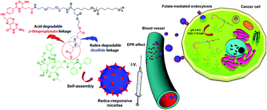
To the best of our knowledge, up to now, amphiphilic and redox and pH dual-responsive polymer–PTX conjugates based on PEG and PCL have not been utilized to construct DDSs possessing synergistic therapeutic and active targeting functions. Aiming at fulfilling the above requirements and also as a continued study to develop functional amphiphilic copolymers for preparing stimuli-responsive drug nanocarriers,35,36,39–42 in the present work, novel folic acid functionalized redox and pH dual-responsive degradable micellar nanoparticles were designed from amphiphilic polymer–PTX conjugates with thiol and acid-labile linkages, FA-PEG-b-P((PTX-SS-CL)-co-CL). The inoculation of β-thiopropionate and disulfide linkers between PTX and polyester block was supposed to release the active PTX into tumor cells in response to an intracellular pH and glutathione level (Scheme 1). The chemical structures and self-assembling properties of these amphiphilic block copolymers were characterized. The FA receptor-mediated cellular uptake, distribution in cells, and antitumor efficacy of the PTX-conjugated micelles were also investigated against tumor cells in detail.
 |
| Scheme 1 Self-assembly and activated intracellular drug delivery and release from redox and pH dual-responsive degradable FA-PEG-b-P((PTX-SS-CL)-co-CL) micelles. | |
Experimental
Reagents and materials
All chemicals were purchased from Sigma-Aldrich or Merck Chemical Companies and used without further purification unless otherwise described. Poly(ethylene glycol) (PEG, Mn = 2000 g mol−1) were purchased from Fluka and dried by azeotropic distillation using anhydrous toluene. Folate-poly(ethylene glycol)-NH2 (FA-PEG-NH2)41 and α-tert-butyl acetate-ε-caprolactone (TBACL)35 were synthesized according to the established literature procedures. For in vitro cytotoxicity test, amniotic epithelial (AE) cells were obtained from elective cesarean. HeLa cells were obtained from Pasteur Institute (Tehran, Iran). Dulbecco's Modified Eagle's Medium (DMEM)/F12, RPMI 1640 and fetal calf serum (FCS) were obtained from GIBCO Invitrogen Corporation. 3-(4,5-Dimethylthiazol-2-yl)-2,5-diphenyl tetrazolium bromide (MTT), dimethyl sulfoxide (DMSO) and epidermal growth factor (EGF) were purchased from Sigma.
Instrumentation
The 1H NMR spectrum of the samples was recorded on a Bruker DRX-300 Avance spectrometer using CDCl3 or DMSO-d6 as the solvents. The gel permeation chromatographic (GPC) system (Waters 2690D) equipped with refractive index detector (Waters 2410) and column of AGILENT PLgel 10 μm 300 × 7.5 mm (500 Å, 103 Å, 104 Å) was used to determine the molecular weights and molecular weight distributions. THF was introduced as the eluent solvent at a flow rate of 0.3 mL min−1. The polystyrene was used as the molecular weight standards. The morphology and size of the self-assembled micelles was performed on a ZEISS EM-900 transmission electron microscopy (TEM) operating at an acceleration voltage of 80 kV. The mean particle size and the size distribution of the self-assembled micelles solution in an aqueous solution (1 mg mL−1) were determined by the DLS using a 90 Plus Particle Size Analyzer (Brookhaven Instruments Corporation). UV-vis spectra were recorded using a Shimadzu UV-2100 spectrophotometer. Fluorescence spectra were recorded on a Varian Cary Eclipse spectrofluorophotometer. All the quantitative results were expressed as mean ± standard deviation (SD) of the mean. Statistical analysis was carried out by means of one-way analysis of variance (ANOVA) followed by the Tukey post-test. p < 0.05 was considered to be statistically significant, and p < 0.01 was considered to be highly significant.
Synthesis of FA-PEG-b-P((PTX-SS-CL)-co-CL). FA-PEG-b-P((PTX-SS-CL)-co-CL) conjugates was synthesized in five steps as show in Scheme 2.
 |
| Scheme 2 Synthetic routes employed for the preparation of FA-PEG-b-P((PTX-SS-CL)-co-CL). | |
Synthesis of FA-PEG-b-P(TBACL-co-CL). The FA-PEG-b-P(TBACL-co-CL) block copolymers were prepared by Sn(Oct)2-catalyzed controlled ring-opening polymerization (CROP) of TBACL and CL using FA-PEG-NH2 as macroinitiator (Scheme 2). Typically, TBACL (2.28 g, 10 mmol), CL (5.70 g, 50 mmol), FA-PEG-NH2 (2.35 g, 1 mmol amine groups) and stannous octoate (0.1 wt% of CL) were charged into a three-necked round bottom flask equipped with a reflux condenser. The reaction mixture was stirred for 24 h at 110 °C under a nitrogen atmosphere. The polymerization was quenched by immersing the flask in a cold water bath. The product was dissolved in a small amount of THF, precipitated in cold methanol and isolated by filtration. The copolymer was dried under vacuum at room temperature until a constant weight was reached (yield = 86.6%).
Synthesis of FA-PEG-b-P((CO2H-CL)-co-CL). In a typical reaction, the amphiphilic copolymer FA-PEG-b-P(TBACL-co-CL) was dissolved in CH2Cl2, and a 5-fold molar excess of TFA (with respect to tert-butyl groups) was added. The solution was stirred vigorously for 48 h at room temperature. Then, the solution was concentrated under reduced pressure to remove the solvent. Finally, the residue was dissolved in a small amount of THF and precipitated with an excess of cold diethyl ether and dried in vacuum at room temperature.
Synthesis of FA-PEG-b-P((HO-CL)-co-CL). The FA-PEG-b-P((HO-CL)-co-CL) was synthesized via a DCC-catalyzed condensation reaction. Briefly, FA-PEG-b-P((CO2H-CL)-co-CL) (0.774 g, 0.5 mmol carboxyl groups), 4-dimethylaminopyridine (DMAP, 0.18 g, 1.5 mmol) and dicyclohexylcarbodiimide (DCC, 0.246 g, 1.2 mmol) in anhydrous CH2CL2 (10 mL) was added dropwise to an anhydrous CH2CL2 solution (10 mL) of excess ethylene glycol (5 mmol) at 0 °C over a period of 1 h under a nitrogen atmosphere. The mixture was warmed to ambient temperature and stirred under N2 flow for 24 h. After the filtration of dicyclohexylurea (DCU), the filtrate was concentrated under reduced pressure to remove the solvent. Finally, the residue was dissolved in a small amount of THF and precipitated with an excess of cold diethyl ether and dried in vacuum at ambient temperature for 24 h.FA-PEG-b-P((CO2H-SS-CL)-co-CL) and FA-PEG-b-P((PTX-SS-CL)-co-CL) was synthesized by the same way used for the preparation of the FA-PEG-b-P((HO-CL)-co-CL) (Scheme 2).
Drug loading and in vitro drug release measurements
The release behavior of the FA-PEG-b-P((PTX-SS-CL)-co-CL) prodrug micelles was investigated using a dialysis method. Typically, 2.5 mL micellar solution of FA-PEG-b-P((PTX-SS-CL)-co-CL) prodrug micelles in phosphate buffer saline (PBS; 0.1 M, pH 5 or 7.4) was placed in a dialysis tube (MWCO = 3500 Da) and then immersed into 30 mL of the PBS or PBS with 10 mM glutathione (GSH) medium and kept in a shaking water bath at 37 °C. At predetermined intervals, 3 mL aliquot of the test solution was withdrawn periodically and replaced with an equal volume of the fresh release medium. The PTX content in FA-PEG-b-P((PTX-SS-CL)-co-CL) prodrug and concentration of the drug in the release samples were measured by UV spectrophotometer (λmax = 228 nm) and calculated in comparison with a standard curve.
In vitro cellular uptaking and cytotoxicity assay
Cellular uptake by HeLa cancer cells was examined using invert fluorescent microscope (IFM) and flow cytometric analyses. HeLa cells were seeded in 24-well plates at a density of 5 × 10⁴ cells per well and incubated in DMEM for 24 h. Then the cells were incubated for 0.5 h, 2 h and 4 h with FITC-labeled FA-PEG-b-P((PTX-SS-CL)-co-CL) micelles solution at the temperature 37 °C. The HeLa cells were gently rinsed with the ice-cold PBS twice. Subsequently, the cells were fixed with formaldehyde for 10 min at room temperature and washed twice with ice-cold PBS again. Then IFM was used to take fluorescent photo. For flow cytometric analyses, the media were piped out and trypsin was used to digest the cell for about 5 min. Next the HeLa cells were rinsed by the ice-cold PBS and centrifuged twice with 800 rpm for 5 min and fluorescence intensity of supernatant was measured.
The in vitro cytotoxicity of the FA-PEG-b-P((PTX-SS-CL)-co-CL) prodrug micelles and free PTX toward HeLa and AE cells was determined by MTT assay. The polymer solutions were prepared in PBS (pH 7.4) and free PTX was dissolved in ethanol/Cremophor-EL (50/50, v/v). Briefly, about 5 × 10⁴ cells per well were seeded in gelatin-coated 24-well plate and incubated overnight in incubator (37 °C, 5% CO2). The growth medium was replaced with fresh medium (200 μL) containing various concentration of test materials. After 48 h, 250 μL of MTT stock solution (5 mg mL−1) was added into each well and the plates were incubated for another 4 h. The MTT formazan crystals were then dissolved with 1 mL DMSO at ambient temperature. After gentle agitation for 10 min, the optical density at 570 nm in each well was measured on a microplate reader. The half-maximal inhibitory concentration (IC50) values were calculated from the dose–effect curves.
Results and discussion
Synthesis and characterization of FA-PEG-b-P((PTX-SS-CL)-co-CL) prodrug
With the aim to develop PEG-b-PCL based amphiphilic block copolymers containing functional reactive carboxyl groups for preparing functional micellar nanovehicles, we utilized a newly developed monomer in our lab, namely TBACL (Scheme 2).35 TBACL contained a protected carboxyl group at α-position, providing the possibility of introducing pendant carboxyl groups onto hydrophobic polyester block of PCL-b-PEG copolymer. As shown in Scheme 2, FA-PEG-b-P((CO2H-CL)-co-CL) including carboxyl pendant groups in hydrophobic PCL segment, were synthesized through controlled ring opening polymerization (CROP) of CL and TBACL using FA-PEG-NH2 as macroinitiator and stannous octoate as catalyst. Subsequent acidolysis deprotection upon selective removal of tert-butyl side group yielded folate-poly(ethylene glycol)-b-poly((α-CH2CO2H-caprolactone)-co-caprolactone) (FA-PEG-b-P((CO2H-CL)-co-CL)) copolymers bearing carboxylic acid functional groups on the PCL block. The chemical structure of the resulting copolymers was characterized by NMR. The hydrogen signals corresponding to EG, CL and TBACL repeating units before and after the acidolysis deprotection were indicated (Fig. 1A and B), confirming the successful synthesis of FA-PEG-b-P(TBACL-co-CL) and FA-PEG-b-P((CO2H-CL)-co-CL). The DP for the P(TBACL-co-CL) block were found to be about 52, by calculating the relative integration of the peak at 3.64 ppm (Fig. 1A, a), which corresponds with the methylene peak of poly(ethylene glycol), and the peak at 4.04 ppm (Fig. 1A, f + f′), which corresponds with the methylene protons in the repeat TBACL and CL units. The integral area ratio of signals f + f′ and h was about 1
:
0.49 (Fig. 1A), which confirmed that the block copolymer consisted of about 89.1% CL and 10.9% TBACL. Comparing the two 1H NMR spectra demonstrates that the peak assigned to the methyl protons from the tert-butyl side groups (h, Fig. 1A) at 1.49 ppm disappeared (Fig. 1B), indicating complete hydrolysis of all the tert-butyl side groups. The presence of the folate end group in block copolymer structure was proved by UV-vis spectroscopy (Fig. S1†). However, the NMR consequence to verify this conclusion was not obtained, because the proton signals of FA molecule are too weak to be detected. The GPC traces of FA-PEG-b-P(TBACL-co-CL) block copolymer exhibit monomodal molecular weight distribution with an Mn of 9100 and an Mw/Mn of 1.17 (Fig. S2†).
 |
| Fig. 1 1H NMR spectra of FA-PEG-b-P(TBACL-co-CL) (A), FA-PEG-b-P((CO2H-CL)-co-CL) (B), FA-PEG-b-P((HO-CL)-co-CL) (C), and FA-PEG-b-P((CO2H-SS-CL)-co-CL) (D) in CDCl3. | |
Introduction of the disulfide functionality, susceptible to reduction cleavage, on the FA-PEG-b-P((CO2H-CL)-co-CL) copolymer was performed in two steps by modification of the carboxyl pendant groups of the P((CO2H-CL)-co-CL) block. Initially, DCC-catalyzed esterification reaction was used to convert the carboxyl pendant groups into hydroxyl functionality in the presence of excess ethylene glycol. The successful synthesis of FA-PEG-b-P((HO-CL)-co-CL) was confirmed by 1H NMR. Fig. 1C shows the representative 1H NMR spectrum of FA-PEG-b-P((HO-CL)-co-CL) block copolymer and three characteristic signals originated from –CH2CH2OH functionality appeared at 3.47, 3.91, and 5.03 ppm. The FA-PEG-b-P((HO-CL)-co-CL) copolymer including hydroxyl pendant groups was then converted to FA-PEG-b-P((CO2H-SS-CL)-co-CL) with redox-responsive disulfide functionality in pendant groups, using a high excess of 3,3′-dithiodipropionic acid (DPA) to avoid cross-linking or branching of the copolymer (Scheme 2). The efficiency of the reactions was confirmed by 1H NMR analysis. According to the 1H NMR result in Fig. 1D, new peaks at 2.65–2.8 ppm, ascribed to –CH2CH2– near the disulfide group on the copolymer, appeared. The molar ratio of conjugated DPA in the FA-PEG-b-P((CO2H-SS-CL)-co-CL) copolymer was calculated by 1H NMR integration using the ratio of the methylene peaks of the DPA (–CH2–CH2–S–S–CH2–CH2–) (Fig. 1D, j and k) versus the methylene peak of poly(ethylene glycol) (Fig. 1D, a). The DPA content in copolymer were calculated to be 0.53 mmol sulfide functional groups per gram of the FA-PEG-b-P(α-HOSS-CL/CL), indicating the complete substitution of hydroxyl groups in the FA-PEG-b-P(α-HOSS-CL/CL) copolymer.
Finally, the PTX was chemically conjugated to a new redox-responsive amphiphilic block copolymer via a DCC-catalyzed esterification reaction of pendant carboxyl groups of FA-PEG-b-P((CO2H-SS-CL)-co-CL) with the hydroxyl group of PTX, using a 1
:
2 molar ratio of CO2H
:
OH (Scheme 2). Due to the successful reaction, the Mn of FA-PEG-b-P((PTX-SS-CL)-co-CL) increased to 15.7 kDa (Fig. S2†) and prodrug had a narrow molecular weight distribution (PDI = 1.19), which might be ascribed to the quantitative reaction of the FA-PEG-b-P((CO2H-SS-CL)-co-CL) copolymer with the PTX. Furthermore, in the 1H NMR spectrum of Fig. 2A, the signals of PTX from 1.1 to 8.1 ppm appeared. The characteristic resonance of 2′-CH proton of conjugated PTX was shifted from 4.77 ppm to 5.54 ppm compared with the 2′-CH proton of free PTX (Fig. 2B), while the resonance of 7-CH at 4.38 ppm remained, indicating that the coupling reaction took place selectively at the 2′-hydroxyl of PTX (Fig. 2A and B). The 7-OH of PTX is less reactive than the 2′-OH owing to its high steric hindrance.32,43 The PTX conjugation efficiency was then determined using UV spectrophotometer by measuring the concentration of PTX in the FA-PEG-b-P((PTX-SS-CL)-co-CL) prodrug as described in the experimental section. UV-vis results indicate the PTX conjugation of the block copolymers was successful. After 24 h of reaction, the conjugation efficiency was measured to be 98%, which prove the quantitative reaction of the carboxyl groups of the FA-PEG-b-P((CO2H-SS-CL)-co-CL) with the PTX.
 |
| Fig. 2 1H NMR spectra of FA-PEG-b-P((PTX-SS-CL)-co-CL) (A) and PTX (B) in CDCl3. | |
Self-assembly properties of FA-PEG-b-P((PTX-SS-CL)-co-CL) copolymers
In general, for micellar DDS, size and stability are key parameters that influence in vivo performance.36,41 Due to the amphiphilic property, FA-PEG-b-P((PTX-SS-CL)-co-CL) could self-assemble into micelles in an aqueous solution with hydrophilic PEG chain as the shell and hydrophobic polyester as the core. Moreover, the chemical conjugation of highly lipophilic PTX molecules with hydrophobic/aromatic interactions between the phenyl rings can more help the self-assembly structures of FA-PEG-b-P((PTX-SS-CL)-co-CL).44 The amphiphilic FA-PEG-b-P((PTX-SS-CL)-co-CL) block copolymer self-assembled into micelles in aqueous solution using the dialysis method and the critical micelle concentration (CMC) of this block copolymers was evaluated by fluorescence spectra using pyrene as a hydrophobic probe. With the presence of micelles, pyrene is primarily solubilized in the hydrophobic core of those micelles. By monitoring fluorescence intensity of probe with different environment around the probe from water to hydrophobic regions of micelles, the CMC value of micelles can be determined.42 As shown in the Fig. 3A, a red shift from 334 to 337 nm was observed when the concentration of FA-PEG-b-P((PTX-SS-CL)-co-CL) increased from 0.0005 to 0.5102 mg mL−1 indicating the micellization of amphiphilic copolymers.35,36,41 The CMC value of micelles was determined as the concentration corresponding to the crossing point of the two tangents, which came out to be 5.21 mg L−1 (Fig. 3B). This small value of CMC also indicated that the micelle structure of FA-PEG-b-P((PTX-SS-CL)-co-CL) may remain stable under highly diluted conditions after being intravenously administered.45
 |
| Fig. 3 Excitation spectra (A) and I337/I334 ratio (B) of pyrene in phosphate buffer solution of FA-PEG-b-P((PTX-SS-CL)-co-CL) at various concentrations. | |
The size distribution and morphology of FA-PEG-b-P((PTX-SS-CL)-co-CL) micelles was characterized by DLS and TEM. The DLS measurement results (Fig. 4A) showed the average hydrodynamic radii (Rh) of the FA-PEG-b-P((PTX-SS-CL)-co-CL) micelles to be 96.5 nm. TEM images confirmed that the FA-PEG-b-P((PTX-SS-CL)-co-CL) micelles were well-dispersed in water in the form of core–shell assemblies with an average diameter of about 85 nm (Fig. 4B). Zeta potentials analyzes of FA-PEG-b-P((PTX-SS-CL)-co-CL) micelles showed negative surface charges (−15.16 ± 1.2 mV), owing to the pendant carboxyl groups on polyester block of copolymer. It has been proved that the slightly negative surface charges will improve blood compatibility and prolonged circulation time of drug nanocarriers because of reduced interactions with blood components.46 The stability of FA-PEG-b-P((PTX-SS-CL)-co-CL) prodrug micelles was investigated by measuring the average size of micelles as a function of standing time in phosphate buffer saline (PBS; 0.1 M, pH 7.4) containing 10% Fetal Bovine Serum (FBS) at 37 °C. As shown in Fig. 4C, the average hydrodynamic radii of the micelles remain approximately unchanged within 6 days. These results indicate that the size and stability of the FA-PEG-b-P((PTX-SS-CL)-co-CL) micelles is proper for efficient intracellular drug delivery.36,41
 |
| Fig. 4 DLS plot (A), TEM image (B) of FA-PEG-b-P((PTX-SS-CL)-co-CL) micelles in phosphate buffer solution (pH 7.4) and the micelles size measured by DLS as a function of standing time in PBS (pH 7.4) containing 10% FBS at 37 °C (C). | |
In vitro release behaviors of FA-PEG-b-P((PTX-SS-CL)-co-CL) micelles
Since PTX is conjugated to the FA-PEG-b-P((PTX-SS-CL)-co-CL) block copolymers backbones through an redox-degradable disulfide linkage (Scheme 1), the FA-PEG-b-P((PTX-SS-CL)-co-CL) micelles are expected to be redox-responsive. Furthermore, FA-PEG-b-P((PTX-SS-CL)-co-CL) micelles may also be pH-responsive owing to existence of the acid-degradable β-thiopropionate functionality in the structure of disulfide linkages (Scheme 1).47 To evaluate the drug release behaviors of the FA-PEG-b-P((PTX-SS-CL)-co-CL) micelles, we investigated the release of PTX from the micelles at pH 7.4 and 5.0 with or without 10 mM GSH. As shown in the Fig. 5, the cumulative release of PTX under normal physiological conditions (pH = 7.4 and T = 37 °C), when the β-thiopropionate and disulfide linkages were stable, was about 6% over the test duration (50 h). While, in the same conditions with 10 mM GSH, more than 79% of PTX was released from FA-PEG-b-P((PTX-SS-CL)-co-CL) micelles (Fig. 5), indicating the redox-responsive drug release property of designed micellar nanovehicles. In contrast with the slow release of PTX at pH 7.4, the drug release rate was significantly increased at pH 5.0, and approximately 43% of PTX was released from FA-PEG-b-P((PTX-SS-CL)-co-CL) micelles at a release time of 50 h, demonstrating the pH-triggered release of PTX (Fig. 5). In support of these results, the fastest and almost completely PTX release (97.3%) was observed at pH 5.0 with 10 mM GSH after 50 h of incubation (Fig. 5). These results demonstrate that the drug release from the FA-PEG-b-P((PTX-SS-CL)-co-CL) micelles had an obvious redox and pH dual-responsive behavior that can effectively decrease the unwanted drug release during blood circulation, so to bring down the PTX-related side effect and intelligent release the drugs under the tumor-relevant conditions, leading to the superior antitumor efficacy of PTX.
 |
| Fig. 5 In vitro PTX release profiles of FA-PEG-b-P((PTX-SS-CL)-co-CL) micelles in PBS at various conditions: pH 7.4, pH 5.0, pH 7.4 with 10 mM GSH and pH 5.0 with 10 mM GSH. Data are presented as mean ± SD (n = 3). | |
In vitro cellular uptake and cytotoxicity studies
In order to visualize the micelles taken by the cells, HeLa cells as an FA-receptor expressing cancer cell line were incubated with FITC-labeled micelles (FA-PEG-b-P((FITC-PTX-SS-CL)-co-CL)) for 0.5 h, 2 h and 4 h, respectively, and examined by flow cytometry and fluorescent microscopy (Fig. 6). FITC was conjugated to the remaining hydroxy groups of PTX in FA-PEG-b-P((PTX-SS-CL)-co-CL). As shown in Fig. 6A, HeLa cells treated with folate-decorated FA-PEG-b-P((FITC-PTX-SS-CL)-co-CL) micelles, at the same time, showed higher fluorescence intensity as compared to the HeLa cells incubated with PTX and control HeLa cells owing to folate receptor overexpression in HeLa cells, indicating that micelles were taken up by the cell. This finding was further confirmed by the fluorescent microscopy analysis. As shown in Fig. 6B, the fluorescence intensity in the cytoplasm of HeLa cells cultured with the FA-PEG-b-P((FITC-PTX-SS-CL)-co-CL) micelles after a 0.5 h incubation at 37 °C was relatively little. When the incubation period increased to 2 and 4 h, the green fluorescence in the cells cytoplasm increased obviously, indicating efficient internalization of micelles.
 |
| Fig. 6 Fluorescence intensities of HeLa cells incubated with FITC-labeled micelles and FITC-labeled PTX (A), and fluorescence microscopy images of HeLa cells incubated with FITC-labeled micelles (B) for 0.5, 2 and 4 h (37 °C) (*p < 0.01 and **p < 0.05). | |
Biocompatibility is a vital factor for the application of drug nanocarriers in the human body. As shown in Fig. 7, the cell viabilities of both AE normal cells and HeLa cancer cells after 48 h incubation with FA-PEG-b-P((OH-SS-CL)-co-CL) copolymer at a concentration ranging from 10 to 500 μg mL−1 have not tangible changes compared with the untreated control cells. These results demonstrate that FA-PEG-b-P((OH-SS-CL)-co-CL) copolymer has good biocompatibility.
 |
| Fig. 7 Viabilities of AE cells and HeLa cells incubated with FA-PEG-b-P((OH-SS-CL)-co-CL) as a function of copolymer concentration for 48 h at 37 °C. Data are presented as mean ± SD (n = 3) (*p < 0.01). | |
The ability of FA-PEG-b-P((PTX-SS-CL)-co-CL) prodrug micelles to inhibit proliferation of HeLa cells as an FA-receptor expressing cancer cell line was investigated using an MTT viability assay and it was compared with free PTX (at equivalent drug concentration in each sample) in the medium without or with 1 μg mL−1 of folic acid. The half-maximal inhibitory concentration (IC50) values were determined after 48 h culture with different formulations dosages from 0.001 to 50 μg mL−1. These results demonstrate an improvement of the toxicity for the FA-PEG-b-P((PTX-SS-CL)-co-CL) prodrug micelles (IC50 = 0.75 μg mL−1) compared to the free PTX (IC50 = 0.87 μg mL−1), in the absence of free folic acid in the cell culture medium (Fig. 8). This can be attributed to the higher uptake of FA-PEG-b-P((PTX-SS-CL)-co-CL) than PTX by HeLa cells via FA-receptors mediated endocytosis and then achieve rapid cleavage of β-thiopropionate and disulfide linkages in response to an intracellular pH and glutathione level once internalized into tumor cells (Scheme 1), thereby improving intracellular PTX release and increasing the antitumor efficacy. It is clear that free PTX on AE normal cells will have a higher toxicity compared to the FA-PEG-b-P((PTX-SS-CL)-co-CL) prodrug micelles, unlike the obtained results for cancer cells. Because the FA-PEG-b-P((PTX-SS-CL)-co-CL) is functionalized with folic acid selectively accumulates in tumor tissues. So we will have a minimal impact on normal cells. However, the cell inhibition with the presence of 1 μg mL−1 of free folic acid is not so high because the targeting is suppressed due to the free folic acid binding (Fig. 9). The data of the cell viabilities in free folic acid competition study indicate the cell inhibition is mainly caused by the cell internalization of the nanoparticles through folate-mediated targeting.
 |
| Fig. 8 Viability of HeLa cells incubated with FA-PEG-b-P((PTX-SS-CL)-co-CL) and free PTX as a function of PTX concentration for 48 h at 37 °C, in the absence of folic acid. Data are presented as mean ± SD (n = 3) (*p < 0.01). | |
 |
| Fig. 9 Viability of HeLa cells incubated with FA-PEG-b-P((PTX-SS-CL)-co-CL) as a function of PTX concentration for 48 h at 37 °C, in the presence of 1 μg mL−1 of folic acid. Data are presented as mean ± SD (n = 3). | |
Conclusions
In conclusion, we have developed a simple and effective polymer–drug conjugate for targeted intracellular delivery of paclitaxel. The PEG-b-PCL based amphiphilic block copolymer scaffold containing functional disulfide linkages, FA-PEG-b-P((OH-SS-CL)-co-CL), was prepared through a well-established CROP method with additional minor modification. And PTX was chemically conjugated to a new redox-responsive amphiphilic block copolymer FA-PEG-b-P((OH-SS-CL)-co-CL) via a DCC-catalyzed esterification reaction. The FA-PEG-b-P((PTX-SS-CL)-co-CL) micelles exhibited a narrow size distribution and appropriate sizes for EPR effect, as determined by DLS and TEM. These micelles exhibit no apparent cytotoxicity to the both cultured AE normal and HeLa cancer cells. The FA-PEG-b-P((PTX-SS-CL)-co-CL) micelles were demonstrated to have high stability, low CMC value, negligible drug leaking in normal conditions and rapid drug release in tumor-relevant reductive and acidic environment, which can decrease non-specific spread of toxic bioactive molecules during circulation in the bloodstream. Flow cytometry and fluorescent microscope studies revealed that the redox and pH dual-responsive micelles could be selectively taken up to HeLa tumor cells via FA-receptors mediated endocytosis. Furthermore, due to the enhancing intracellular uptake and drug release in tumor intracellular environment, the cytotoxicity of FA-PEG-b-P((PTX-SS-CL)-co-CL) micelles improved compared to the free PTX, confirming the relation between such redox and pH dual-sensitive feature and therapeutic efficacy of this carrier. All these results suggested that these dual-sensitive prodrug micelles hold great promise for targeted intracellular transport of potent hydrophobic anticancer drugs, thereby enhance the therapeutic efficacy.
Acknowledgements
This work was supported by the “Iran National Science Foundation: INSF”.
Notes and references
- K. C. Nicolaou and R. A. Valiulin, Org. Biomol. Chem., 2013, 11, 4154–4163 Search PubMed.
- J. S. Sohn, J. I. Jin, M. Hess and B. W. Jo, Polym. Chem., 2010, 1, 778–792 RSC.
- Z. Zhang, L. Mei and S.-S. Feng, Expert Opin. Drug Delivery, 2013, 10, 325–340 CrossRef CAS PubMed.
- Z. Xie, H. Guan, X. Chen, C. Lu, L. Chen, X. Hu, Q. Shi and X. Jing, J. Controlled Release, 2007, 117, 210–216 CrossRef CAS PubMed.
- E. Markovsky, H. Baabur-Cohen, A. Eldar-Boock, L. Omer, G. Tiram, S. Ferber, P. Ofek, D. Polyak, A. Scomparin and R. Satchi-Fainaro, J. Controlled Release, 2012, 161, 446–460 CrossRef CAS PubMed.
- J. Nicolas, S. Mura, D. Brambilla, N. Mackiewicz and P. Couvreur, Chem. Soc. Rev., 2013, 42, 1147–1235 RSC.
- R. Dong, Y. Zhou, X. Huang, X. Zhu, Y. Lu and J. Shen, Adv. Mater., 2015, 27, 498–526 CrossRef CAS PubMed.
- P. P. Deshpande, S. Biswas and V. P. Torchilin, Nanomedicine, 2013, 8, 1509–1528 CrossRef CAS PubMed.
- A. M. Jhaveri and V. P. Torchilin, Front. Pharmacol., 2014, 5, 77 Search PubMed.
- M. Yokoyama, J. Drug Targeting, 2014, 22, 576–583 CrossRef CAS PubMed.
- Z. Ahmad, A. Shah, M. Siddiq and H.-B. Kraatz, RSC Adv., 2014, 4, 17028–17038 RSC.
- A. Gothwal, I. Khan and U. Gupta, Pharm. Res., 2015, 33, 18–39 CrossRef PubMed.
- B. P. K. Reddy, H. K. S. Yadav, D. K. Nagesha, A. Raizaday and A. Karim, J. Nanosci. Nanotech., 2015, 15, 4009–4018 CrossRef PubMed.
- L. Bildstein, C. Dubernet and P. Couvreur, Adv. Drug Deliv. Rev., 2011, 63, 3–23 CrossRef CAS PubMed.
- V. Delplace, P. Couvreur and J. Nicolas, Polym. Chem., 2014, 5, 1529–1544 RSC.
- M. Chang, F. Zhang, T. Wei, T. Zuo, Y. Guan, G. Lin and W. Shao, J. Drug Targeting, 2016, 24, 475–491 CrossRef CAS PubMed.
- F. Meng, W. E. Hennink and Z. Zhong, Biomaterials, 2009, 30, 2180–2198 CrossRef CAS PubMed.
- Q. Zhang, N. Re Ko and J. Kwon Oh, Chem. Commun., 2012, 48, 7542–7552 RSC.
- X. Pang, Y. Jiang, Q. Xiao, A. W. Leung, H. Hua and C. Xu, J. Controlled Release, 2016, 222, 116–129 CrossRef CAS PubMed.
- S. Mura, J. Nicolas and P. Couvreur, Nat. Mater., 2013, 12, 991–1003 CrossRef CAS PubMed.
- B. Deng, P. Ma and Y. Xie, Nanoscale, 2015, 7, 12773–12795 RSC.
- M. Kanamala, W. R. Wilson, M. Yang, B. D. Palmer and Z. Wu, Biomaterials, 2016, 85, 152–167 CrossRef CAS PubMed.
- R. Cheng, F. Meng, C. Deng and Z. Zhong, Nano Today, 2015, 10, 656–670 CrossRef CAS.
- S. Kaur, C. Prasad, B. Balakrishnan and R. Banerjee, Biomater. Sci., 2015, 3, 955–987 RSC.
- K. C. Remant Bahadur, B. Thapa and P. Xu, Mol. Pharm., 2012, 9, 2719–2729 CrossRef PubMed.
- Y.-J. Pan, Y.-Y. Chen, D.-R. Wang, C. Wei, J. Guo, D.-R. Lu, C.-C. Chu and C.-C. Wang, Biomaterials, 2012, 33, 6570–6579 CrossRef CAS PubMed.
- W. Chen, P. Zhong, F. Meng, R. Cheng, C. Deng, J. Feijen and Z. Zhong, J. Controlled Release, 2013, 169, 171–179 CrossRef CAS PubMed.
- S. Yu, C. He, J. Ding, Y. Cheng, W. Song, X. Zhuang and X. Chen, Soft Matter, 2013, 9, 2637–2645 RSC.
- M. Cai, K. Zhu, Y. Qiu, X. Liu, Y. Chen and X. Luo, Colloids Surf., B, 2014, 116, 424–431 CrossRef CAS PubMed.
- S. Wang, H. Wang, Z. Liu, L. Wang, X. Wang, L. Su and J. Chang, Nanoscale, 2014, 6, 7635–7642 RSC.
- X.-Q. Yi, Q. Zhang, D. Zhao, J.-Q. Xu, Z.-L. Zhong, R.-X. Zhuo and F. Li, Polym. Chem., 2016, 7, 1719–1729 RSC.
- S. Lv, Z. Tang, D. Zhang, W. Song, M. Li, J. Lin, H. Liu and X. Chen, J. Controlled Release, 2014, 194, 220–227 CrossRef CAS PubMed.
- Q. N. Bui, Y. Li, M.-S. Jang, D. P. Huynh, J. H. Lee and D. S. Lee, Macromolecules, 2015, 48, 4046–4054 CrossRef CAS.
- L. Wang, Y. Wang, Q. Jin, F. Jia, H. Wang and J. Ji, Polymer, 2015, 76, 237–244 CrossRef CAS.
- S. J. Tabatabaei Rezaei, V. Amani, M. R. Nabid, N. Safari and H. Niknejad, Polym. Chem., 2015, 6, 2844–2853 RSC.
- S. J. Tabatabaei Rezaei, H. S. Abandansari, M. R. Nabid and H. Niknejad, J. Colloid Interface Sci., 2014, 425, 27–35 CrossRef CAS PubMed.
- S. V. Lale, A. Kumar, S. Prasad, A. C. Bharti and V. Koul, Biomacromolecules, 2015, 16, 1736–1752 CrossRef CAS PubMed.
- B. Yu, H. C. Tai, W. Xue, L. J. Lee and R. J. Lee, Mol. Membr. Biol., 2010, 27, 286–298 CrossRef CAS PubMed.
- H. S. Abandansari, M. R. Nabid, S. J. T. Rezaei and H. Niknejad, Polymer, 2014, 55, 3579–3590 CrossRef CAS.
- S. J. T. Rezaei, M. R. Nabid, H. Niknejad and A. A. Entezami, Int. J. Pharm., 2012, 437, 70–79 CrossRef CAS PubMed.
- S. J. Tabatabaei Rezaei, M. R. Nabid, H. Niknejad and A. A. Entezami, Polymer, 2012, 53, 3485–3497 CrossRef CAS.
- M. R. Nabid, S. J. Tabatabaei Rezaei, R. Sedghi, H. Niknejad, A. A. Entezami, H. A. Oskooie and M. M. Heravi, Polymer, 2011, 52, 2799–2809 CrossRef CAS.
- J. Zou, F. Zhang, S. Zhang, S. F. Pollack, M. Elsabahy, J. Fan and K. L. Wooley, Adv. Healthcare Mater., 2014, 3, 441–448 CrossRef CAS PubMed.
- P. M. Valencia, M. H. Hanewich-Hollatz, W. Gao, F. Karim, R. Langer, R. Karnik and O. C. Farokhzad, Biomaterials, 2011, 32, 6226–6233 CrossRef CAS PubMed.
- K. T. Oh and E. S. Lee, Polym. Adv. Technol., 2008, 19, 1907–1913 CrossRef CAS.
- J.-Z. Du, X.-J. Du, C.-Q. Mao and J. Wang, J. Am. Chem. Soc., 2011, 133, 17560–17563 CrossRef CAS PubMed.
- L. Qiu, Q. Liu, C.-Y. Hong and C.-Y. Pan, J. Mater. Chem. B, 2016, 4, 141–151 RSC.
Footnote |
† Electronic supplementary information (ESI) available: UV-vis absorption spectra of: FA-PEG-b-P(TBACL-co-CL) and FA (Fig. S1). GPC trace of FA-PEG-b-P(TBACL-co-CL) and FA-PEG-b-P((PTX-SS-CL)-co-CL) (Fig. S2). See DOI: 10.1039/c6ra11824k |
|
This journal is © The Royal Society of Chemistry 2016 |
Click here to see how this site uses Cookies. View our privacy policy here.