A novel approach towards hydrazine sensor development using SrO·CNT nanocomposites

Mohammed M. Rahman*ab, Mohammad Musarraf Hussainab and Abdullah M. Asiriab
aChemistry Department, Faculty of Science, King Abdulaziz University, Jeddah 21589, P.O. Box 80203, Saudi Arabia. E-mail: mmrahman@kau.edu.sa; mmrahmanh@gmail.com; Fax: +966-026952292; Tel: +966-59-6421830
bCenter of Excellence for Advanced Material Research (CEAMR), King Abdulaziz University, Jeddah 21589, P.O. Box 80203, Saudi Arabia

Received 4th May 2016 , Accepted 24th June 2016

First published on 28th June 2016


Abstract

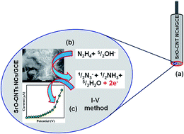
Strontium oxide nanoparticle decorated carbon nanotube nanocomposites (SrO·CNT NCs) were prepared in alkaline medium using a wet-chemical technique at low temperature. The SrO·CNT NCs were investigated using Fourier transform infrared (FT-IR) spectroscopy, UV/visible spectroscopy, field emission scanning electron microscopy (FESEM) coupled with electron dispersive spectroscopy (EDS), X-ray photoelectron spectroscopy (XPS), and X-ray powder diffraction (XRD) methods. A selective hydrazine sensor on a glassy carbon electrode (GCE) was fabricated with a thin-layer of the NCs using conducting coating binders. Improved electrochemical responses, a higher sensitivity including a large dynamic range, and long-term stability towards hydrazine were acquired using the fabricated SrO·CNT NCs/GCE sensor. The calibration curve was found to be linear (r2: 0.7311) over a wide range of hydrazine concentrations (0.2 nM to 0.2 M). The detection limit and sensitivity were calculated as 0.036 nM and ∼26.37 μA mM−1 cm−2 respectively. In this approach, hydrazine was detected using a current vs. voltage (IV) method and a SrO·CNT NC modified GCE electrode with very high sensitivity compared to various nanomaterials. The synthesis of SrO·CNT NCs using a wet-chemical process is a unique way of establishing sensor based nanocomposites for toxic and carcinogenic agents.


1. Introduction

Hydrazine is one of the most generally used compounds found in industrial and environmental samples, and it has toxic effects in the environment and human beings. It is extensively utilized in antioxidants, rocket fuels, catalysts, emulsifiers, corrosion inhibitors, reducing agents, pesticides, and plant growth regulators.1,2 It is a neurotoxin, and produces carcinogenic and mutagenic effects causing damage to the lungs, liver, and kidneys, respiratory tract infections and long-term effects on the central nervous system.3 In the pharmaceutical industry, it has much significance that hydrazine is acknowledged as a carcinogenic and hepatotoxic chemical that affects the liver and brain.4 Electro-oxidation of hydrazine is the basis of an established fuel cell, owing to a high capacity and no contamination. In addition to this, hydrazine is widely used as a high-energy propellant in rockets and spacecrafts by military and aerospace industries.5 Owing to the aforementioned applications and effects in industry, the environment and pharmacology, it is very important to fabricate a reliable, cheap and effective sensor for the efficient detection of toxic hydrazine. Among several detection methodologies, electrochemical IV methods offer an opportunity for portable, cheap and rapid techniques. Therefore, numerous chemically modified electrodes based on different nanostructure materials, semiconductor doped or undoped nanomaterials, transition metal oxides and electrocatalytic moieties (electron-mediator species) have been developed for the detection of hydrazine and have been reported in the literature.6–9 Recently, scientists have demonstrated the use of semiconductor nanostructures as electron mediators to modify electrodes for the electrochemical detection of toxic hydrazine.10,11

Safety is a major concern for the environment and health, which is a vital issue to consider from the perspective of using chemical sensors for the identification of poisonous chemicals through a well-established method. Due to the high active surface area and size to volume ratio in comparison with conventional nanocomposites at the micro- to nano-range, nanostructure coupled nanocomposite materials are very efficient, stable, and sensitive. Nanostructures of metal oxides having good properties, used in the fabrication of chemical sensors, and having a high active surface area, high porosity, permeability, a quantum confinement effect and stability are receiving a great deal of attention at present.12 Having the properties of a fast response, large surface area, lower carrier charge, meso-porosity and portability, sensor-based metal oxide conjugated carbon nanotube composites are widely employed for the monitoring of chemical constituents, air/water contaminants, and toxic chemical agents in the environment.13 The separation of hazardous materials from industrial waste water is one of the vital issues in environmental and biological science. Different techniques have been reported for the removal of carcinogenic molecules from industrial waste water. But some issues still remain unsettled, such as efficient removal of the hazardous agents, the re-usability of the NC materials, and the need for preparation of green NCs facilely and at a lower cost. The meso-porous nature of the NC material allows facile recycling without major deterioration of the sensor efficiency and potentiality. A good absorption/adsorption power of the hybrid NC along with other properties such as easy separation, an environmentally friendly composition and re-usability, makes it a suitable sensor for the removal of target toxic and hazardous chemicals from industrial as well as environmental waste.

Hydrazine is a colorless inorganic compound that has antioxidant and reducing properties. It has wide applications in various fields such as aircraft fuels, antioxidants, blowing agents, catalysis, corrosion inhibition, emulsifiers, explosives, fuel cells, heat stabilizers, insecticides, metal film production, oxygen scavengers, pharmaceutical intermediates, photographic development, pesticides, plastic polymers, plant growth regulators, plutonium extraction, rocket propellants, and textile dyes. Beyond these applications, hydrazine is a toxic agent and carcinogen responsible for blood abnormalities, cell mutagenicity, comas, dizziness, hepatoxicity, nausea, neurotoxic activity, pulmonary edema, temporary blindness and throat infections, and its effects include damage to the brain, DNA, eyes, kidneys, liver, lungs, nose, nervous system and respiratory system.14–18 Different conventional analytical methods have been used for the determination of hydrazine including amperometry, chromatography, capillary electrophoresis, chemi-luminescence, colorimetry, coulometry, electrochemical techniques, fluorescence, mass spectroscopy, potentiometry, spectrophotometry, and titrimetry.19–22 But most of the techniques are too complex for on-site analysis, costly and time consuming, as well as not being an economical approach. Therefore, from the perspective of the environmental and biological consequences, it is necessary to develop a selective and sensitive analytical technique for the detection and determination of hazardous hydrazine. The detection of hydrazine using a fabricated electrode with metal oxide-conjugated CNT nanocomposites is easy with a better analytical performance including higher sensitivity, a lower potential, lower cost, and real time analysis. In this approach, to enhance the sensor performance with a glassy carbon electrode, SrO nanoparticle decorated CNTs have been used for selective hydrazine detection, which is a suitable nanocomposite for electrode modification and support in biosensor applications because of the high accessible surface area of the nanocomposite, low-electrical resistance, a higher conductive surface, extremely high mechanical strength, outstanding charge-transport characteristics, and higher chemical stability compared to other materials. Reliable IV electrochemical methods offer practical advantages including operation simplicity, satisfactory sensitivity, a wide linear concentration range, lower cost of the instrument, possibility of miniaturization, suitability for real-time detection, and less sensitivity to matrix effects in comparison with separation and spectral methods.

In previous studies, SrO has been used as a coating material for carbon nanotubes,23 in additive coloration,24 as a doping agent,25 in aldol addition, and kinetic, paramagnetic resonance, IR, Mass, and UV/Vis spectroscopic studies,26–28 as a cement forming ion,29 as a heterogeneous catalyst,30 as an isomerization effect moiety,31 in the preparation of thin films,32 in the deposition of magnetron sputtering,33 and for photocatalytic activity.34 Hydrazine is extremely toxic and usually has serious effects on health and the environment, so its detection using a reliable method is immediately required, for example with SrO·CNT NCs using a GCE electrode. The detection of hydrazine using prepared SrO·CNT NC-deposited thin films on GCEs is studied in detail. An easy coating method for construction of the SrO·CNT NC thin-layer within conducting binding agents is executed for preparation of the films on a GCE. In this approach, mesoporous SrO·CNT NC fabricated films with conducting binders are utilized towards detection of the target carcinogenic analyte using a reliable IV method. It is confirmed that the fabricated hydrazine sensor is unique and noble research work has been conducted for ultra-sensitive hydrazine recognition using active SrO·CNT NCs on a GCE in a short response-time. In this work, the synthesis and electrochemical characterization of SrO·CNT NCs and screening for the determination of hydrazine using the fabricated SrO·CNT NCs are reported.

2. Experimental section

2.1. Materials and methods

The used chemicals of analytical grade, acetone, 4-aminophenol, ammonium hydroxide, benzaldehyde, bisphenol A, carbon nanotubes, disodium phosphate, ethanol, hydrazine, melamine, methanol, 4-methoxyphenol, monosodium phosphate, nafion (5% ethanolic solution), 2-nitrophenol, sodium hydroxide and strontium chloride, were purchased from Sigma-Aldrich Company. FTIR and UV/Vis spectra of the dried SrO·CNT NCs were recorded using a Thermo scientific NICOLET iS50 FTIR spectrometer (Madision, WI, USA) and a 300 UV/Visible spectrophotometer (Thermo scientific) respectively. The XPS measurements were carried out with a K-α spectrometer (Thermo scientific, K-α 1066) with an excitation radiation source (AlKα, beam spot size = 300.0 μm, pass energy = 200.0 eV, pressure 10−8 torr) for calculation of the binding energies (eV) among Sr, C and O. The arrangement, morphology and particle size of the SrO·CNT NCs were investigated using field emission scanning electron microscopy (FESEM, JEOL, JSM-7600F, Japan). An X-ray diffraction (XRD) examination was performed under ambient conditions to identify the crystalline pattern of the SrO·CNT NCs. IV measurements were conducted to detect hydrazine at a selected potential using a Keithley electrometer (6517A, USA) under normal conditions using the fabricated SrO·CNT NCs.

2.2. Synthesis of SrO·CNT nanocomposites

A wet-chemical procedure is a primary stage in the preparation of powder materials using a liquid phase, which is a typical solid-state synthesis technique and widely used in the synthesis of doped or undoped nanomaterials. In this process, the products are obtained as smaller grains along with a shorter duration of phase formation. Strontium chloride (SrCl2·6H2O), carbon nanotubes (CNT) and sodium hydroxide (NaOH) were used in the preparation of the SrO·CNT NCs as active reacting agents. According to this method, CNT (1.0 wt%, 0.25 μg) were added under continuous stirring to dissolved SrCl2·6H2O (0.1 M, 2.67 g) in distilled water (100 mL) in an Erlenmeyer flask (250.0 mL), the pH of the resultant mixture was adjusted to 10.13 with addition of NaOH and then the mixture was subjected to continuous stirring at 80.0 °C. The precipitate was washed thoroughly with water and acetone subsequently after the continuous stirring (6 hours) and kept for drying at room temperature. The resultant gray products (SrO·CNT NCs) were dried in a furnace for 24 hours at 60.0 °C and then used for characterization (elemental, morphological, optical and structural). Strontium oxide nanoparticles (SrO) without CNT were also prepared using a similar method. Possible reaction mechanisms for the formation of the SrO·CNT NCs are shown as follows.
 
NaOH(s) → Na+(aq) + OH(aq) (i)
 
SrCl2 → Sr2+(aq) + 2Cl(aq) (ii)
 
2Na+(aq) + 2OH(aq) + Sr2+(aq) + 2Cl(aq) → Sr(OH)2(aq) + 2NaCl(aq) (iii)
 
2Sr(OH)2(aq) → 2SrO(s)↓ + 2H2O(aq) (iv)
 
SrO + dispersed CNT → SrO·CNT(s) (v)

2.3. Fabrication of a GCE with SrO·CNT NCs

At room temperature, Na2HPO4 (0.2 M), NaH2PO4 (0.2 M) and distilled water (200.0 mL) were used in the preparation of a phosphate buffer solution (PBS, 0.1 M, pH = 7). Conducting binding agents (5% nafion) and ethanol were used to fabricate the glassy carbon electrode (GCE, surface area = 0.0316 cm2) with SrO·CNT NCs. The fabricated electrode was then kept at room temperature for 6 hours to allow formation of a uniform film with complete drying. As for the working, it was fabricated with NCs on a flat GCE. The target analyte, hydrazine was used as a stock solution with different concentrations (0.2 nM to 0.02 M) to identify the detection limit and the volume of PBS was kept constant (10.0 mL) throughout the whole process. During the analysis, a hydrazine solution (0.25 μL) was added onto the NCs-electrode from lower to higher concentration (0.2 nM to 0.2 M). The sensitivity and detection limit (DL) of the NCs were calculated from the slope and the ratio of noise to slope (3N/S) at the linear dynamic range of the calibration curve respectively. As a constant current source for the two assembled electrodes (working and counter), a Keithley electrometer was used. The fabricated SrO·CNT NC electrode was examined in a liquid phase system to determine the target hydrazine analyte.

3. Results and discussion

3.1. Evaluation of the optical and structural properties

Optical properties are some of the prime criteria for assessment of the catalytic activity of the fabricated grey SrO·CNT NCs. According to the UV/visible light principle, the outer electrons of the atom absorb radiant energy and make transitions to high energy levels. Due to optical absorption, a spectrum and the band-gap energy of the metal oxide were obtained. The UV/Vis spectra of the SrO NPs and SrO·CNT NC solution were recorded in the visible range (400–800 nm) and broad absorption bands at around 296.8 and 320.0 nm were found (Fig. 1a and c) respectively. Based on the maximum-band absorption, the band-gap energy of the SrO NPs and SrO·CNT NCs was calculated using eqn (v), and according to the direct band-gap rule [Tauc’s equation, (vi)], curves of (αhv)2 vs. hv were plotted and then extrapolated to the x-axis. From the extrapolated curve, the band-gap energy of the SrO NPs and SrO·CNT NCs was found to be 2.2 and 2.1 eV respectively (Fig. 1b and d). Where, Ebg = band gap energy, λmax = maximum absorption wavelength, α = absorption coefficient, A = a constant related to the effective mass of the electrons, r = 0.5 (direct transition), h = Planck’s constant, and v = frequency. There were no additional peaks found in the spectra associated with impurities or structural defects, which indicated a crystalline nature of the prepared SrO NPs and SrO·CNT NCs.
 
Ebg = 1240/λmax (eV) (vi)
 
(αhv)1/r = A(hvEbg) (vii)

image file: c6ra11582a-f1.tif
Fig. 1 (a and b) UV/Vis spectra and (c and d) band-gap energy plots for the SrO NPs and SrO·CNT NCs respectively.

The fabricated grey SrO·CNT NCs were also characterized from the perspective of atomic and molecular vibrations to determine their functional properties. In this regards, a FT-IR spectrum was recorded in the region of 4000–400 cm−1 under normal conditions. The FT-IR spectra of the CNT, SrO NPs, and SrO·CNT NCs (Fig. 2a) show peaks at 3282 (s, br), 2105 (w), 1552 (w), 1354 (m), 1103 (s), 852 (m) and 605 (m) cm−1, which denoted the presence of O–H (stretching), –C[triple bond, length as m-dash]C– (stretching), –C[double bond, length as m-dash]C– (stretching), C–H (rocking), Sr–O–Sr (stretching), C–H and Sr[double bond, length as m-dash]O (stretching) respectively in the SrO·CNT NCs.35 The observed peak at 605 cm−1 shows the formation of metal–oxide (Sr–O) bonds, which indicates the configuration of the SrO NPs, and this peak also exists for the SrO·CNT NCs.


image file: c6ra11582a-f2.tif
Fig. 2 (a) FT-IR spectra and (b) XRD patterns of the CNT, SrO NPs, and SrO·CNT NCs.

XRD analysis was performed to study the crystal pattern of the prepared CNT, SrO NPs, and SrO·CNT NCs. The observed potential peaks were found at 2θ values of 25(100), 32(110), 42(111), 46(200), 56(210), 58(211), 66(220), 72(221), and 78(310) degrees (Fig. 2b). All the observed peaks in the spectra were assigned using a JCPDS file (no. 001-1113, 6-520, 75-0263 and 01-073-0661). The strong peaks denoted the crystalline nature and purity of the NCs. The crystalline pattern is indicative of a metal–oxygen framework. From these parameters it was also concluded that a significant amount of crystalline SrO is present in the doped SrO·CNT NCs.36–38

3.2. Evaluation of the morphological and elemental properties

FESEM is one of the perfect techniques used to examine the morphology of nanocomposites, including nanostructure materials. The morphology and elemental properties of the prepared CNT, SrO NPs, and SrO·CNT NCs were investigated using FESEM and EDS respectively. Using FESEM, the typical shapes of the CNT, SrO NPs and SrO·CNT NCs were recorded and are presented in Fig. 3a–d (low to high magnification images). The magnified images indicate that the SrO NPs are aggregated, with a bright contrast, and are well dispersed on the surface of the CNTs (Fig. 3c and d). The conductance of the nanocomposite is increased with the adsorption/incorporation of SrO NPs onto/into the CNTs, which directly correlates with the calculation of the band-gap energy (Ebg) of the SrO·CNT nanocomposites.
image file: c6ra11582a-f3.tif
Fig. 3 FESEM magnified images (a) CNT, (b) SrO NPs, and (c and d) SrO·CNT NCs.

From EDS observations, the atomic percentages of the pure CNT (Fig. 4a and b) and pure SrO NPs (Fig. 4b and c) were measured and included in this study. On the basis of the EDS analysis, carbon (C), oxygen (O), and strontium (Sr) exist in the prepared grey SrO·CNT NCs and they consist of C(21.60), O(21.44) and Sr(56.96) wt% respectively (Fig. 4e and f).


image file: c6ra11582a-f4.tif
Fig. 4 FESEM images and elemental analysis: (a and b) CNT, (c and d) SrO NPs, and (e and f) SrO·CNT NCs.

According to the FESEM coupled with EDS, no other peaks related to impurities were observed, which indicated that the NCs are composed of C, O and Sr. A comparison of the CNT, SrO NPs, and SrO·CNT NCs with respect to the weight (%) was made and the results are presented in Table 1.

Table 1 Comparison from the perspective of weight (%)
Materials Weight (%)
C O Sr
CNT 94.55
SrO NPs 27.35 72.65
SrO·CNT NCs 21.60 21.44 56.96


3.3. Evaluation of binding energy

The chemical nature of the elements present in the NCs could be indicated using a quantitative spectroscopic technique, XPS. XPS spectra can be recorded by irradiation of the nanocomposite material with an X-ray beam and the kinetic energy, as well as the electron number of the sample, could be measured simultaneously.12 On the basis of the XPS spectra, carbon, oxygen and strontium were present in the synthesized SrO·CNT NCs. A comparison of the spectra of the CNT, SrO NPs, and SrO·CNT NCs is included in Fig. 5a. The spin orbit of strontium Sr 3d5/2 and Sr 3d3/2 is denoted by the peaks at 133.0 and 137.0 eV respectively, which indicates that strontium (Sr2+) is present in the NCs (Fig. 5b). The C 1s and O 1s spectra show a main peak at 283.0 and 535.0 eV, which denoted that carbon (C) and oxygen (O2−) are also present in the prepared SrO·CNT NCs (Fig. 5c and d).36,39 A comparison of the binding energies among the CNT, SrO NPs and SrO·CNT NCs is presented in Table 2.
image file: c6ra11582a-f5.tif
Fig. 5 Study of X-ray photoelectron spectroscopy. (a) Full spectra, (b) Sr 3d5/2 and Sr 3d3/2 levels, (c) C 1s level and (d) O 1s level of the SrO·CNT NCs, SrO NPs and CNT acquired with MgKα1 radiation. X-ray beam-spot size was 400.0 μm; pass-energy was 200.0 eV; pressure was less than 10−8 Torr.
Table 2 Comparison of the elemental binding energies of the CNT, SrO NPs, and SrO·CNT NCs
Materials Binding energies (eV)
Sr2+ C 1s O 1s
CNT 285.0
SrO NPs 133.0, 137.0 537.0
SrO·CNT NCs 133.2, 139.0 283.0 535.0


3.4. Applications: detection of hydrazine using SrO·CNT nanocomposites

The potential application of the SrO–CNT NCs assembled onto a GCE as a chemical sensor (especially for hydrazine analytes in a buffer system) was investigated for measuring and detecting the target chemical. Use of the SrO–CNT NCs/GCE as a chemical sensor is in the initial stage and no other reports are available. SrO–CNT NCs/GCE sensors have advantages such as stability in air, non-toxicity, chemical inertness, electro-chemical activity, being simple to assemble, and an easy fabrication. As in the case of hydrazine sensors, the current response using an IV method of the SrO–CNT NCs/GCE considerably changes when an aqueous hydrazine analyte is adsorbed. The SrO–CNT NCs/GCE is applied for fabrication of a chemi-sensor, where hydrazine is detected as the target analyte. The fabricated-surface of the SrO–CNT NCs/GCE sensor was prepared using conducting binders (5% ethanolic nafion solution) on the GCE surface. IV signals of the hydrazine chemical sensor, with a thin-film-containing SrO–CNT NCs/GCE, were anticipated as a function of current versus potential. The resultant electrical responses to the target hydrazine were investigated using a simple and reliable IV technique using the SrO–CNT NCs/GCE. The holding time of the electrometer was set to 1.0 s. A significant amplification in the current response with an applied potential was noticeably confirmed. A simple and possible reaction mechanism is shown in Fig. 6 for detection in the presence of SrO–CNT NCs/GCE surfaces using a IV method. Using the SrO–CNT NCs/GCE, the electrons are released in the presence of hydrazine by adsorbing reduced oxygen, which improved and enhanced the current response against the potential during the IV measurements under ambient conditions. According to the previous reports, an electrochemical reaction for the electro-catalytic oxidation of hydrazine (reaction (viii)) has been proposed by Hu et al.3,40–42
 
N2H4 (SrO–CNT NCs) + [/]OH → ½N−3 + ½NH3 + [/]H2O + 2e (viii)

image file: c6ra11582a-f6.tif
Fig. 6 Schematic mechanism of hydrazine detection using a IV method. (a) GCE fabrication with SrO·CNT NCs and conducting nafion-coating binders; (b) mechanism of hydrazine detection on the SrO·CNT NC surfaces; (c) hydrazine detection with the IV method using a SrO·CNT NC modified GCE. PBS: 0.1 M; potential range: 0–1.5 V; delay time: 1 s; surface area of GCE: 0.0316 cm2; method: IV.

NC materials have been reported earlier as chemical sensors using various electroanalytical methods.43–46 The significant application of SrO·CNT NCs assembled onto an electrode as a chemical sensor has been employed for the detection of chemicals that are environmentally toxic. The SrO·CNT NC sensors possess lots of typical advantages including being biologically safe, chemical stability, stability in air, high electrochemical activity, nontoxicity, a large surface area and being easy to assemble. The current response using the IV method for the SrO·CNT NCs is considerably changed during adsorption of hydrazine as the target agent. For the potential range (0.1 to 1.5 V), the current responses for the uncoated GCE and the GCE coated with SrO·CNT NCs on the working electrode surface are presented in Fig. 7a.


image file: c6ra11582a-f7.tif
Fig. 7 (a) Uncoated and coated electrode with the SrO·CNT NCs, (b) absence and presence of hydrazine with the SrO·CNT NC electrode, (c) concentration variation of the hydrazine, and (d) calibration curve of the SrO·CNT NCs fabricated on the GCE surface.

In Fig. 7a it is exhibited that the current response was slightly affected by the coated electrode in comparison to the bare GCE. The target molecule hydrazine (0.2 nM to 0.2 M) was added dropwise to the SrO·CNT NC modified electrode and the changes in the current response were recorded for without (blue-dotted) and with (black-dotted) the analyte (Fig. 7b). Due to the presence of the NCs, a significant enhancement of the current was achieved with hydrazine, which give a higher surface area with better coverage for improved absorption and adsorption of the target molecule (hydrazine) onto the porous NCs surfaces. The responses (current vs. voltage) of the SrO·CNT NC modified electrode were recorded for different concentrations (0.2 nM to 0.2 M) of hydrazine, which indicated the changes of the current of the fabricated electrode as a function of hydrazine concentration under normal conditions (Fig. 7c), and it was also apparent that the current responses increased regularly from a lower to higher concentration of the target agent (relative standard deviation, RSD: 3.8%; N = 7). For determination of the probable analytical limit, a broad range of analyte concentrations (0.2 nM to 0.2 M) were used for measurements from the lower to higher potential (0.1 to 1.5 V). Using the various concentrations of hydrazine, a linear calibration and magnified calibration curve at 0.5 V were plotted (Fig. 7d). The linear dynamic range (0.2 nM to 0.2 M), regression coefficient (r2: 0.7311), detection limit (0.036 ± 0.02 nM) and sensitivity (∼26.37 μA mM−1 cm−2; at signal to noise ratio of 3) were calculated from the calibration curve (Fig. 7d).

The resistance value of the SrO·CNT NC modified GCE chemical sensors could be decreased with enhancement of the active surface area, which is an important criterion of the nanocomposite particles.47,48 These reactions are conducted in a bulk-system/air–liquid interface/neighboring atmosphere owing to the small carrier concentration, which increases the magnitude of the resistance. The hydrazine sensitivity towards the SrO·CNT NCs is ascribed to a greater lack of oxygen, which leads to enhancement of the oxygen adsorption. The larger the amount of oxygen adsorbed on the SrO-doped nanocomposite-sensor surface, the higher the oxidizing potentiality would be and the faster the oxidation of hydrazine would be. The activity of hydrazine would have been extremely high in contrast to other toxic chemicals with the surface under indistinguishable conditions.49,50 A selectivity investigation was performed with different chemicals such as 2-nitrophenol, 4-aminophenol, 4-methoxyphenol, acetone, benzaldehyde, bisphenol A, ethanol, hydrazine, melamine and ammonium hydroxide (Fig. 8a). Clear IV responses are observed in the magnified view of the selected potential area in the inset of Fig. 8a. Hydrazine showed the maximum current response to the SrO·CNT NC fabricated electrode and therefore it was clearly shown that the sensor was most selective towards hydrazine compared with the other chemicals. The IV response of the SrO·CNT NC coated electrode sensor was measured for up to 2 weeks for determination of the reusability or reproducibility and the long-term stability. It was marked that the current response was not significantly changed after washing for each experiment with the fabricated SrO·CNT NC electrode substrate (Fig. 8b). The sensitivity remained almost similar to the initial value for up to two weeks and after that the responses of the fabricated electrode decreased gradually. Under different conditions, a series of seven successive measurements of a hydrazine solution (0.2 μM) yielded good reproducible responses with the SrO·CNT NC electrode (relative standard deviation, RSD: 2.8%; N = 7; Run-1 to Run-7). The small % RSD may be due to mass variation of the coating material, the SrO·CNT NCs, on the GCE working electrode. When the same working electrode was used in different solutions of the same concentration, even under identical conditions, the current response decreased slightly. This is because after each run the total number of active sites of the SrO·CNT NCs decreases slightly. A control experiment was also conducted with a 0.2 μM hydrazine concentration with different fabricated electrodes (SrO·CNT NCs/GCE, SrO NPs/GCE, and CNT/GCE) and a significant increase of the current response was noticeable for the SrO·CNT NCs compared with SrO and CNT (Fig. 8c).


image file: c6ra11582a-f8.tif
Fig. 8 IV responses of the SrO·CNT NC coated electrode for hydrazine sensing: (a) selectivity (inset: magnified view of selected region), (b) reproducibility study, (c) control experiment, and (d) sensor response time.

The responses of the NC sensor with respect to storage time were determined for measurement of the long term storage stability. The storage stability measurements of the SrO·CNT NC electrode sensor were conducted under the standard conditions and the sensitivity remained at almost 90% for several days as the initial response. From the above experiment, it was clearly revealed that the fabricated sensor might be used without any significant deterioration of the sensitivity for up to several weeks. The sensor current response (10 s) was also measured against the response time to produce a current–time plot using an electrometer and the results are presented in Fig. 8d. It is clearly shown that the current response is stable and saturated after 10 s of response time, which indicates that the sensor is saturated. The response time and the limit of the dynamic response (0.2 nM to 0.2 M) of the electrode sensor were obtained using the practical concentration variation graph. The SrO·CNT NCs unusual regions dispersed on the surface could improve the ability of the CNT NCs to absorb more O2 species, giving a higher resistance in the PBS system. In previous studies, hydrazine has been determined using different chemical techniques. Here, we report the determination of hydrazine using a current vs. voltage method. The analytical performances of different electrochemical procedures for the determination of hydrazine are given in Table 3.14,16,17,19,51–60

Table 3 A comparison study of the analytical performances of different electrochemical methods for hydrazine determinationa
Electrode Method Medium pH DL (μM) LDR (μM) Sensitivity (μA mM−1 cm−2) Ref.
a BDD: boron-doped diamond, CA: chronoamperometry, CGA: chlorofenic acid, CM: curcumin, CNTs: carbon nanotubes, CV: cyclic voltametry, FePC: iron pathalocyamine, DPV: differential pulse voltametry, HPIMBD: 4-((2-hydroxyphenylimino)methyl)benzene-1,2-diol, MWCNT: multiwalled carbon nanotubes, NCs: nanocomposites, NiHCF: nickel hexacyanoferrate, NFs: nanofibers, NPs: nanoparticles, NSs: nanosheets, OSWV: ostero young square wave voltametry, PPy: polypyrrole, PBS: phosphate buffer solution, LSV: linear sweep voltametry.
HPIMBD/MWCNT/GCE Amperometry PBS 7.0 1.1 4.0–32.9 1.6 × 10⁻⁵ 16
MnO2/GO/GCE Amperometry PBS 7.0 0.16 1.007 17
Ag/ZIF-8/CPE Amperometry NaOH 1.57 6–5000 5.446 19
TiO2–Pt NFs/GCE Amperometry PBS 7.0 0.142 1.0 mM 4.42 51
CM-MWCNT modified GC Amperometry PBS 8.0 1.4 2–44 2.29 52
NiHCF modified graphene Amperometry NaNO3 7 1.0 2.4–8200 1.13 53
C@ZnO nanorod array Amperometry PBS 8.0 0.1 0.1–3.8 54
CGA modified GC CA PBS 7.5 50–11[thin space (1/6-em)]000 55
PtNPs/TiO2NSs/GCE CA + CV PBS 7.0 2.0 20–900 1.874 14
AuNPS/PDTYP/MWCNTS/GCE CV PBS 8.0 0.6 2.0–130 56
O-AP modified GC CV PBS 9.0 0.5 2.0–20.0 0.016 57
FePc/Au CV + OSWV PBS 7.0 5.0, 11.0 13–92 1.62 × 10⁻⁵ 58
Au/PPy/GC DPV PBS 7.0 0.20 5–200 1.26 59
Pd NPS decorated BDD LSV PBS 7.0 2.6 27.2–85 0.60 60
SrO·CNT NCs/GCE IV PBS 7.0 0.036 nM 0.2 nM to 0.2 M 26.37 This work


3.5. Analysis of real samples

Real samples (industrial effluent, PC bottle from SAFA, PVC food packaging bag, sea water and tap water) were analyzed in order to validate the proposed IV method using the SrO·CNT NCs/GCE. A standard addition method was applied to determine the concentration of hydrazine in the real samples. A fixed amount (∼25.0 μL) of each sample was analyzed in PBS (10.0 mL, 0.1 M) using the fabricated SrO·CNT NCs/GCE. The results obtained, including regarding the quantity of hydrazine in the industrial effluent, PC bottle from SAFA company, PVC food packaging bag, sea water and tap water samples, apparently established that the proposed IV technique is satisfactory, reliable, and suitable for analyzing real samples with the assembled SrO·CNT NCs/GCE (Table 4).
Table 4 Measured hydrazine concentration with different real samples
Real samples Calibrated concentration range Measured current (μA) Respective concentration (nM)
Industrial effluent   3.7 ∼0.04 ± 0.02
PC bottle from SAFA   3.4 ∼0.03 ± 0.02
PVC food packaging bag 0.2 nM to 200.0 mM 4.7 ∼0.05 ± 0.02
Sea water   5.9 ∼0.06 ± 0.02
Tap water   9.5 ∼0.10 ± 0.02


4. Conclusion

SrO·CNT NCs were prepared using active reducing agents through a wet-chemical method at low temperature, which is easy, cost effective, efficient and simple. The electrochemical characteristics of the NCs were recorded using FTIR, FESEM, EDS, XPS, XRD and UV/Vis instruments. A simple fabrication method was used to fabricate a SrO·CNT NC electrode by chemical coating. A sensitive and selective sensor for hydrazine was prepared successfully based on an electrode embedded with SrO·CNT NCs and having conducting binders. The electrochemical parameters of the fabricated hydrazine sensor were good, in view of the detection limit, linear dynamic response, sensitivity and short response time. The SrO·CNT NC electrode was recognized as having a higher sensitivity (26.37 μA mM−1 cm−2) and lower detection limit (0.036 nM). Finally, a well-established technique might be introduced from this novel approach for the development of an efficient selective chemical sensor for biologically and environmentally toxic agents.

Acknowledgements

Center of Excellence for Advanced Materials Research (CEAMR), King Abdulaziz University, Jeddah is highly acknowledged.

References

  1. S. Amlathe and V. K. Gupta, Analyst, 1988, 113, 1481 RSC.
  2. A. Umar, M. M. Rahman and Y. B. Shim, J. Nanosci. Nanotechnol., 2009, 9, 4686 CrossRef CAS PubMed.
  3. A. Umar, M. M. Rahman, S. H. Kim and Y. B. Hahn, Chem. Commun., 2008, 166 RSC.
  4. E. H. Vernot, J. D. MacEwen, R. H. Bruner, C. C. Haus and E. R. Kinkead, J. Appl. Toxicol., 1985, 5, 1050 CrossRef CAS.
  5. K. Y. Amada, K. Y. Asuda, N. F. Ara, Z. Siroma, H. T. Anaka, Y. Miyazaki and T. Kobayashi, Electrochem. Commun., 2003, 5, 892 CrossRef.
  6. S. V. Guerra, L. T. K. Ubota, C. R. Xavier and S. Nakagaki, Anal. Sci., 1999, 15, 1231 CrossRef CAS.
  7. J. Ding, S. Zhu, T. Zhu, W. Sun, Q. Li, G. Wei and Z. Su, RSC Adv., 2015, 5, 22935 RSC.
  8. J. Ding, W. Sun, G. Wei and Z. Su, RSC Adv., 2015, 5, 35338 RSC.
  9. X. Zhao, P. Zhang, Y. Chen, Z. Su and G. Wei, Nanoscale, 2015, 7, 5080 RSC.
  10. A. Salimi, L. Miranzadeh and R. Hallaj, Talanta, 2008, 75, 147 CAS.
  11. H. R. Zare and N. Nasirrizadeh, Electrochim. Acta, 2007, 52, 4153 CrossRef CAS.
  12. M. M. Rahman and A. M. Asiri, RSC Adv., 2015, 5, 63252 RSC.
  13. J. Q. Xu, J. J. Han, Y. Zhang, Y. A. Sun and B. Xie, Sens. Actuators, B, 2008, 132, 334 CrossRef CAS.
  14. X. Yue, W. Yang, M. Xu, X. Liu and J. Jia, Talanta, 2015, 144, 1296 CrossRef CAS PubMed.
  15. A. Safavi and M. Tohidi, Anal. Methods, 2012, 4, 2233 RSC.
  16. H. R. Zare, Z. Shekari, N. Nasirizadeh and A. A. Jafari, Catal. Sci. Technol., 2012, 2, 2492 CAS.
  17. J. Lei, X. Lu, W. Wang, X. Bian, Y. Xue, C. Wang and L. Li, RSC Adv., 2012, 2, 2541 RSC.
  18. M. Sun, J. Guo, Q. Yang, N. Xiao and Y. Li, J. Mater. Chem. B, 2014, 2, 1846 RSC.
  19. A. S. Maybodi, S. Ghasemi and H. G. Rad, Sens. Actuators, B, 2015, 220, 627 CrossRef.
  20. A. K. Mahapatra, R. Maji, K. Maiti, S. K. Manna, S. Mondal, S. S. Ali, S. Manna, P. Sahoo, S. Mandal, M. R. Uddin and D. Mandal, RSC Adv., 2015, 5, 58228 RSC.
  21. M. M. Rahman, H. B. Balkhoyor and A. M. Asiri, RSC Adv., 2016, 6, 29342 RSC.
  22. M. M. Rahman, B. M. Abu-Zied, M. M. Hasan, A. M. Asiri and M. A. Hasnat, RSC Adv., 2016, 6, 48435 RSC.
  23. F. Jin, Y. Liu and C. M. Day, Appl. Phys. Lett., 2007, 90, 143114 CrossRef.
  24. B. P. Johnson and E. B. Hensley, Phys. Rev., 1969, 180, 931 CrossRef CAS.
  25. N. J. Lakhkar, E. A. B. Neel, V. Salih and J. C. Knowles, J. Mater. Sci.: Mater. Med., 2009, 20, 1339 CrossRef CAS PubMed.
  26. G. Zhang, H. Hattori and K. Tanabe, Appl. Catal., 1988, 36, 189 CrossRef CAS.
  27. J. A. Cape and E. A. Coomes, J. Chem. Phys., 1960, 32, 210 CrossRef CAS.
  28. A. J. Shuskus, J. Chem. Phys., 1964, 41, 1885 CrossRef CAS.
  29. S. Deb and J. W. Nicholson, J. Mater. Sci.: Mater. Med., 1999, 10, 471 CrossRef CAS PubMed.
  30. T. Seki, K. Akutsu and H. Hattori, Chem. Commun., 2001, 11, 1000 RSC.
  31. M. Mohri, K. Tanabe and H. Hattori, J. Catal., 1974, 32, 144 CrossRef CAS.
  32. B. Roy, J. D. Perkins, T. Kaydanova, D. L. Young, M. Taylor, A. Miedaner, C. Curtis, H.-J. Kleebe, D. W. Reddy and D. S. Ginley, Thin Solid Films, 2008, 516, 4093 CrossRef CAS.
  33. F. Jin, Y. Liu, C. M. Day and S. A. Little, Carbon, 2007, 45, 587 CrossRef CAS.
  34. J.-K. Han, S.-M. Choi and G.-H. Lee, Mater. Lett., 2007, 61, 3798 CrossRef CAS.
  35. F. G. Correa and J. B. Martinez, Mater. Sci.-Pol., 2014, 32, 682 Search PubMed.
  36. T. X. Wang and W. W. Chen, Mater. Lett., 2008, 62, 2865 CrossRef CAS.
  37. S. Sultana, R. Uddin, M. Z. Khan, K. Umar, A. S. Ahmed and M. Shahadat, J. Mol. Struct., 2015, 1098, 393 CrossRef CAS.
  38. B. Mari, K. C. Singh, M. Moya, I. Singh, H. Om and S. Chand, Opt. Mater., 2012, 34, 1267 CrossRef CAS.
  39. X. Liu and H. Bai, Mater. Chem. Phys., 2011, 127, 21 CrossRef CAS.
  40. C. G. Hu, W. L. Wang, S. X. Wang, W. Zhu and Y. Li, Diamond Relat. Mater., 2003, 12, 1295 CrossRef CAS.
  41. S. B. Khan, M. M. Rahman, H. M. Marwani, A. M. Asiri and K. A. Alamry, J. Taiwan Inst. Chem. Eng., 2014, 45, 2770 CrossRef CAS.
  42. A. S. Sarycheva, A. A. Semenova, E. Y. Parshina, N. A. Brazhe, A. Y. Polyakov, A. Y. Kozmenkova, A. V. Grigorieva, G. V. Maksomov and E. A. Goodilin, Mater. Lett., 2014, 121, 66 CrossRef CAS.
  43. B. Dong, B. L. He, J. Huang, G. Y. Gao, Z. Yang and H. L. Li, J. Power Sources, 2008, 175, 266 CrossRef CAS.
  44. M. M. Rahman, A. Khan and A. M. Asiri, RSC Adv., 2015, 5, 71370 RSC.
  45. T. Ahmad, I. A. Wani, O. A. Al-Hartomy, A. S. Al-Shihri and A. Kalam, J. Mol. Struct., 2015, 1084, 9 CrossRef CAS.
  46. W. Lu, J. Shu, Z. Wang, N. Haung and W. Song, Mater. Lett., 2015, 154, 33 CrossRef CAS.
  47. M. Faisal, S. B. Khan, M. M. Rahman, A. Jamal, A. M. Asiri and M. M. Abdullah, Appl. Surf. Sci., 2011, 258, 672 CrossRef CAS.
  48. W. Zhou, Y. Zhou, Y. Liang, X. Feng and H. Zhou, RSC Adv., 2015, 5, 50505 RSC.
  49. Y. Zhang, Z. Cui, L. Li, L. Guo and S. Yang, Phys. Chem. Chem. Phys., 2015, 17, 14656 RSC.
  50. J. Liu, H. Chen, Z. Lin and J. M. Lin, Anal. Chem., 2010, 82, 7380 CrossRef CAS PubMed.
  51. Y. Ding, Y. Wang, L. Zhang, H. Zhang, C. M. Li and Y. Lei, Nanoscale, 2011, 3, 1149 RSC.
  52. L. Zheng and J. F. Song, Sens. Actuators, B, 2009, 135, 650 CrossRef CAS.
  53. S. J. R. Prabakar and S. S. Narayanan, J. Electroanal. Chem., 2008, 617, 111 CrossRef CAS.
  54. J. Liu, Y. Lie, J. Jiang and X. Huang, Dalton Trans., 2010, 39, 8693 RSC.
  55. S. M. Golabi and H. R. Zare, J. Electroanal. Chem., 1999, 465, 168 CrossRef CAS.
  56. A. R. Fakhari, H. Ahmar, H. Hosseni and S. K. Movahed, Sens. Actuators, B, 2015, 213, 82 CrossRef CAS.
  57. H. M. Nassef, A.-E. Radi and C. K. O’Sullivan, J. Electroanal. Chem., 2006, 592, 139 CrossRef CAS.
  58. K. I. Ozoemena and T. Nyokong, Talanta, 2005, 67, 162 CrossRef CAS PubMed.
  59. J. Li and X. Lin, Sens. Actuators, B, 2007, 126, 527 CrossRef CAS.
  60. C. B. McAuley, C. E. Bnaks, A. O. Siman, T. G. J. Jones and R. G. Compton, Analyst, 2006, 131, 106 RSC.

This journal is © The Royal Society of Chemistry 2016