Preparation of broccoli-like ferromagnetic cobalt microstructures with superior coercivity via an aqueous reduction strategy

Huanhuan Xiao, Zhiqiang Fu, Keling Chen, Qin Long, Yi Deng* and Kenan Xie
School of Chemical Engineering, Sichuan University, Chengdu 610065, China. E-mail: 18210357357@163.com; Fax: +86 28 85464466; Tel: +86 28 85464466

Received 30th April 2016 , Accepted 8th July 2016

First published on 8th July 2016


Abstract

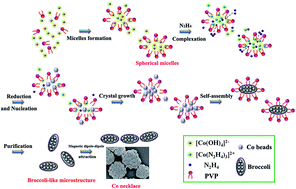
Controlled synthesis of novel hierarchical cobalt (Co) microstructures with extraordinary magnetic performances is a promising strategy for the development of magnetic metals for industrial purposes. Unlike the majority of experiments necessitating organic solvents at high temperatures or pressures, here broccoli-like Co microstructures were successfully prepared in aqueous solution under ambient temperature with the assistance of polyvinylpyrrolidone (PVP) for the first time. The synthetic process was associated with the chemical reduction of Co with pure water as solvent, hydrazine hydrate as reductant, and no complexant, nucleator, or external magnetic force was employed. The Co spheroids with broccoli shape were constructed by several beads with the length ranging from 0.67 μm to 1.22 μm, and they slightly self-assembled into a necklace with adjacent Co spheroids via spontaneous magnetic dipole–dipole attraction. The results indicated that the temperature, and the concentration of PVP and NaOH played an imperative role in the morphology and size of the ultrafine Co micro-aggregates in the present approach. More importantly, the broccoli-like Co entities exhibited a decreased saturation magnetization of 25.6 emu g−1 but an enhanced coercivity of 499.2 Oe mainly due to its anisotropic structure and smaller size, and could hold great potential for technological applications such as high-density data storage, and permanent magnetic materials.


1. Introduction

In recent years, ferromagnetic metals, including iron (Fe), nickel (Ni), and cobalt (Co), have attracted considerable attention in the scientific research with potential applications in catalysis, magnetic recording media, electromagnetic wave absorption, ferrofluids, and magnetic targeted drug delivery because of their outstanding magnetic attributes.1–5 Among these magnetic materials, Co is recognized as a strong candidate due to its high Curie temperature, good wear-resistance, and structure-dependent electronic and magnetic properties.6,7 Previous literature has proved that hexagonal close packed (hcp) Co with an intrinsic anisotropic structure displays superior coercivity and therefore is preferred for permanent magnetic applications.8 While the face-centered cubic (fcc) phase possessing low coercivity can serve as a soft magnetic material.9 So far, a wide variety of Co nanoparticles or micro-aggregates with different hierarchical architectures, such as nanowires,10,11 nanofibers,12 hollow spheres,13,14 micro-chains,9,15 dendrites,16,17 and flower-like structures,18,19 have been successfully synthesized via different methods including thermal decomposition of organometallic precursors, soft/hard template-mediated routes, hydro/solvothermal methods, and wet chemical reductive procedures.

Among these methods, wet chemical reductive synthesis (also called solution-phase metal salt reduction) has received more attention owing to its well-known inherent advantages, such as scalability, simplicity of operation, low energy consumption, high purity, and better control over size and shape,20–22 therefore it has been well-developed to fabricate several types of hierarchical Co micro-/nano-structures with the assistance of a complexing agent, or using another metal as the heterogeneous nucleator, or under an external magnetic field. For instance, Li et al. reported that uniform Co microspheres with the size of about 2–5 μm constructed by the assembly of nanoplatelets were synthesized through an aqueous chemical reductive route with the help of glycerin and citric acid.23 Flowery assembly of hcp Co nanorods were facilely attained in propylene glycol solution as solvent using ruthenium (Ru) as the heterogeneous nucleating agent and hexadecylamine as the structure-directing agent.24 Recently, our group have developed a highly ordered Co nanowires with uniform linear morphology using magnetic force in the presence of edetate disodium (EDTANa2) complexing agent.25 Although organic solvents, nucleating agent, and complexant are conductive to well crystallization and dispersibility of products, it is extensively accepted that complicated posttreatment process is needed to remove the additives and organics, and the introduction of metal as nucleation agent would seriously lead to the impurity of product. Thus, direct one-step synthesis of hierarchical Co microstructures on a large scale without any initiator, complexant, or organic solvent is still tough challenging.

Herein, based on these considerations, we applied an aqueous reduction strategy to prepare a broccoli-like Co microstructure at room temperature with polyvinylpyrrolidone (PVP) as an assistant, and no complexant or nucleator was added in the present work. The simple one-pot fabrication under ambient temperature can resist the structural collapse when the system faces heating.26 To our best knowledge, this is the first report on preparation of broccoli-like Co powders via the aqueous reduction strategy with a high yield. The as-prepared Co broccoli-like product exhibited an interesting ferromagnetic behavior with an enhanced coercivity of 499.2 Oe. The Co micro-entities with superior coercivity could bode good applications for magnetic sensors and devices in the future.

2. Materials and methods

2.1. Materials

Cobalt chloride (CoCl2·6H2O, 99 wt%), sodium hydroxide (NaOH), hydrazine hydrate (N2H4·H2O, 80 vol%), polyvinylpyrrolidone K30 (PVP, (–CH(NCH2CH2CH2CO)CH2–)n, Mw = 29[thin space (1/6-em)]000–35[thin space (1/6-em)]000) were purchased from Chengdu KeLong Reagent Co., Ltd. (China). All other chemicals were of analytical reagent grade and were used as received without further purification. All aqueous solutions were prepared with de-ionized water (D.I. water).

2.2. Preparation of cobalt microstructures

In a typical experiment, 4 g of NaOH (10 mol L−1) was put into a glass bottle of 50 mL capacity, completely dissolved in 10 mL of D.I. water, and then the solution was allowed to cool to room temperature. Another 5 mL of CoCl2·6H2O (0.05 mol L−1) with 2 wt% PVP as capping agent was introduced with intensive stirring. Subsequently, 1 mL of N2H4·H2O worked as reductant was added dropwise to the above solution for reduction of Co2+. The bottle was sealed and the mixed solution was continued stirring by a mechanical stirrer at 25 °C (room temperature) for 30 min in a thermostatic water bath. After reaction, the resulting mixture was black in color as a result of reduction of pink [Co(N2H4)3]2+ complex (II) to metallic Co(0). Finally, the black precipitate was collected from the solution using a magnet, and ultrasonically washed with D.I. water and absolute ethanol at least three times respectively to remove organics on the surface of particles. To investigate the formation mechanism of Co microstructures, a series of controlled experiments were carried out by altering the amount of NaOH, PVP, temperature, and reaction time, respectively, while kept other synthetic parameters and procedures the same as those of typical reaction. All the as-prepared products were dried through vacuum freeze-drying before characterization. The specific synthetic condition of each sample was listed in Table 1.
Table 1 Synthesizing conditions for preparing cobalt particles with PVP as surfactant
Sample name CoCl2 (mol L−1) Temperature (°C) PVP (2 wt%) NaOH (mol L−1)
CoT70-NP-1 0.05 70 No 10
CoT60-NP-1 0.05 60 No 10
CoT50-NP-1 0.05 50 No 10
CoT25-NP-1 0.05 25 No 10
CoT70-YP-1 0.05 70 Yes 10
CoT60-YP-1 0.05 60 Yes 10
CoT50-YP-1 0.05 50 Yes 10
CoT25-YP-1 0.05 25 Yes 10
CoT25-NP-2 0.05 25 No 15
CoT25-YP-2 0.05 25 Yes 15


2.3. Characterization

The crystalline phase and purity of the as-obtained samples were examined by X-ray diffraction analysis (XRD, Philips X'Pert Pro MPD) using Cu target as radiation source (λ = 0.154249 nm). The diffraction angles (2θ) were set between 20° and 95°, with an incremental step size of 4° min−1. The phase identification was achieved by comparing the sample diffraction pattern with standard cards in ICDD-JCPDS database. Fourier-transform infrared spectrometry (FTIR, Nicolet 750, USA) was used to identify the functional groups of the products detected in the form of pellets (KBr pellet). The spectra were recorded from 1200 cm−1 to 4000 cm−1. The morphological structure and elemental analysis of these Co microstructures were characterized by a field emission scanning electron microscope (FESEM, JSM-7500F, JEOL, Japan) equipped with X-ray energy dispersive spectroscopy (EDS). All samples were coated by gold for 1 min before SEM observation. In addition, the average diameter and particle size distribution were analyzed by Nano Measurer 1.2 image analysis software (Jie Xu, Fudan University, Shanghai, China). Transmission electron microscopy (TEM) image and high-resolution TEM (HRTEM) image were taken on a Tecnai G2 TEM (FEI, USA) with an accelerating voltage of 200 kV. All quantitative data expressed as mean ± standard deviations were derived from experiments.

2.4. Magnetic measurement

The magnetic properties, such as saturation magnetization (Ms) and coercivity (Hc), for the samples in the powder form was conducted at room temperature using a vibrating sample magnetometer (VSM, Lakeshore 7410, USA) with a maximum magnetic field of 30 kOe.

3. Results and discussion

3.1. Effect of temperature, PVP and NaOH concentration on the morphology and size of Co microstructures

To shed light on the formation of different Co microstructures, a series of controlled reactions were performed by changing some reaction parameters, such as the temperature, and the concentration of NaOH and PVP.
3.1.1. Reaction temperature. Firstly, the morphology and size of the samples synthesized under different temperature were examined by SEM with different magnifications, which were displayed in Fig. 1. It was obvious that the reaction temperature had a great impact on the morphology and size of the products. For these ultrafine Co products without PVP, when the experiment was conducted at 25 °C, the petal-shaped Co particles (0.51–0.72 μm) assembled into uniform flower architecture with the average diameter of 1.19 μm and a narrow distribution (Fig. 1d and S1d). Once the synthesis was carried out at 50 °C, rough Co irregular spheres and a small amount of smooth spheres coexisted with diameter ranging from 1.11 μm to 2.09 μm. Fig. 1b showed that the Co product obtained at 60 °C were self-assembled by wheatear-like nanosheets with the average length of 4.03 μm and the average width of 1.02 μm. If the temperature rose up to 70 °C, the energy was more sufficient. The chemical reaction would be more intense and the growth rate of the particles was greatly accelerated, so it was difficult to separate the nucleation step from crystal growth. As displayed in Fig. 1a, the Co bead was composed of more nanoparticles with larger size, contributing to increased the diameter and wider distribution of the final polygonal microstructure at 70 °C. These special Co morphologies might result from the different temperatures, which could change the absorbing of hydroxyl ion (OH) on different Co crystal plane, thus leading to various growth rate of each surfaces.9 Clearly, Co powders produced at 25 °C exhibited smaller size and better distribution than those grown at higher temperature (50–70 °C) (Fig. 1 and S1). Such phenomena were also observed for the samples prepared with the assistance of PVP capping agent (from 3.18 ± 0.23 μm at 70 °C to 0.96 ± 0.11 μm at 25 °C) (Fig. 2 and S2). Therefore, the morphology and size of final products varied greatly depending on the reaction temperature.
image file: c6ra11198j-f1.tif
Fig. 1 SEM images of the obtained cobalt powders in the absence of PVP fabricated under different temperatures: (a) CoT70-NP-1, (b) CoT60-NP-1, (c) CoT50-NP-1, and (d) CoT25-NP-1.

image file: c6ra11198j-f2.tif
Fig. 2 SEM images of the cobalt products with the assistance of PVP synthesized at different temperatures: (a) CoT70-YP-1, (b) CoT60-YP-1, (c) CoT50-YP-1, and (d) CoT25-YP-1.
3.1.2. Adding PVP. The PVP molecule was another important issue to influence the formation of Co microstructures. We could see that irregular polygonal Co particles with average diameter of 6.73 μm were appeared when no PVP was employed (Fig. 1a), and these particle tended to aggregate at 70 °C. While uniform polygonal Co powders with smaller size were dominant with the help of PVP at the same temperature. The morphologies of the products changed from polygonal structure at high temperature of 70 °C to flower-like assemblies under 50 °C using similar amount of PVP in the samples of different temperatures. Interestingly, a large amount of broccoli-like Co composed of many nanosphere (about 300–500 nm) with a size of 0.96 ± 0.11 μm were produced, possessing a narrow size distribution (Fig. 2d and S2d) under 25 °C when 2 wt% PVP was used. Simultaneously, these adjacent particles slightly combined together to form a Co necklace due to the intrinsic magnetic dipole–dipole attraction in pure water system, because the low viscosity of pure water allowed their movement. Meanwhile, the rough surface of CoT25-YP-1 also had a strong tendency to combine other Co crystals in order to reduce such high surface energy. When no PVP was employed, only flowery Co microstructures were dominant under 25 °C (Fig. 1d). In addition, it was obvious that smaller Co particles, more homogeneous morphologies, and narrower distribution of products were formed in the presence of PVP compared with the Co counterparts without PVP at each temperature. These advantages may attribute to the addition of PVP, because it plays a crucial role in dispersion, stabilizing and structure orientation of nanomaterials.21,27,28 The influence of PVP content on the morphology of Co microstructures was also examined through SEM as shown in Fig. S4. It was easy to understand that an enormous amount of irregular flowery Co products were generated with 1% PVP, because low concentration of PVP could not form micelles in the solution. When 5% PVP was used, the concentration of surfactant was so high that Co particles formed with PVP as template were closely connected, and especially some of the neighboring particles would grow together leading to the larger size with average diameter of 1.99 ± 0.28 μm (Fig. S4b), much bigger than that of CoT25-YP-1 (0.96 ± 0.11 μm). Hence, 2% PVP concentration was beneficial to formation of broccoli-like Co with appropriate size. PVP can effectively afford protection from collision and agglomeration of Co particles, when it absorbs on the surface of the polycrystals by five-membered nitrogen-containing heterocyclic side chain and interaction between the C–N/C[double bond, length as m-dash]O and Co, resulting in large steric hindrance.29 Moreover, PVP may be selective absorption on the facets of Co crystals via van der waals force in the initial nucleation.30 So it can hinder kinetic control growth rates of these facets, prevent its spread, and slow down or even restrict its growth. Consequently, the results showed that the presence of the PVP strongly affects the nucleation and growth rates of the Co polycrystals and therefore determining the particle size and the morphology.
3.1.3. The concentration of NaOH. In addition to the reaction temperature and PVP, the dosage of NaOH was also found to be crucial for the formation of the hierarchical broccoli-like Co configuration. In the strong base solution, Co2+ can coordinate with OH to form blue [Co(OH)4]2− complex before the chemical reduction process. If no NaOH was employed, the Co2+ ions could not be reduced to Co particles in absence of OH, but forming pink [Co(N2H4)3]2+ complex. As shown in Fig. 3. The SEM results revealed that the size of Co particles increased with the increasing of NaOH concentration no matter whether PVP was used. It is understandable that the reaction rate is accelerated by increasing the NaOH concentration, and the higher reaction rate favors the formation of larger structure in a short time. To be specific, for the samples without PVP, flower-like Co microstructures as the main product were achieved. When 15 mol L−1 NaOH was introduced, these Co micro-flowers had a mean size of about 2.88 ± 0.25 μm and possessed a wider size distribution (Fig. 3b and S3a) than that for CoT25-NP-1 prepared under 10 mol L−1 NaOH condition (Fig. 3a and S1d). From a magnified image of a randomly selected flower shown in the inset, it could be clearly seen that each Co flower was composed of dozens of spear-like petals attached to a mutual center. The petals grew radially from the spherical core with diameters of 300–400 nm at the center and length of 1.13–1.91 μm from the center to the tip. With regard to the CoT25-YP-2, the product was dominated by bigger Co spheroids with some size variations to some extent (2.24 ± 0.25 μm) compared with CoT25-YP-1. The oversize of spheroids was adverse to their movement under their spontaneous magnetic dipole–dipole force. These results demonstrated that low temperature, and appropriate concentration of PVP and NaOH jointly contributed to the orientation growth of the broccoli-like Co crystals.
image file: c6ra11198j-f3.tif
Fig. 3 SEM images of the as-prepared cobalt microstructures synthesized at room temperature with different NaOH dosages in the presence/absence of PVP capping agent: (a) CoT25-NP-1, (b) CoT25-NP-2, (c) CoT25-YP-1, and (d) CoT25-YP-2.
3.1.4. TEM of Co broccoli. The microstructure of CoT25-YP-1 sample was further investigated by TEM and high-resolution TEM (HRTEM) as shown in Fig. 4. A hierarchical Co sample with broccoli morphology was clearly seen in Fig. 4a. The surface of the micro-aggregate was not smooth, and the size of each Co particle was about 1 μm, almost consistent with what observed from the SEM images. The HRTEM image taken on the edge of the Co broccoli was displayed in Fig. 4b. The interplanar spacing was measured to be 0.20 nm and 0.22 nm, corresponding to the (002) and (100) crystal planes of hcp Co, respectively.
image file: c6ra11198j-f4.tif
Fig. 4 TEM (a) and HRTEM (b) images of the broccoli-like Co microstructure.

3.2. Chemical component analysis

Here we chose the microspheres prepared with/without PVP under room temperature as examples to characterize the chemical structures and magnetic properties of the broccoli-like Co microcrystals. To examine that Co metal was the primary phase via hydrazine hydrate reduction route, we performed powder XRD analysis. Fig. 5 displayed the corresponding XRD patterns of the ultrafine samples. It could be see that both the diffraction peaks of CoT25-NP-1 and CoT25-YP-1 were well indexed with the reflections of fcc Co (PDF standard cards, JCPDS 15-0806, space group Fm[3 with combining macron]m) and hcp Co (PDF standard cards, JCPDS 05-0727, space group P63/mmc). Four characteristic peaks of fcc Co at 2θ value of 44.43°, 51.52°, 75.94°, and 92.22° corresponding to Miller indices (111), (200), (220) and (311) were observed, respectively, with the cell parameter of a = 3.545 Å, b = 3.545 Å, and c = 3.545 Å. The recorded diffraction peaks at 2θ = 41.68°, 44.43°, 47.53°, and 75.94° were well assigned to the (100), (002), (101), and (110) planes of hcp Co phase, respectively. The cell parameter was a = 2.503 Å, b = 2.503 Å, and c = 4.061 Å. The XRD results indicated that the as-synthesized samples were a mixture of hcp and fcc Co whether there was PVP used or not. However, it was obvious that sharper and higher diffraction peaks was present in PVP-capped CoT25-YP-1 samples, implying an increase of crystalline degree. Next, the average crystalline grain sizes were estimated from the XRD patterns according to the Scherrer formula D = λk/(β[thin space (1/6-em)]cos[thin space (1/6-em)]θ) (where D is the average crystallite size, k is particle shape factor, λ is the X-ray wavelength 0.1542 nm, β denotes the angular line width of half-maximum intensity, and θ represents the Bragg's angle) with the values of 26.3 nm and 20.7 nm for CoT25-NP-1 and CoT25-YP-1, respectively. They were obviously smaller than the sizes of the Co beads in FESEM micrograph, which implied that each bead consisted of several crystal grains. No characteristic peaks due to the impurities of cobalt oxides or hydroxides could be detected, indicating that high purity of Co could be obtained in the present synthetic route in water solution without chelating agent and initiator under room temperature.
image file: c6ra11198j-f5.tif
Fig. 5 XRD patterns of the cobalt particles prepared in aqueous reduction process at room temperature.

FTIR spectrum was helpful to know whether the PVP has been removed from product. Fig. S5 showed the FTIR spectra of CoT25-YP-1 before and after rinse. The basic hydroxyl group OH of H2O at 3410–3551 cm−1 and C[double bond, length as m-dash]O stretching vibration at 1624 cm−1 were found for the product before rinse. The bands at around 1448 cm−1 and 1385 cm−1 were attributed to C–C in-plane vibration and C–N stretch band, respectively, proving the presence of PVP polymer.31,32 However, only the typical peaks of adsorbed H2O at about 3431 cm−1 and 1633 cm−1 were observed in Co product after rinse, suggesting that PVP has been removed completely. To further verify the formation of Co particles, EDS analysis were applied to characterize their chemical composition. As shown in Fig. 6, except for trace amounts of carbon and oxygen, Co was the prominent element with weight fraction (wt%) >87% for the two groups. The presence of carbon and oxygen might be ascribed to the adsorption of CO2 in the air by the surface of Co. Furthermore, EDS data showed that Co content including weight fraction (94.98 wt%) and atom fraction (83.24 at%) of crystals assisted by PVP (Fig. 6b) was much higher than that of CoT25-NP-1 samples without PVP (87.19 wt%, and 60.40 at%), which confirmed that the PVP-capped Co particles were comprised of Co in relatively high purity, consistent with the result from XRD analysis. PVP molecules absorbed around the surface of particles, and could protect the surface oxidation of Co, resulting in the enhancement of product purity.


image file: c6ra11198j-f6.tif
Fig. 6 EDS spectra of the ultrafine cobalt microstructures synthesized in aqueous reduction process at room temperature: (a) CoT25-NP-1, and (b) CoT25-YP-1.

3.3. Proposed formation mechanism of broccoli-like Co microstructure

In order to understand the formation mechanism of such interesting broccoli-like Co structure, time-dependent experiments were carried out at 25 °C with different reaction intervals and the shape evolution of products was revealed in Fig. 7. At the reaction time of 9 min, the product was composed of irregular spherical microparticles and poor-formed grape-like aggregates. Primary Co nanocrystals were initially generated through reduction and conventional nucleation process, and these nanocrystals tended to join each other with the assistance of magnetic dipole interactions and thermodynamic driving force in order to reduce the magnetic anisotropic energy and the surface energy. When the reaction was prolonged to 12 min, snowflake-like and grape-like microspheres with varied diameters coexisted in the system, and it seemed that the grape shapes composed of many microbeads became to convert to snowflake-like particles as shown in Fig. 7b. As reaction proceeded, the loosely microbeads became compact gradually through Ostwald ripening and the interface between microbeads nearly disappeared. Well-organized Co broccoli emerged as the main product through further growth and ripening process if the reaction time was continuously increased to 15 min. At last, by prolonging the time to 18 min we could see some chain-like Co necklaces due to the dipole magnetism of these broccoli.
image file: c6ra11198j-f7.tif
Fig. 7 SEM images of CoT25-YP-1 product prepared at 25 °C for different reaction times: (a) 9, (b) 12, (c) 15, and (d) 18 min, respectively.

PVP, an organic polymer containing both hydrophilic and hydrophobic groups, similar to other common surfactants such as sodium dodecyl benzenesulfonate (SDBS) and cetyltrimethyl ammonium bromide (CTAB), has been widely used in the solution-phase reduction of many kinds of metals and their compounds, where it is mainly considered as a steric stabilizer or capping agent with the main purpose for protecting crystals from agglomeration, besides, it also plays an active role in self-assembly of product.9,33,34 Typically, the formation of Co broccoli-like micro-spheroids involves in multiple steps of complexation, reduction, nucleation and assembly in the water system. The chemical equations and Nernst equation of the whole reduction reaction can be formulated as follows:

 
Co2+ + 4OH = [Co(OH)4]2− (1)
 
[Co(OH)4]2− + 2N2H4 = [Co(N2H4)2]2+ + 4OH (2)
 
[Co(N2H4)2]2+ + N2H4 = [Co(N2H4)3]2+ (3)
 
[Co(N2H4)3]2+ + N2H4 + 2OH = Co↓ + 4NH3↑ + 2N2↑ + H2↑ + 2H2O (4)
 
image file: c6ra11198j-t1.tif(5)
where E is the electromotive force difference between the electrodes in a cell; Eθ is the standard reduction potential for the cell; R, T, and F denote the gas constant (8.314 J K−1 mol−1), temperature, and Faraday constant (9.6437 × 10⁴ J V−1 mol−1), respectively; n represents the number of moles of electrons transferred through the external circuit in the balanced equation; [OH] and [Co2+] represents the concentration of OH and Co ions in the reaction, respectively. Per the Nernst equation, E increases with the increase of the concentration of Co2+ and OH, and the reaction temperature. The larger the value of E is, the easier the reaction is.

Based on our obtained results above, herein, the possible self-assembly mechanism of broccoli-like Co assisted by PVP surfactant under ambient temperature was illustrated in Fig. 8a. In the beginning, hydroxyl ions (OH) coordinated with Co2+ ions to generate blue [Co(OH)4]2− complexes in the solution. The PVP molecules self-assembled to spheroid micelles in the water,35,36 and the Co nuclei were wrapped and possibly absorbed onto the surface of spherical micelles in order to reduce the surface Gibbs free energy. Besides, the metallic ions could be anchored or chelated on the side chain of PVP molecule through the steric stabilization and electrostatic interaction between the quaternary amine (+) or C–O(−) groups of the pyrrolidone rings and the metallic ions (Fig. 8b), therefore, PVP can act as a stabilizer for the dissolved metallic salts. Subsequently, with the introduction of N2H4·H2O into the reaction system, a portion of N2H4 ceaselessly replaced the OH anions and coordinated with Co2+ cations to form pink [Co(N2H4)3]2+ complexes, and the rest of them as reducing agent entered into the mini-reactor and converted [Co(N2H4)3]2+ to small Co nanoparticles with the help of OH. Co as well as its compounds was preferred to form micro-spheres according to the previous literatures.23,37 These micro-spheres tended to aggregate due to their high surface energy and magnetic dipole–dipole attraction. At the same time, the PVP spherical micelles limited the range of their growth, which led to the self-assembly of microbeads to broccoli-like spheroids after reaching equilibrium condition.


image file: c6ra11198j-f8.tif
Fig. 8 (a) Schematic drawings of the Co broccoli formation mechanism assisted by PVP (2%), and (b) a proposed interactions between PVP and Co ion precursors.

Conversely, without PVP or low concentration of PVP, the crystal grew in all directions and finally produced a flowery morphology without the confine of micelles at room temperature. On the other hand, it was difficult to generate intact PVP micelles under higher temperature condition, thereby reducing the capping function on the surface of the metallic ions. Moreover, the temperature might change the reaction rate and the adsorption of OH on the surface of Co particles, resulting in various shapes.

3.4. Magnetic property

The magnetic hysteresis measurement (MH curve) of the broccoli-like Co obtained from the typical synthesis was conducted at room temperature in the applied magnetic field sweeping from −30 kOe to 30 kOe. In order to compare with the micro-spheroids, magnetic properties of Co microcrystal with flower-like morphology prepared without PVP corresponding to the sample of Fig. 3a were also given. Fig. 9 showed that the both resulting Co powders displayed ferromagnetic nature with the saturation magnetization (Ms) and coercivity (Hc) values of 147.8 emu g−1, 147.9 Oe, and 25.6 emu g−1, 499.2 Oe, respectively. Compared to the flower-like particles, the saturation magnetization of the broccoli-like micro-spheroids showed a reduction. Moreover, the saturation magnetization value of the broccoli-like Co sample was found to be much lower than that of bulk counterpart (about 168 emu g−1) originated from the presence of surfaces pinning disorder and inevitably minute surface anti-ferromagnetic oxidation in the product. On the other hand, the coercivity value was 499.2 Oe with an open hysteresis loop in the low field region for CoT25-YP-1, which was remarkably enhanced compared to that of Co bulk with a few tens of oersteds (Oe). In addition, such coercivity was still higher than those of various Co polycrystals with different shapes, including flowery (293 Oe),38 dendritic (208 Oe),39 micro-spherical (382.9 Oe),40 hollow spherical (202 Oe),35 chain-like (139.5 Oe),20 and nanowire (250 Oe)41 Co powders reported from other groups. Overall, the properties of the as-prepared Co microcrystals were listed in Table 2. It is well-known that the magnetic properties of materials strongly depend on the shape, size, crystalline, phase, defect structures, orientations, and so on.40,42,43 The Co ultrafine ferromagnetic particles with high anisotropic shape of broccoli-like assemblies could increase the coercivity in comparison with the corresponding bulk material.44 Moreover, according to the fanning mode of magnetization reversal per the “chain of spheres” model:45,46
 
image file: c6ra11198j-t2.tif(6)
where μ and R are the dipole moment and the diameter of each sphere. The expression in parentheses takes into account the dipolar interaction between the magnetic spheres in the chain based on the assumption of fanning mode. In particular, Kn accounts for the dipolar interaction between each every pair of the magnetic particles, and Ln, between each odd-numbered and even-numbered pairs. The number (n) is for the sphere numbering in the chain. Hence, in general the coercivity (Hc) is inversely proportional to the diameter of sphere (R) according to the model formula. Compared with the Co product synthesized without PVP (1.19 ± 0.10 μm), the broccoli-like Co powder had a smaller particle size with 0.96 ± 0.11 μm, resulting in much higher coercivity. Furthermore, it is also reported that ferromagnetic particles with high degree of crystalline possess enhanced coercivity.47 As per XRD result, the degree of crystalline for PVP-capped CoT25-YP-1 was higher than that for CoT25-NP-1 without PVP. Hcp phase in Co particles also could improve coercivity of cobalt particles due to larger magnetocrystalline anisotropy energy.8 Therefore, the superior coercivity for Co broccoli was a comprehensive consequence of its anisotropic structure, small particle size, highly crystalline, and crystal phase. Because a higher coercivity is an important issue for high-density information storage, further experiments are currently underway to improve the coercivity of these microspheres in our laboratory.

image file: c6ra11198j-f9.tif
Fig. 9 Magnetic hysteresis loop of the cobalt powders measured at room temperature and the hysteresis loop at low field: (a) CoT25-NP-1, and (b) CoT25-YP-1.
Table 2 Properties of all obtained ultrafine cobalt powders prepared in aqueous solution
Sample name Morphology Particle size (μm) Ms (emu g−1) Hc (Oe)
CoT70-NP-1 Polygonal 6.73 ± 0.75
CoT60-NP-1 Wheat-like 4.03 ± 0.54
CoT50-NP-1 Irregular spherical 1.65 ± 0.17
CoT25-NP-1 Flower-like 1.19 ± 0.10 147.8 147.9
CoT70-YP-1 Polygonal 3.18 ± 0.23
CoT60-YP-1 Irregular 2.22 ± 0.26
CoT50-YP-1 Flower-like 1.48 ± 0.15
CoT25-YP-1 Broccoli-like 0.96 ± 0.11 25.6 499.2
CoT25-NP-2 Flower-like 2.88 ± 0.25
CoT25-YP-2 Broccoli-like 2.24 ± 0.25


4. Conclusion

In summary, a novel broccoli-like Co microstructure has been successfully synthesized through a facile aqueous solution reduction using hydrazine as reducing agent in the presence of PVP under room temperature. No complexing agent or organic solvent was employed during the synthetic process. The SEM results showed that the Co broccoli with the length ranging from 0.67 μm to 1.22 μm was assembled by many beads (about 300–500 nm), and these adjacent particles slightly self-assembled to Co necklace due to the intrinsic magnetic dipole–dipole interaction. The reaction temperature, adding PVP, and NaOH concentration were the fundamental factors influencing the formation of Co microcrystals. The possible formation mechanism of broccoli-like Co configuration was proposed with the help of PVP as steric stabilizer based on a series of controlled experiments. This kind of ferromagnetic Co powders possessed an enhanced coercivity value of 499.2 Oe compared with bulk counterpart, therefore, having a promising use in magnetic recording devices and other related micro-devices.

Acknowledgements

This work was supported by the financial support from scientific research foundation of Sichuan University.

References

  1. C. B. Murray, S. Sun, H. Doyle and T. A. Betley, MRS Bull., 2001, 26, 985 CrossRef CAS.
  2. K. Nagaveni, J. Mater. Chem., 2002, 12, 3147 RSC.
  3. J. I. Martin, J. Nogues, L. Kai, J. L. Vicent and I. K. Schuller, J. Magn. Magn. Mater., 2003, 256, 449 CrossRef CAS.
  4. J. Ahmed, S. Sharma, K. V. Ramanujachary, S. E. Lofland and A. K. Ganguli, J. Colloid Interface Sci., 2009, 336, 814 CrossRef CAS PubMed.
  5. X. Wang, G. Shi, F.-N. Shi, G. Xu, Y. Y. Qi, D. Li, Z.-D. Zhang, Y. Zhang and H. You, RSC Adv., 2016, 6, 40844 RSC.
  6. C. T. Black, C. B. Murray, R. L. Sandstrom and S. Sun, Science, 2000, 290, 1131 CrossRef CAS PubMed.
  7. S. Wen, X. C. Zhao, Y. Liu, J. W. Cheng and H. Li, RSC Adv., 2014, 4, 40456 RSC.
  8. D. P. Dinega and M. G. Bawendi, Angew. Chem., Int. Ed., 1999, 38, 1788 CrossRef CAS.
  9. Y. J. Zhang, Q. Yao, Y. Zhang, T. Y. Cui, D. Li, W. Liu, W. Lawrence and Z. D. Zhang, Cryst. Growth Des., 2008, 8, 3206 CAS.
  10. H. Q. Cao, Z. Xu, H. Sang, D. Sheng and C. Y. Tie, Adv. Mater., 2001, 13, 121 CrossRef CAS.
  11. M. I. Irshad, N. M. Mohamed, M. Z. Abdullah, M. S. M. Saheed and J. Sorte, RSC Adv., 2016, 6, 14266 RSC.
  12. N. A. M. Barakat, B. Kim, S. J. Park, Y. Jo, M. H. Jung and H. Y. Kim, J. Mater. Chem., 2009, 19, 7371 RSC.
  13. H. Yoshikawa, K. Hayashida, Y. Kozuka, A. Horiguchi, K. Awaga, S. Bandow and S. Iijima, Appl. Phys. Lett., 2004, 85, 5287 CrossRef CAS.
  14. H. Yang, C. Shen, N. Song, Y. Wang, T. Yang, H. Gao and Z. Cheng, Nanotechnology, 2010, 21, 375602 CrossRef PubMed.
  15. M. Liu, L. Chen, C. Lin, L. Zhang and H. Song, J. Colloid Interface Sci., 2013, 410, 116 CrossRef CAS PubMed.
  16. R. Sivasubramanian and M. V. Sangaranarayanan, Mater. Chem. Phys., 2012, 136, 448 CrossRef CAS.
  17. R. P. Panmand, R. H. Patil, B. B. Kale, L. K. Nikam, M. V. Kulkarni, D. K. Thombre, W. N. Gade and S. W. Gosavi, RSC Adv., 2014, 4, 4586 RSC.
  18. Z. An, J. Zhang and S. Pan, CrystEngComm, 2010, 12, 500 RSC.
  19. Y. Zhu, Q. Yang, H. Zheng, W. Yu and Y. Qian, Mater. Chem. Phys., 2005, 91, 293 CrossRef CAS.
  20. H. Chen, R. Zhou, C. Xu, G. Zhao and Y. Liu, Mater. Res. Bull., 2013, 48, 2399 CrossRef CAS.
  21. Y. Hou, A. Hiroshi Kondoh and T. Ohta, Chem. Mater., 2005, 17, 3994 CrossRef CAS.
  22. Z. Liu, S. Li, Y. Yang, S. Peng, Z. Hu and Y. Qian, Adv. Mater., 2003, 15, 1946 CrossRef CAS.
  23. Y. Li, J. Zhao, X. Su, Y. Zhu, Y. Wang, L. Tang and Z. Wang, Colloids Surf., A, 2009, 336, 41 CrossRef CAS.
  24. Q. Liu, X. Guo, Y. Li and W. Shen, J. Phys. Chem. C, 2009, 113, 3436 CAS.
  25. T. Qiwei, H. Junqing, Z. Yihan, Z. Rujia, C. Zhigang, Y. Shiping, L. Runwei, S. Qianqian, H. Yu and L. Xiaogang, J. Am. Chem. Soc., 2013, 135, 8571 CrossRef PubMed.
  26. X. Wang, F. Yuan, P. Hu, L. Yu and L. Bai, J. Phys. Chem. C, 2008, 113, 8773 Search PubMed.
  27. A. A. Umar and M. Oyama, Cryst. Growth Des., 2006, 6, 818 CAS.
  28. B. Çelik, Y. Yıldız, H. Sert, E. Erken, Y. Koşkun and F. Şen, RSC Adv., 2016, 6, 24097 RSC.
  29. N. Soltani, E. Saion, M. Erfani, K. Rezaee, G. Bahmanrokh, G. P. Drummen, A. Bahrami and M. Z. Hussein, Int. J. Mol. Sci., 2012, 13, 12412 CrossRef CAS PubMed.
  30. G. Y. Huang, X. U. Sheng-Ming, L. I. Lin-Yan and X. J. Wang, Trans. Nonferrous Met. Soc. China, 2014, 24, 3739 CrossRef CAS.
  31. A. Baykal, N. Bıtrak, B. Ünal, H. Kavas, Z. Durmus, Ş. Özden and M. S. Toprak, J. Alloys Compd., 2010, 502, 199 CrossRef CAS.
  32. D. Cho, D. Choi, R. C. Pawar, S. Lee, E. H. Yoon, T. Y. Lee and C. S. Lee, Mater. Chem. Phys., 2014, 148, 859 CrossRef CAS.
  33. X. Shi, C. Xin, X. Chen, S. Zhou, S. Lou, Y. Wang and Y. Lin, Chem. Eng. J., 2013, 222, 120 CrossRef CAS.
  34. C. Jiang and Y. Wang, Mater. Chem. Phys., 2009, 113, 531 CrossRef CAS.
  35. C. Xu, D. Nie, H. Chen, G. Zhao and Y. Liu, Mater. Lett., 2013, 110, 87 CrossRef CAS.
  36. F. S. Diana, S. H. Lee, P. M. Petroff and E. J. Kramer, Nano Lett., 2003, 3, 891 CrossRef CAS.
  37. R. Xu, T. Xie, A. Y. Zhao and Y. Li, Cryst. Growth Des., 2007, 7, 1904 CAS.
  38. X. Liu, R. Yi, Y. Wang, G. Qiu, N. Zhang and X. Li, J. Phys. Chem. C, 2006, 111, 163 CrossRef.
  39. H. Chen, C. Xu, X. Zhou, Y. Liu and G. Zhao, Mater. Res. Bull., 2012, 47, 4353 CrossRef CAS.
  40. R. Zhou, H. Chen, G. Liu, G. Zhao and Y. Liu, Mater. Lett., 2013, 112, 97 CrossRef CAS.
  41. S. Gayen, M. K. Sanyal, B. Satpati and A. Rahman, Appl. Phys. A, 2013, 112, 775 CrossRef CAS.
  42. V. F. Puntes, K. M. Krishnan and A. P. Alivisatos, Science, 2001, 291, 2115 CrossRef CAS PubMed.
  43. S. Sun and C. B. Murray, J. Appl. Phys., 1999, 85, 4325 CrossRef CAS.
  44. G. Dumpich, T. P. Krome and B. Hausmanns, J. Magn. Magn. Mater., 2002, 248, 241 CrossRef CAS.
  45. W. Zhou, K. Zheng, L. He, R. Wang, L. Guo, C. Chen, X. Han and Z. Zhang, Nano Lett., 2008, 8, 1147 CrossRef CAS PubMed.
  46. I. S. Jacobs and C. P. Bean, Phys. Rev., 1955, 100, 1060 CrossRef.
  47. A. Ramazani, M. A. Kashi and G. Seyedi, J. Magn. Magn. Mater., 2012, 324, 1826 CrossRef CAS.

Footnote

Electronic supplementary information (ESI) available. See DOI: 10.1039/c6ra11198j

This journal is © The Royal Society of Chemistry 2016
Click here to see how this site uses Cookies. View our privacy policy here.