Monitoring bicosomes containing antioxidants in normal and irradiated skin

Estibalitz Fernández*a, Sarah Hostachyb, Christophe Sandtc, Gelen Rodríguezd, Helene C. Bertrandb, Sylvain Clèdeb, Mercedes Cócerad, Alfonso de la Mazaa, François Lambertb, Clotilde Policarb and Olga Lópeza
aInstitute of Advanced Chemistry of Catalonia (IQAC-CSIC), Jordi Girona 18-26, 08034 Barcelona, Spain. E-mail: efptqt@cid.csic.es; Fax: +34 932 045 904; Tel: +34 934 006 100
bÉcole Normale Supérieure – PSL Research University, Département de Chimie Sorbornne Universités – UPMC UNIV Paris 06, CNRS UMR 7203 LBM, 24 rue Lhomond, 75005 Paris, France
cSynchrotron SOLEIL, SMIS beamline, L'Orme des Merisiers, 91190 Saint-Aubin, France
dBicosome S.L., Jordi Girona 18-26, 08034, Barcelona, Spain

Received 29th April 2016 , Accepted 22nd July 2016

First published on 25th July 2016


Abstract

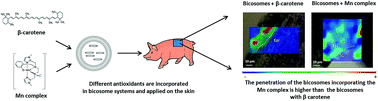
This study evaluates the penetration of bicosome systems incorporating two different antioxidants into normal skin and skin exposed to ultraviolet-visible radiation (UV-VIS) by Fourier-transform infrared microspectroscopy (FT-IR) using synchrotron radiation. Bicosomes are phospholipid assemblies based on mixtures of discoidal lipid structures protected by spherical lipid vesicles able to incorporate different molecules. In the current work, the antioxidants incorporated in these systems were β-carotene and a Mn complex as a superoxide dismutase (SOD) mimic. Additionally, a rhenium tri-carbonyl derivative was incorporated in the bicosome systems in order to map their penetration following the tag specific carbonyl signal by FT-IR microspectroscopy. The characterization of bicosome systems using the dynamic light scattering technique (DLS) showed a modification in the size of the systems containing β-carotene (Bcβ) or MnII complex (BcMn). After skin permeation, FT-IR results indicated a higher and deeper penetration of the BcMn system than the Bcβ system into the skin. Likely, the different physicochemical properties of both antioxidants could be responsible for this effect. Moreover, the penetration of both bicosome systems in irradiated skin was lower in comparison with the normal skin. This fact could be a consequence of the alteration of water transport in the skin during the irradiation process. In conclusion, these results indicated the effectiveness of bicosome systems as skin carriers, and provide information to protect skin under radiation using antioxidants.


1. Introduction

The absorption of different active compounds into skin can be an efficient strategy to protect normal skin and to treat impaired skin.1,2 However, the efficacy of topically applied compounds on the skin mainly depends on their incorporation in this tissue. Factors such as the strong barrier function of the skin or solar exposure can affect the absorption of these active compounds in the tissue.3,4 The use of lipid vehicles to facilitate the incorporation of active compounds in the skin is frequently used to address this problem.5

Bicosomes are phospholipid assemblies based on mixtures of discoidal lipid structures with diameters of approximately 10–20 nm and a thickness of 5.4 nm (bicelles) protected by spherical lipid vesicles with diameters of approximately 150–250 nm.6 The encapsulation of bicelles in spherical vesicles forming bicosomes is a good strategy to maintain the discoidal structure of this lipid carrier. Besides, bicosomes combine the advantages of disks and spherical vesicles. The bilamellar structure of both aggregates forming bicosomes has been studied in previous works and is represented in Fig. 1.7 Bicelles have recently demonstrated a great potential as carriers and as modifiers of skin permeability, and lipid vesicles have been used in dermatological applications for decades as delivery systems of different actives.5,7–10 The combination of discoidal and vesicular assemblies forming bicosomes potentiates the effects of both nanostructures on the cutaneous tissue, and allows a two-step process of interaction with the skin difficult to achieve with other nanostructures.6,11


image file: c6ra11170j-f1.tif
Fig. 1 Bicosome structure: discoidal bicelles encapsulated in spherical vesicles.

In this sense, bicosomes are able to interact with the skin on the surface (by means of the external vesicle) and inside the tissue (by means of the internal bicelles).6,12

Additionally, bicosomes have demonstrated to be more effective in comparison with bicelles avoiding free radical formation in the skin and preservating the antioxidant β-carotene under radiation.12,13

The purpose of this work was to study the penetration of bicosomes incorporating β-carotene (Fig. 2A) and a MnII complex C16–enPI2–Mn (Fig. 2B) into pig skin by Fourier-transform infrared microspectroscopy (FT-IR) using synchrotron radiation source. The β-carotene has demonstrated antioxidant activity in previous works.2,14 Moreover, the MnII moiety (enPI2–Mn) was shown to be a mimic of superoxide dismutase (or SOD mimics), with an anti-superoxide activity characterized both out of any cellular context and in cells.15 In the present work, this moiety was conjugated to a lipophilic C16 alkyl chain to form the complex C16–enPI2–Mn.


image file: c6ra11170j-f2.tif
Fig. 2 (A) β-Carotene and (B) C16–enPI2–Mn complex.

The β-carotene is a lipophilic molecule, while the C16–enPI2–Mn has hydrophilic and lipophilic sites in its structure. The hydrophilic character of the C16–enPI2–Mn comes from the enPI2–Mn moiety, and the lipophilic property comes from the C16 alkyl chain. Thus, these two antioxidants could provide to bicosomes different character; lipophilic character incorporating β-carotene and amphiphilic character in the case of C16–enPI2–Mn. They also show a different reactivity, the SOD mimics being active on reactive oxygen species such as superoxide, whereas the β-carotene is known to quench peroxyl radicals.16 In the present study the penetration of bicosomes incorporating β-carotene (Bcβ system) and bicosomes incorporating the C16–enPI2–Mn (BcMn system) was also studied in irradiated skin in order to evaluate the influence of the radiation in the permeation of both bicosome systems.

The distribution of bicosome systems through the different layers of normal and irradiated skin was evaluated by monitoring the IR signal of a rhenium tri-carbonyl derivative C12ReCO3(fac-[Re(CO)3Cl(2-(1-dodecyl-1H-1,2,3,triazol-4-yl)-pyridine)]) attached to the lipid systems (Fig. 3). Indeed, this probe displays two IR bands around 1920 cm−1 and 2020 cm−1 for the E vibration and A1 vibration respectively, and it was used for FT-IR microspectroscopy bio-imaging in cells and tissues17,18 This lipophilic derivative has also demonstrated to be a good probe for skin penetration studies by FT-IR microspectroscopy18 and it has been used in previous works for evaluating the penetration of bicosome systems.6 In one of these studies, the penetration of bicosomes tagged by C12ReCO3 was compared to the penetration of this derivative dissolved in a solution of the classical permeation enhancer dimethyl sulfoxide (DMSO). The results showed a higher penetration of the C12ReCO3 incorporated in the bicosomes. This fact demonstrated the properties of our bicosome systems as penetration enhancer and vector for skin delivery.


image file: c6ra11170j-f3.tif
Fig. 3 C12Re(CO)3 derivative (fac-[Re(CO)3Cl(2-(1-dodecyl-1H-1,2,3,triazol-4-yl)-pyridine)]).

2. Materials and methods

2.1. Chemicals

Bicosome systems were formed using 1,2-dipalmitoyl-sn-glycero-3-phosphocholine (DPPC) and DHPC (1,2-dihexanoyl-sn-glycero-3-phosphocholine) purchased from Avanti Polar Lipids (Alabaster, USA), cholesterol (CHOL) obtained from Sigma-Aldrich (Sto Louis, MO, USA) and Lipoid S-100, whose main component (>94%) is phosphatidylcholine (PC), purchased from Lipoid GmbH (Ludwigshafen, Germany). Chloroform was purchased from Merck and purified water was obtained by an ultra-pure system, Milli-Q plus 185 (Millipore, Bedford, USA). β-Carotene was purchased by Sigma-Aldrich (St. Louis, MO, USA).

The C12Re(CO)3 derivative was synthesized by the École Normale Supérieure (Paris, France) as described previously.17

Reagents and chemicals for the synthesis of the C16–enPI2–Mn were purchased from Sigma-Aldrich unless otherwise stated. Analytical HPLC was performed on a Dionex Ultimate 3000 equipped with a variable wavelength detector, using an ACE 5 C8-300 column. The results were analyzed using Chromeleon software. Hexadecanal19,20 and N-(2-hydroxy-benzyl)-N,N′-bis[2-(N-methylimidazolyl)methyl]ethane-1,2-diamine] (enPI2) ligand19,20 were synthesized as previously described.

1H and 13C NMR spectra were recorded on a Bruker Avance 300 using solvent residuals as internal references. The following abbreviations are used: singlet (s), doublet (d), doubled doublet (dd), triplet (t), doubled triplet (td) and multiplet (m). TLC analysis was carried out on silica gel (Merck 60F-254) with visualization at 254 and 366 nm. Preparative flash chromatography was carried out with Merck silica gel (Si 60, 40–63 μm).

2.1.1. Synthesis of the MnII–SODmimic (C16–enPI2–Mn).
Synthesis of C16–enPI2 ligand (N-(2-hydroxy-benzyl)-N′-hexadecyl-N,N′-bis-(2-(N-methylimidazolyl)methyl)ethane-1,2-diamine). enPI2 (73.8 mg, 0.21 mmol, 1.0 equiv.) and hexadecanal (50.0 mg, 0.21 mmol, 1.0 equiv.) were dissolved in EtOH (absolute, 3 mL) and the solution was stirred at room temperature for 2 h. NaBH3CN (19.7 mg, 0.31 mmol, 1.5 equiv.) and trifluoroacetic acid (TFA) (32.0 mL, 0.42 mmol, 2.0 equiv.) were added and the solution was stirred at room temperature for 2 h. A 1 M aqueous solution of NaOH (3.0 mL) was added, EtOH was evaporated and dichloromethane (DCM) was added. The pH was adjusted to pH 9 by addition of a 1 M aqueous HCl solution. The aqueous phase was extracted three times with DCM; the organic phase was dried over Na2SO4, filtered and evaporated. The residue was purified by column chromatography on silica gel (gradient DCM/MeOH 95[thin space (1/6-em)]:[thin space (1/6-em)]5 to 5[thin space (1/6-em)]:[thin space (1/6-em)]5) to afford C16–enPI2 as a colourless oil (79.0 mg, 66%).

Rf (DCM/MeOH 95[thin space (1/6-em)]:[thin space (1/6-em)]5) = 0.10; 1H-NMR (300 MHz, CDCl3): δ (ppm). 7.14 (dtapp, J = 1.5, 7.5 Hz, 1H), 6.96 (dd, J = 1.5, 7.5 Hz, 1H), 6.92 (d, J = 1.2 Hz, 1H), 6.85 (d, J = 1.2 Hz, 1H), 6.79 (dd, J = 1.2, 7.5 Hz, 1H), 6.76 (d, J = 1.2 Hz, 1H), 6.72 (m, 2H), 3.59 (s, 4H), 3.56 (s, 3H), 3.53 (s, 2H), 3.39 (s, 3H), 2.63 (s, 4H), 2.28 (m, 2H), 1.24 (m, 28H), 0.86 (t, J = 6.6 Hz, 3H); 13C-NMR (75 MHz, CDCl3): δ (ppm). 157.4, 145.1, 144.4, 130.0, 129.0, 127.1, 126.6, 122.6, 121.51, 121.47, 119.0, 116.4, 56.4, 54.0, 50.5, 50.4, 50.1, 49.3, 33.0, 32.5, 32.0, 29.8–29.7 (10 CH2), 29.6, 29.4, 27.5, 26.1; HPLC (0 to 100% ACN in 30 min – column C8A): rt 24.60 (84%).

C16–enPI2–Mn was prepared by combining a stock solution of the C16–enPI2 ligand (1 mL at 20 mM in H2O/ACN 1[thin space (1/6-em)]:[thin space (1/6-em)]1) with an aqueous stock solution of MnCl2 (1 mL at 20 mM, 1.0 equiv.) and 1 mL of 2-[4-(2-hydroxyethyl)piperazin-1-yl]ethanesulfonic acid (HEPES) buffer (100 mM, pH 7.5). The resulting solution was lyophilised to give a powder, which was re-dissolved in 2 mL of H2O to afford a 10 mM [C16–enPI2–Mn]Cl in a 50 mM HEPES solution.

2.2. Preparation of bicosomes

Two different bicosome systems tagged with the C12Re(CO)3 derivative were formed: Bcβ system and BcMn system.

Bicelles with β-carotene were prepared by mixing appropriate amount of DPPC, DHPC, C12Re(CO)3 derivative and β-carotene in a chloroform solution. Once the components were mixed, the chloroform was evaporated with a rotary evaporator and the obtained lipid film was hydrated. The obtained water dispersion was subjected to several cycles of sonication and freezing until the sample became transparent.

Bicelles with the C16–enPI2–Mn were prepared following the same procedure, but this complex was added in a water solution.

To prepare the bicosome systems appropriate amounts of PC and CHOL were mixed in chloroform. The chloroform was removed with a rotary evaporator until a lipid film was obtained. Finally, the film was hydrated with the previously prepared bicelle dispersions.

The concentration of each ingredient in the bicosome systems was the following (w/v%): DPPC 4.25%, DHPC 0.75%, C12Re(CO)3 derivative 1%, β-carotene/C16–enPI2–Mn 0.01%, PC: 8% and CHOL 2%.

2.3. Size measurement of bicosomes by dynamic light scattering (DLS)

The hydrodynamic diameter (HD) was determined by means of DLS using a Zetasizer nano ZS90 (Malvern Instruments, UK). The particle sizes were determined by detection and analysis of the scattered light from the 632 nm He/Ne laser beam. Non-invasive back-scatter technology was used to minimize multiple scattering effects. The detection of the scattered light was performed at an angle of 173°. Measurements were carried out at 25 °C in triplicate for each bicosome system. Values presented in Table 1 are the mean values of HD and the standard deviations (SD) from the triplicate. The intensity percentages of the proportion of scattered light are also presented.
Table 1 Mean values and standard deviation (SD) of hydrodynamic diameters (HD) of the different bicosome systems. Proportions of the scattered intensity corresponding to the different populations in the size distribution curves at 25 °C for each lipid system are also shown
Bicosome system HD (nm) ± SD (peak 1) % int ± SD (peak 1) HD (nm) ± SD (peak 2) % int ± SD (peak 2)
a significant difference p < 0.05.
BcRe 16 ± 6 25 ± 6 253 ± 78a 71 ± 2
Bcβ 15 ± 4 18 ± 4 154 ± 39a 76 ± 4
BcMn 10 ± 1 17 ± 4 371 ± 21a 82 ± 2


A statistical analysis has been performed in the values obtained by DLS. Wilcoxon Rank–Sum test was carried out using STAT/SE 12.0 software. Statistical differences were considered for p values below 0.05.

2.4. Skin preparation and irradiation procedure

The skin from the back of Landrace large white pigs weighing around 40 kg was obtained from the Veterinary Faculty of the Universitat Autònoma of Barcelona, Spain. The skin was washed with tap water and dermatomed to 500 ± 50 μm thicknesses (Dermatome GA630, Aesculap, Tuttlingen, Germany). Then, the skin was cut into two pieces and one of them was subjected to irradiation. The exposure was performed using a light source simulating solar radiation (Suntest CPS+, Atlas, USA) at 500 W m−2 for 30 min (90 J cm−2). The radiation range was from 310 nm to 800 nm (2% Ultraviolet B, 18% Ultraviolet A, 72% Visible light and 8% Infrared A). This irradiation intensity (500 W cm−2) is the equivalent to the solar exposure for two days in June in Catalonia. The irradiation was performed with a ventilation system. The maximum temperature reached in the simulator was 35 °C.

2.5. Skin treatment with bicosomes

The normal and irradiated skin samples were cut into pieces of an area of 25 mm2 and then were treated with 10 μl of Bcβ and BcMn systems. The treatment was performed overnight at room temperature (20–25 °C) on a Petri plate on wet filter paper. Then, the plate was covered with paraffin. Under these conditions, the drying of the skin was avoided. The relative humidity inside the Petri plate was approximately 90%. During the treatment, the hydration and temperature of the skin samples remained constant. After the treatment, the skin pieces were cleaned with water. The treatment was performed using three skin pieces for each lipid system.

Finally, all skin samples were covered firstly with aluminium paper and then with Optimal Cutting Temperature compound (OCT). The aluminum paper was used to avoid the direct contact between the skin and the OCT. Next, skin samples were frozen in liquid N2 before cutting into transverse 6 μm thick sections. The cuts were performed using a Cryostat CM3050 (Leica Biosystems Nussloch, Germany). The different skin sections were placed on CaF2 circular windows (1 cm in diameter).

Considering that a small fraction of the bicosome systems could flow over the edges of the skin during the treatment, the slices from the edges of the tissue were removed. Only skin slices from the central part of the pieces were used in the FT-IR microspectroscopy.

2.6. FT-IR experiments

IR microspectroscopy was performed using synchrotron radiation at the SMIS beamline in synchrotron SOLEIL. Synchrotron radiation was employed because it provides a high spectral quality and produces high contrast chemical imaging. A Thermo Scientific Continuum XL IR microscope coupled to a Thermo Scientific FT-IR Nicolet 5700 spectrometer (Thermo electron corporation, Madison, Wisconsin, USA) was employed in these experiments. The microscope is equipped with a 32X/NA 0.65 Schwarzschild objective and matching condenser, a mercury–cadmium–telluride (MCT) type A narrow band detector cooled by liquid nitrogen and an X–Y–Z motorised stage.

Optical micrographs were produced from the microscope in the visible image mode to define the sampling positions. The spectra were acquired at room temperature in transmission mode, in the 3600–1000 cm −1 range, and 128 scans were averaged at 4 cm−1 resolution. Spectral maps were recorded with an aperture of 10 × 10 μm2 and steps of 10 μm in X and Y.

The spectra and mappings obtained by FT-IR technique generated information about the penetration of the C12Re(CO)3 derivative attached to bicosomes. Thus, the presence of the C12Re(CO)3 derivative in the skin allowed to predict the location and distribution of bicosome systems inside the tissue.

2.7. Data processing

To calculate the relative concentration of the C12Re(CO)3 derivative in the skin from FT-IR maps, the ratio between the areas under the curves (AUC) of A1 vibration of the C12Re(CO)3 and amide II skin vibration were calculated.6 Amide II vibration was used as internal reference (skin reference), thus, the AUCC12Re(CO)3,A1/AUCamide II ratio indicates the relative concentration of the C12Re(CO)3 in the skin.

The different areas of the skin (SC and epidermis) were visually differentiated in each map and the ratio AUCC12Re(CO)3,A1/AUCamide II was calculated in each step of the map (spectral maps were recorded with steps of 10 μm). Then, the mean value of all the ratios was calculated and from each map a mean value of the ratio AUCC12Re(CO)3,A1/AUCmide II in SC and in Epi was obtained. This procedure was performed in all the maps obtained for skin treated with Bcβ and BcMn systems using Omnic-Atlus software. Finally, from all the means obtained in SC and Epi in each bicosome system, the median value was calculated for each skin layer. These median values of the ratios AUCC12Re(CO)3,A1/AUCamide II are presented in Table 2.

Table 2 The median value of the AUCRe(CO)3,A1/AUCamide II ratio in SC and Epi for normal and irradiated skin treated with different bicosome systems
Skin sample SC ratio (×10−2) Epi ratio (×10−2)
Normal skin + Bcβ 1.72 0.35
Normal skin + BcMn 2.57 1.85
Irradiated skin + Bcβ 0. 83 0.24
Irradiated skin + BcMn 0.77 0.37


The quantification of the C12Re(CO)3 derivative in the skin allows monitoring accurately the localization and distribution of bicosomes inside the skin.

3. Results

3.1. Characterization of bicosome systems

The sizes as HD of the different bicosome systems obtained by DLS at 25 °C are shown in Table 1. For comparative purposes the size of bicosomes only with the C12Re(CO)3 derivative was also measured.

In all cases, two populations with different HDs and light scattered intensities were found. As commented in the introduction section, bicosomes are formed by discoidal nanostructures, bicelles, encapsulated in spherical vesicles. Some non-encapsulated bicelles could be found in this dispersion. Thus, in the bicosome systems the small size population (peak 1) detected by DLS corresponds to non-encapsulated discoidal structures (free bicelles) and the large population (peak 2) corresponds to the external vesicle encapsulating discoidal bicelles.

The HDs for free bicelles with the C12Re(CO)3 derivative and for the corresponding external vesicle (BcRe) were approximately 16 nm and 253 nm, and their intensity percentages were 25% and 71%, respectively. The Bcβ system showed HDs of approximately 15 nm (18%) and approximately 154 nm (76%). Finally, the HDs for the BcMn system were approximately 9 nm for free bicelles and 371 nm for external vesicle, and the intensity percentages were 17% and 82%, respectively.

The statistical analysis showed no significant differences between the values of peak 1 (non-encapsulated bicelles), thus these values can be equally considered. Concerning the values of peak 2 (the external vesicle encapsulating discoidal bicelles), significant differences were detected between all the samples. In this sense, the size of BcRe, Bcβ and BcMn can be considered as different.

3.2. Penetration of bicosomes inside the skin

IR spectra of normal and irradiated skin samples were recorded in the 3600–1000 cm−1 range. Fig. 4 shows the spectrum for normal skin treated with bicosomes tagged with the C12Re(CO)3 derivative. The spectra obtained for irradiated skin treated with bicosome systems displayed similar patterns to the spectra obtained for normal skin treated with these systems (data not shown). The spectrum shows bands characteristics of skin amides about 3300 cm−1 corresponding to NH vibrations (amide A), at 1660 cm−1 corresponding to CO vibration (amide I) and at 1550 cm−1 corresponding to CN vibrations (amide II). The characteristic vibration of CH3, CH2 stretching of lipids is around 2960 and 2920 cm−1 respectively.17 From a spectroscopic point of view the C12Re(CO)3 derivative shows a specific vibrational signature with two characteristic bands. The first one (E-band at 1920 cm−1) corresponds to asymmetric stretching vibrations and the second (A1-band at 2020 cm−1) comes from symmetric stretching vibrations.17 These vibrations do not interfere with the IR signals from the skin and consequently, the C12Re(CO)3 derivative can be detected in the skin by FT-IR without any interferences.6,18
image file: c6ra11170j-f4.tif
Fig. 4 IR spectra of normal skin treated with bicosomes tagged with C12Re(CO)3. From left to right; NH vibration of polypeptides and proteins of skin (amide A), CH3 and CH2 stretching vibration of skin and bicosome lipids, symmetric and asymmetric stretching vibrations of CO from C12Re(CO)3 molecule (A1 at 2020 cm−1 and E at 1920 cm−1), CO vibration of proteins of skin (amide I) and CN vibration of proteins of skin (amide II).

The FT-IR maps obtained for normal and irradiated skin samples treated with the Bcβ and BcMn systems are shown in Fig. 5A–D (in order to obtain a better image of the skin, the images of Fig. 5 without the superimposition maps are showed in ESI material). The sections of the micrographs display the skin surface including the SC and the Epidermis (Epi). These figures reflect the distribution of the band A1 of the C12Re(CO)3 derivative across the different regions of the skin. The colour scale, from blue to red, represents the location of the C12Re(CO)3 derivative attached to bicosomes. Red areas concentrate the highest amount of the C12Re(CO)3 derivative (hotspots) while in blue areas no amount of this molecule is detected. Yellow and green represent areas with intermediate amounts. Hence, the detection of the C12Re(CO)3 derivative in the skin brings the distribution of bicosome systems through the tissue. The use of the C12Re(CO)3 derivative as a probe to follow bicosome systems into the skin has been well established in a previous work.6 This lipophilic molecule allows estimating the penetration and distribution of the systems incorporating the studied antioxidants.


image file: c6ra11170j-f5.tif
Fig. 5 IR maps of A1 vibration obtained for normal and irradiated skin treated with different bicosomes. (A) Normal skin treated with Bcβ system, (B) normal skin treated with BcMn system, (C) irradiated skin treated with Bcβ system and (D) irradiated skin treated with BcMn system. The scale goes from blue to red, indicating no amount of C12Re(CO)3 derivative in blue colour and high amount of this molecule in red colour. SC: stratum corneum, Epi: epidermis.

The image of normal skin treated with the Bcβ system shows the main signal intensity of the C12Re(CO)3 derivative located in the SC, the superficial layer of the skin (Fig. 5A). The map obtained for normal skin treated with the BcMn system shows the main signal intensity in the SC and in the Epi (Fig. 5B).

The maps obtained for irradiated skin treated with the bicosome systems are also shown in Fig. 5C and D. As in the case of normal skin, the irradiated skin treated with the Bcβ system shows the main signal intensity of the C12Re(CO)3 derivative located in the SC (Fig. 5C). In the case of the BcMn system (Fig. 5D), the main intensity of the BcMn system is localized in the SC. This result contrasts with the result obtained in normal skin treated with the BcMn system (Fig. 5B), where a high signal intensity of this lipid system is also observed in the Epi.

Semi-quantitative analysis was performed by calculating the median of the AUCC12Re(CO)3, A1/AUCamide II ratio in the SC and the Epi (see Experimental section) and these results are shown in Table 2. To overcome problems that could be associated with different thickness of the skin, and assuming that amides are homogeneously distributed, amide II vibration was used to normalize the AUC obtained from the C12Re(CO)3 derivative. This quantification was performed in all the maps obtained by FT-IR for each bicosome system (between 4 and 8 maps for each bicosome system).

The relative concentrations of the C12Re(CO)3 derivative in SC and Epi in normal skin treated with the BcMn system were higher than the relative concentrations of this molecule detected in normal skin treated with the Bcβ system. Thus, the penetration of the BcMn system was higher than penetration of the Bcβ system.

Otherwise, the relative amounts of the C12Re(CO)3 derivative in both skin compartments were lower in irradiated skin with respect to those obtained for non-irradiated skin in both bicosome systems. In this case these relative concentrations were similar for bicosome system.

4. Discussion

4.1. Size of bicosome systems

In the formation of bicosome systems, the lipid film of PC and CHOL is hydrated with the initial solution of preformed bicelles. At this step, a portion of the lipids of the bicelles (and also C12Re(CO)3 derivative and both antioxidants) could be incorporated in the external membrane of the bicosome. Conversely, another fraction of the lipids from the external bilayer of the bicosome could be incorporated between the long chain phospholipid molecules (DPPC) in the membrane of bicelles. Therefore, the location of the C12Re(CO)3 derivative and both antioxidants, would be distributed in the free bicelles and in the external vesicle of bicosome systems.
4.1.1. Dimensions of the free bicelles (small size population). Although the size of the different non-encapsulated bicelles is similar, likely the location of both antioxidants could be different in the bilayer of these discoidal structures due to the different characteristics between both molecules. The following figure (Fig. 6) represents the putative location of both antioxidants in bicelle membrane.
image file: c6ra11170j-f6.tif
Fig. 6 Putative location of the different molecules in the bicelle structure. β-Carotene (orange) and the C16–enPI2–Mn (red).

Considering the planar geometry and the low solubility in water of β-carotene, the location of this antioxidant in the lipophilic region of the bilayer is expected. Some works suggest that the β-carotene molecules could be located perpendicular to the alkyl chains of phospholipids deep into the lipid bilayer21,22 as Fig. 6 shows.

The C16–enPI2–Mn is soluble in water and hence the location of this complex out of bicelle structure could be expected. However, the different size between the bicosome systems with the C12Re(CO)3 derivative and bicosomes containing the C16–enPI2–Mn together with this derivative (Table 1) indicates an interaction between the MnII complex and bicelle membrane. The unique alkyl chain present in the C16–enPI2–Mn could give to this molecule an amphiphilic character and consequently, the location of the MnII complex would be more favoured between the lipid molecules of bicelles.

4.1.2. Dimensions of the external vesicles encapsulating bicelles (large size population). The incorporation of β-carotene in bicosomes promoted a decrease in the size of the external vesicle of bicosome systems, while, the incorporation of the C16–enPI2–Mn entailed an increase in the HD of the large size (Table 1). This fact could be related to the partial location of both antioxidants in the external bilayer of bicosome systems. The C16–enPI2–Mn is an amphiphilic molecule and the incorporation of this antioxidant could happen between the hydrophilic and lipophilic region of the bilayer, that is, between polar heads and lipophilic chains of phospholipids (as in bicelle membrane). Thus, the C16–enPI2–Mn should be located parallel with respect to the alkyl chain of lipids. This fact would entail the separation between the lipid molecules promoting an increase in the diameter of the external vesicle.

Considering that the location of the C16–enPI2–Mn in the membrane of the external vesicle is the same as the location in the membrane of non-encapsulated bicelles (described in 4.1.1.), an increase in the diameters of these discoidal structures could be expected. Nevertheless, the diameter of free bicelles is the same regardless the incorporation of the C16–enPI2–Mn. Likely, the inclusion of the C16–enPI2–Mn in the flat bilayer of bicelles parallel to the alkyl chains should not severely affect the diameter of the discoidal structure of bicelles. However, the same type of inclusion in a curved membrane, i.e., in the external membrane of bicosomes, could flatten this bilayer, which would decrease the curvature radius of bicosomes, and the diameter of the spherical vesicle would increase,31 as our DLS results have shown.

Concerning the location of β-carotene in the external bilayer, the presence of this antioxidant provides a decrease in the diameter of Bcβ. Probably, the incorporation of β-carotene together with the C12Re(CO)3 derivative is responsible of the decrease of Bcβ system in current study, although the mechanism that induces this decrease is not yet fully known. In this sense, it is necessary more experiments to clarify this point.

4.2. Distribution of Bcβ and BcMn systems in normal and irradiated skin

In a previous work we demonstrated the interaction of the C12Re(CO)3 derivative with bicosomes and the penetration of this molecule in the skin.6 Those results allowed us to assume that the C12Re(CO)3 derivative remains linked to lipid aggregates. Consequently, the use of C12Re(CO)3 tag for monitoring the bicosomes with different actives through skin provides new information about the penetration of this system into the tissue.

The most common route for delivery of molecules in the skin is the intercellular route.23 To understand the penetration of bicosome systems into the skin by this route, the structure of the SC and the structure of these systems should be considered. The penetration mechanism using bicosome systems involve a multi-step process of the interaction with the skin, which has been demonstrated in previous works.6,12 The dimensions of the intercellular spaces of the SC, where intercellular penetration occurs, are approximately 6–10 nm, thus, only systems with dimensions around or smaller than 6–10, nm would be able to pass through these spaces. Considering the morphology of this lipid system, the external spherical vesicles with a size of approximately 200 nm are not be able to penetrate through the SC by a intercellular route, and they remain on the skin surface in a similar way as described for other lipid vesicles.24 However, in contact with the skin, these external vesicles burst and the encapsulated bicelles are released from inside and due to their small dimensions are able to penetrate through the narrow interlamellar spaces of the SC.7,25 Once incorporated into the SC, bicelles could mix with SC lipids or could reach the Epi. Next, bicelles increase in size due to the natural hydration gradient of the skin. This increase involves a transition from bicelles to vesicles and promotes the retention of bicelle components into the skin.7,25

Besides, the non-encapsulated bicelles would also penetrate with the same mechanism. Therefore, the bicelles are the responsible of the penetration of the C12Re(CO)3 derivative into the skin, and as commented before, we assume that the C12Re(CO)3 derivative remains linked to lipid aggregates.

The use of bicosome system encapsulating bicelles with the external vesicle incorporates an advantage to the skin, which is a superficial treatment to this tissue. That is, bicosomes are able to interact with the skin on the surface (by means of the external vesicle) and inside the tissue (by means of the internal bicelles). Additionally, bicosomes have demonstrated to be more effective in comparison with bicelles avoiding free radical formation in the skin and preservating the antioxidant β-carotene under radiation.

The different sizes observed for the bicelles forming the Bcβ and BcMn systems (15 ± 4 and 10 ± 1 nm respectively), could be a reason to explain the higher penetration of BcMn system. However, considering that both bicelles have the same thickness (5 nm), and that SC and the bicelles are deformable structures, the size difference does not enough to explain the higher concentration of the BcMn system into the skin. The different characteristics of both antioxidants could be the main reason of this fact.

β-Carotene is a lipophilic molecule and the C16–enPI2–Mn is an amphiphilic molecule. This fact could not only influence the size of the bicelles but also the interaction with the skin. The SC and Epi are environments with different physicochemical properties that would influence their interactions with both lipid systems.26 The SC is highly enriched in lipids compared to the Epi, but the intercellular route through the SC provides hydrophilic and lipophilic sites.27 Therefore, while the Bcβ system preferably passes through the lipophilic areas inside the skin, the BcMn system could be located in the hydrophilic and lipophilic pathways of this tissue. The Bcβ systems could integrate more readily in the lipophilic regions of the SC explaining why it is 5 times more concentrated in the SC than in the Epi. On the other hand, the BcMn is only 1.4 times more concentrated in the SC than in the Epi but is five times more concentrated than the Bcβ in the Epi. The Bcβ system appears to be preferentially retained in the SC, impairing its penetration to the Epi. The amphiphilic BcMn system seems to pass more freely through the SC to accumulate in the more hydrated Epi.

Considering its possibilities of interaction with the different skin compartments, the deeper penetration and higher local concentration of the BcMn system observed in our measurements seems consistent. Moreover, the amphiphilic character of the BcMn system would facilitate the distribution of this system to the deepest areas of the skin with more hydrated environments, that is, the Epi.

The overall penetration efficiency would result from the balance between the physicochemical properties of the antioxidants (mainly lipophilicity/amphiphilicity), rheological, chemical and hydration properties of the skin compartments, bicelles size being of less importance. Since bicelles sizes cannot completely explain the distributions observed in this study, our results suggest that the efficiency of the exchange between the bicelles and the skin is the dominating factor behind the deeper penetration and higher concentration of the amphiphilic molecule.

The penetration of both bicosome systems was lower in irradiated skin than in normal skin. The electromagnetic radiation (310–800 nm) used in our experiments is based on ultraviolet (UV), visible light (VIS) and infrared A (IR-A). The negative effect induced by radiation in this range is mainly associated to UV exposure, however, VIS and IR can also affect the skin.28,29 The damage caused by IR-A has been mainly associated to an increase in the temperature in the range of 40–45 °C.29,30 In our experiments the irradiation procedure was performed with a ventilation system, and the skin temperature was maintained at 35 °C to avoid the possible damage produced by IR-A radiation.

Concerning the visible light, some authors have demonstrated the reversible damage induced by blue light (380–495 nm) in vivo after 100 J cm−2.28 Previous works showed the formation of free radicals with a combination of UV and VIS light ex vivo, and the influence of UV radiation always dominated this process.31

Therefore, we assume that the main damage produced to the skin samples come from the radiations in the UV range and that the possible heat damage coming from VIS light would be negligible with respect the UV radiation.

It is know that UV radiation can modify skin structure, which can affect the skin permeation.32,33 The irradiation does not necessarily increase the permeation of substances in this tissue.4,34 In fact, our results demonstrated the opposite effect. Some studies have demonstrated that UVA radiation can alter the skin barrier characteristics causing the loss of endogenous water, and consequently, the alteration of water transport inside the skin.4,32,35 This fact can alter the passage of hydrophilic molecules or aqueous systems. For instance, Duracher and co-workers observed a decrease in permeation of an aqueous formulation in irradiated skin and attributed this reduction to the dryness of skin induced by radiation.4 Thus, although bicosome systems are formed by lipids, these structures are dispersed in 85% water, and consequently, the penetration of these lipid systems could be restricted. Additionally, this may explain why the BcMn system penetration, which is heavily depending on the amphiphilic character of the antioxidant as seen before, is even more heavily affected than the Bcβ system penetration.

Other studies suggest the creation of new polar sites, such as, –COOH and –NH2 under UV radiation improving the permeation of aqueous systems.32 Nevertheless, considering the smaller penetration in irradiated skin of our experiments, this effect would be negligible to explain our results. Therefore, we consider the alteration of water transport as the main reason of the low penetration of both bicosome systems in irradiated skin in vitro.

5. Conclusions

Bicosomes are able to incorporate both antioxidants and are useful carriers for skin applications.

The physicochemical properties of the actives incorporated in bicosome systems are an important factor to estimate its penetration in the skin. The hydrophilic actives would be more favoured to penetrate in the skin than the lipophilic actives.

Therefore, the modification of the hydrophilicity or lipophilicity of the actives and of the bicosome should be considered to obtain the required distribution of these actives into the skin.

The radiation applied makes skin more impermeable (in vitro experiments). Therefore, in order to achieve the required concentration of these antioxidants into the skin, the application of these actives before the solar exposition should be considered.

Abbreviations

AUCArea under the curve
BcβBicosomes containing β-carotene
BcMnBicosomes containing the C16–enPI2–Mn
BcReBicosomes containing the C12ReCO3
C16–enPI2–MnManganese(II)-N-(2-hydroxy-benzyl)-N′-hexadecyl-N,N′-bis-(2-(N-methylimidazolyl)methyl)ethane-1,2-diamine
CHOCholesterol
C12ReCO3fac-[Re(CO)3Cl(2-(1-dodecyl-1H-1,2,3,triazol-4-yl)-pyridine)]
DCMDichloromethane
DHPC1,2-Dihexanoyl-sn-glycero-3-phosphocholine
DLSDynamic light scattering
DPPC1,2-Dipalmitoyl-sn-glycero-3-phosphocholine
EpiEpidermis
FT-IRFourier-transform infrared microspectroscopy
HDHydrodynamic diameter
HEPES2-[4-(2-Hydroxyethyl)piperazin-1-yl]ethanesulfonic acid
HPLCHigh performance liquid chromatography
IRInfrared light
NMRNuclear magnetic resonance
PCPhosphatidylcholine
SCStratum corneum
SDStandard deviation
SODSuperoxide dismutase
TLCThin layer chromatography
UVUltraviolet light
VISVisible light

Acknowledgements

This work has supported by funds from CTQ 2013-44998 P.

References

  1. M. Darvin, L. Zastrow, W. Sterry and J. Lademann, Skin Pharmacol. Physiol., 2006, 19, 238–247 CrossRef CAS PubMed.
  2. M. E. Darvin, J. W. Fluhr, M. C. Meinke, L. Zastrow, W. Sterry and J. Lademann, Exp. Dermatol., 2011, 20, 125–129 CrossRef CAS PubMed.
  3. L. Norlén, J. Invest. Dermatol., 2001, 117, 830–836 CrossRef PubMed.
  4. L. Duracher, L. Blasco, A. Abdel Jaoued, L. Vian and G. Marti-Mestres, Photochem. Photobiol., 2009, 85, 1459–1467 CrossRef CAS PubMed.
  5. Z. E. Suntres, J. Toxicol., 2011, 152474–152489 Search PubMed.
  6. E. Fernández, G. Rodríguez, S. Hostachy, S. Clède, M. Cócera, C. Sandt, F. Lambert, A. de la Maza, C. Policar and O. López, Colloids Surf., B, 2015, 131, 102–107 CrossRef PubMed.
  7. G. Rodríguez, L. Barbosa-Barros, L. Rubio, M. Cócera, F. Fernández-Campos, A. Calpena, E. Fernández, A. De La Maza and O. López, J. Biomed. Nanotechnol., 2015, 11, 282–290 CrossRef.
  8. Y. Malam, M. Loizidou and A. M. Seifalian, Trends Pharmacol. Sci., 2009, 30, 592–599 CrossRef CAS PubMed.
  9. D. D. Verma, S. Verma, G. Blume and A. Fahr, Eur. J. Pharm. Biopharm., 2003, 55, 271–277 CrossRef CAS PubMed.
  10. R. R. Sawant and V. P. Torchilin, Soft Matter, 2010, 6, 4026–4044 RSC.
  11. G. Rodriguez, G. Soria, E. Coll, L. Rubio, L. Barbosa-Barros, C. Lopez-Iglesias, A. M. Planas, J. Estelrich, A. de la Maza and O. Lopez, Biophys. J., 2010, 99, 480–488 CrossRef CAS PubMed.
  12. E. Fernández, G. Rodríguez, M. Cócera, L. Barbosa-Barros, C. Alonso, C. López-Iglesias, T. Jawhari, A. de la Maza and O. Lopez, Phys. Chem. Chem. Phys., 2015, 17, 18710–18721 RSC.
  13. E. Fernández, L. Fajarí, G. Rodríguez, C. López-Iglesias, M. Cócera, L. Barbosa-Barros, A. de la Maza and O. Lopez, RSC Adv., 2014, 4, 53109–53121 RSC.
  14. W. Stahl and H. Sies, Skin Pharmacol. Appl. Skin Physiol., 2002, 15, 291–296 CrossRef CAS.
  15. A. S. Bernard, C. Giroud, H. Y. Ching, A. Meunier, V. Ambike, C. Amatore, M. G. Collignon, F. Lemaitre and C. Policar, Dalton Trans., 2012, 41, 6399–6403 RSC.
  16. T. A. Kennedy and D. C. Liebler, J. Biol. Chem., 1992, 267, 4658–4663 CAS.
  17. S. Clede, F. Lambert, C. Sandt, Z. Gueroui, M. Refregiers, M. A. Plamont, P. Dumas, A. Vessieres and C. Policar, Chem. Commun., 2012, 48, 7729–7731 RSC.
  18. S. Clède, N. Delsuc, C. Laugel, F. Lambert, C. Sandt, A. Baillet-Guffroy and C. Policar, Chem. Commun., 2015, 51, 2687–2689 RSC.
  19. R. S. Lankalapalli, J. T. Eckelkamp, D. Sircar, D. A. Ford, P. V. Subbaiah and R. Bittman, Org. Lett., 2009, 11, 2784–2787 CrossRef CAS PubMed.
  20. F. Cisnetti, A. S. Lefèvre, R. Guillot, F. Lambert, G. Blain, E. Anxolabéhère-Mallart and C. Policar, Eur. J. Inorg. Chem., 2007, 4472–4480 CrossRef CAS.
  21. D. C. Liebler, S. P. Stratton and K. L. Kaysen, Arch. Biochem. Biophys., 1997, 338, 244–250 CrossRef CAS PubMed.
  22. H. Tsuchihashi, M. Kigoshi, M. Iwatsuki and E. Niki, Arch. Biochem. Biophys., 1995, 323, 137–147 CrossRef CAS PubMed.
  23. M. H. Lee, B. Lim, J. W. Kim, E. J. An and D. Lee, Soft Matter, 2012, 8, 1539–1546 RSC.
  24. R. H. Muller, R. D. Petersen, A. Hommoss and J. Pardeike, Adv. Drug Delivery Rev., 2007, 59, 522–530 CrossRef CAS PubMed.
  25. G. Rodriguez, L. Barbosa-Barros, L. Rubio, M. Cocera, C. Lopez-Iglesias, A. de la Maza and O. Lopez, Colloids Surf., B, 2011, 84, 390–394 CrossRef CAS PubMed.
  26. P. Elias and K. Feingold, Skin barrier, Taylor and Francis group, LCC, New York, 2006 Search PubMed.
  27. H. A. Benson, Curr. Drug Delivery, 2005, 2, 23–33 CrossRef CAS.
  28. S. Vandersee, M. Beyer, J. Lademann and M. E. Darvin, Oxid. Med. Cell. Longevity, 2015, 2015, 579675–579681 Search PubMed.
  29. M. Y. Akhalaya, G. Maksimov, A. Rubin, J. Lademann and M. Darvin, Ageing Res. Rev., 2014, 16, 1–11 CrossRef CAS PubMed.
  30. M. Darvin, S. Haag, M. Meinke, L. Zastrow, W. Sterry and J. Lademann, Skin Pharmacol. Physiol., 2010, 23, 40–46 CrossRef CAS PubMed.
  31. R. Haywood, Photochem. Photobiol., 2006, 82, 1123–1131 CrossRef CAS PubMed.
  32. D. J. McAuliffe and I. H. Blank, J. Invest. Dermatol., 1991, 96, 758–762 CrossRef CAS PubMed.
  33. H. Wefers, B. C. Melnik, M. Flur, C. Bluhm, P. Lehmann and G. Plewig, J. Invest. Dermatol., 1991, 96, 959–962 CrossRef CAS PubMed.
  34. C.-F. Hung, C.-L. Fang, S. A. Al-Suwayeh, S.-Y. Yang and J.-Y. Fang, J. Dermatol. Sci., 2012, 68, 135–148 CrossRef CAS PubMed.
  35. S. L. Jacques, D. J. McAulife, I. H. Blank, D. Maes and L. Dunn, Photochem. Photobiol., 1987, 45, 94S CrossRef.

Footnote

Electronic supplementary information (ESI) available. See DOI: 10.1039/c6ra11170j

This journal is © The Royal Society of Chemistry 2016