
Wet-chemical preparation of barium magnesium orthophosphate, Ba2Mg(PO4)2:Eu2+, nanorod phosphor with enhanced optical and photoluminescence properties†
Hee Jo Songa,
Chan Woo Leea,
Sung Won Hwangb and
In Sun Cho *b
*b
aDepartment of Materials Science & Engineering, Seoul National University, Seoul 151-744, South Korea
bDepartment of Materials Science & Engineering and Energy Systems Research, Ajou University, Suwon 443-749, South Korea. E-mail: insuncho@ajou.ac.kr; Fax: +82-31-219-1613; Tel: +82-31-219-2468
First published on 20th June 2016
Abstract
We report a wet-chemical synthesis of yellow-emitting nanorod phosphors based on a Eu2+-doped Ba2Mg(PO4)2 (BMP:Eu) orthophosphate compound, and its enhanced optical and photoluminescence properties. A phase-pure BMP:Eu nanorod phosphor was prepared, for the first time, by using a two-step procedure: preparation of an amorphous nanorod precursor by a solution method (pH and reaction-temperature control) and a subsequent post-annealing process. The BMP:Eu phosphor prepared under optimal conditions (5.0 mol% of Eu2+, reacted at a pH 9, and annealed at 800 °C in a reducing atmosphere) consisted of ∼350 nm (length) × ∼60 nm (diameter) nanorods. This phosphor exhibited superior photoluminescence and optical properties compared to a micro-sized bulk BMP:Eu phosphor that was prepared by using a conventional solid-state reaction method: the nanophosphor, exhibited a higher light absorption in the n-UV region and higher transmittance of longer-wavelength light (yellow, ∼600 nm), owing to its smaller/uniform size, nanorod morphology, and high dispersivity in epoxy. The nanophosphor is therefore well-suited for white-LED applications.
Introduction
In recent years, white light-emitting diodes (w-LEDs) have emerged as a potential replacement for conventional light sources, such as incandescent and fluorescent lamps, due to their low power consumption, high energy-conversion efficiency, long lifetime, and environmental friendliness.1–6 Most w-LEDs are fabricated by combining a blue-emitting LED chip with a yellow-emitting Y3Al5O12:Ce3+ (YAG:Ce) phosphor, and are referred to as phosphor-converted (pc) w-LEDs.7–9 However, these w-LEDs have a poor color rendering index (CRI) and a low stability of color temperature, and hence new w-LED systems based on an ultraviolet (UV) or near-UV (n-UV) LED chip and multi-phosphors (more than two types), have been developed in order to overcome these drawbacks and even allow more flexible design.10–12 Until now, a number of phosphors that are suitable for the n-UV system were developed (BaMgAl10O17:Eu2+, Sr3SiO5:Ce3+, Sr5(PO4)3Cl:Eu2+, LiCaPO4:Eu2+, Ba2MgSi2O7:Eu2+, SrSi2O2N2:Eu2+, SrAl2O4:Eu2+, Ca(Si,Al)12−x(O,N)16:Eu2+, and SrMg2(PO4)2:Eu2+–Mn2+) and some of them demonstrated a higher CRI and light color tolerance for w-LEDs.13–15 However, the development of new phosphors that can be excited by n-UV LEDs and emit visible, especially long-wavelength (yellow, orange or red) light, is still limited for practical applications.15Nanophosphors, based on solid inorganic materials, have received considerable attention during the past few years due to their unique chemical, physical and optical characteristics apart from their bulk phosphor counterparts.16–20 For example, YAG:Ce3+ nanophosphor prepared by a salted sol–gel (SSG) method at 800 °C exhibited much higher photoluminescence (PL) intensity than their bulk counterpart and their emission wavelength was red-shifted by reducing the phosphor size and doping concentration.21 It was also observed that for Sr4Al14O25:![[thin space (1/6-em)]](https://www.rsc.org/images/entities/char_2009.gif) Eu2+ nanophosphor with in the size range of 10–20 nm, the energy transfer between Eu2+ centers was reduced by more than 50%, indicating much higher doping concentration can be achieved without concentration quenching effect.22 Furthermore, it is reported that bulk-sized YAG:Ce pc-LEDs have an overall light conversion efficiency of only 30% due to the emission loss arising from multiple scattering effects by large and irregular phosphor particles.23 The Rayleigh scattering intensity of a particle exhibits a sixth-power dependence on the phosphor particle size.24 Therefore, it is considered that nanophosphors should be well-suited in terms of PL intensity, emission wavelength, and light scattering for use in the pc-LEDs. However, there has been limited reports on the synthesis and design of nanophosphors that exhibit desirable luminescence performance, especially for multi-component phosphors so far.25,26
Eu2+ nanophosphor with in the size range of 10–20 nm, the energy transfer between Eu2+ centers was reduced by more than 50%, indicating much higher doping concentration can be achieved without concentration quenching effect.22 Furthermore, it is reported that bulk-sized YAG:Ce pc-LEDs have an overall light conversion efficiency of only 30% due to the emission loss arising from multiple scattering effects by large and irregular phosphor particles.23 The Rayleigh scattering intensity of a particle exhibits a sixth-power dependence on the phosphor particle size.24 Therefore, it is considered that nanophosphors should be well-suited in terms of PL intensity, emission wavelength, and light scattering for use in the pc-LEDs. However, there has been limited reports on the synthesis and design of nanophosphors that exhibit desirable luminescence performance, especially for multi-component phosphors so far.25,26
Barium magnesium orthophosphate, Ba2Mg(PO4)2, one of the most efficient host materials for Eu2+ activator, has a monoclinic crystal structure that is described by the P21/n (Z = 4) space group.27 In its crystal lattice, two different Ba2+ sites (7- and 8-coordinated) can be substituted by the Eu2+ activator, thus enabling broad emission. Z. Wu et al., reported that Eu2+-doped Ba2Mg(PO4)2 (denoted as BMP:Eu) phosphor constitutes an excellent new-phosphor candidate because this phosphor emits strong yellow light after being excited by n-UV LEDs (350–420 nm).28 Recently, Q. Hu and P. Zaifa et al., reported a detailed crystal structure and photoluminescence mechanism of Ba2Mg(PO4)2:Eu2+ and its solid solution phosphors by using a XRD and Rietveld refinement.29,30 However, there was no report on the synthesis of the BMP:Eu nanophosphor, especially for well-defined nanorod morphology.
In this study, we synthesized Ba2Mg(PO4)2:Eu2+ (BMP:Eu) nanorod phosphors (that produces strong yellow emission), for the first time, by using a novel wet-chemical process. The amorphous precursor of the BMP:Eu nanorods was firstly prepared by controlling the pH and reaction temperature, and then phase-pure crystalline BMP:Eu nanorod phosphor was synthesized by post-annealing under a reducing atmosphere. This two-step process enables to maintain the nanorod morphology. The photoluminescence properties of the phosphor were investigated and compared with those of a micro-sized bulk BMP:Eu phosphor that was prepared by using a conventional solid-state reaction method. The nanorod phosphor exhibited higher photoluminescence and optical properties compared to its bulk BMP:Eu counterpart; for example, the nanophosphor exhibited a higher light absorption in the n-UV region and higher transmittance of emitted yellow light owing to its smaller/uniform size, nanorod morphology, and high dispersivity in epoxy.
Experimental
Preparation of BMP:Eu nanophosphors
Ba2−2xMg(PO4)2:Eu2x2+ (x = 0.01, 0.03, 0.05, 0.07, 0.09) nanophosphors were synthesized through a wet-chemical process. The starting materials were Ba(NO3)2 (99%, Aldrich), Mg(NO3)2·6H2O (99%, Aldrich), (NH4)2HPO4 (99%, Junsei), and Eu2O3 (99.9%, High Purity Chemicals) in molar ratios of Ba:Eu:Mg:P = 2 − 2x:2x:1:2. In the typical procedure, stoichiometric amounts of Ba(NO3)2, Mg(NO3)2·6H2O, and Eu2O3 were completely dissolved in deionized (DI) water (100 mL) by adding HNO3 solution (solution A). (NH4)2HPO4 was separately dissolved in DI water (100 mL) via magnetic stirring (solution B). Solution B was then slowly added to the solution A under magnetic stirring. The pH of the mixed solution was adjusted to 7–11 by adding sodium hydroxide (NaOH) and then heated to 90 °C, under magnetic stirring, for 6 h in a water bath. The reaction products were centrifuged, repeatedly washed with DI water and ethanol, and dried by using a freeze-drying method. Afterwards, the dried powders were annealed for 2 h at temperatures ranging from 600–800 °C, under a flowing H2 (5%) + N2 (95%) gas mixture (flow rate: 500 mL min−1). During the post-annealing, a rapid heating rate of 10 °C min−1 was used to minimize fusion/aggregation of powders. For the sake of comparison, micro-sized bulk BMP:Eu phosphors were prepared from BaCO3 (99.9%, High Purity Chemicals), MgO (99.9%, High Purity Chemicals), (NH4)2HPO4 (99%, Junsei), and Eu2O3 (99.9%, High Purity Chemicals), by using a solid-state reaction method. These materials were mixed, for 24 h, with ZrO2 balls in an ethanol medium. The resultant mixture was dried and subsequently annealed at 1200 °C for 2 h under a flowing H2 (5%) + N2 (95%) gas mixture (flow rate: 500 mL min−1).31
Preparation of BMP:Eu nanophosphor/epoxy composites
A dispersion agent (DISPERBYK-110, BYK-Chemie), methyl ethyl ketone (MEK), and toluene were mixed with epoxy for 30 min. The hardener and hardening accelerator, which were melted at 60 °C for times ranging from 10–20 min, were mixed with the epoxy resin. A portion (10 vol%) of phosphor powder was added to the resin, via 4 h of continuous sonication and stirring. The phosphor–epoxy resin mixture was tape-cast and annealed for 1 h at 50 °C, 70 °C, and 140 °C, respectively, and subsequently cured at 180 °C for 1 h.Characterization
The particle size and morphology were determined by using a field emission scanning electron microscope (FE-SEM; LSM-6330F, JEOL), and a high-resolution transmission electron microscope (HR-TEM; JEM-3000F, JEOL). In addition, the crystal structures and constituent phases of the prepared BMP:Eu phosphors were determined via X-ray diffraction (XRD; M18XHF-SRA, MAC Science Instruments), using CuKα radiation and selected-area electron diffraction (SAED). The XRD scans were performed at a power of 40 kV and 200 mA, for 2θ ranging from 15–50°, and SAED was conducted by using a device coupled to the TEM machine. The photoluminescence (PL) properties were measured by using a fluorescence spectrometer (LS55, PerkinElmer Instrument). Furthermore, the light absorbance and transmittance of the phosphors and BMP:Eu/epoxy composite film were measured via ultraviolet-visible light (UV-vis) spectroscopy (U-3501, Hitachi).Results & discussion
Ba2−2xMg(PO4)2:Eu2x2+ (x = 0.05, 5 mol%) precursor powders were prepared at 90 °C by using a wet-chemical method. Fig. 1a–c show the SEM images of the as-prepared BMP:Eu precursors obtained at pH of 7, 9, and 11, respectively. The images reveal significant differences in the morphology of these precursors. For example, the precursor obtained at a pH of 7 (denoted as BMP7) consisted of agglomerated nanoparticles (Fig. 1a). The morphology of the precursor changed with increasing addition of sodium hydroxide and hence the sample obtained at a pH of 9 (i.e., BMP9) consisted of nanorods (Fig. 1b). The morphology of the powder obtained at a pH of 11 (i.e., BMP11) was similar to that of BMP9. However, these nanorods are highly aggregated and significantly smaller (Fig. 1c) than their BMP9 counterparts. Fig. 1d shows the XRD patterns of the BMP7, BMP9, and BMP11. In the case of the BMP7, the reflection peaks were all indexed to the pyrophosphate Ba2P2O7 (JCPDS no. 30-0144). The patterns corresponding to the BMP9 and BMP11, in contrast, exhibited broader peaks that are consistent with the formation of amorphous phases. The precipitation of Ba2+ and Mg2+ ions is strongly dependent on the pH value of the reaction mixture and favored at high pH value.32,33 At lower pH (<9), more soluble magnesium phosphate complexes such as Mg(H2PO4)2 is precipitated, i.e., less precipitation of Mg-complex in the reaction product and thus forming Ba2P2O7 phase. Therefore, a stoichiometric amorphous phase, (i.e., Ba:Mg:P = 2:1:2) composed of nanorods is preferentially formed in the case of high-pH reaction mixtures.
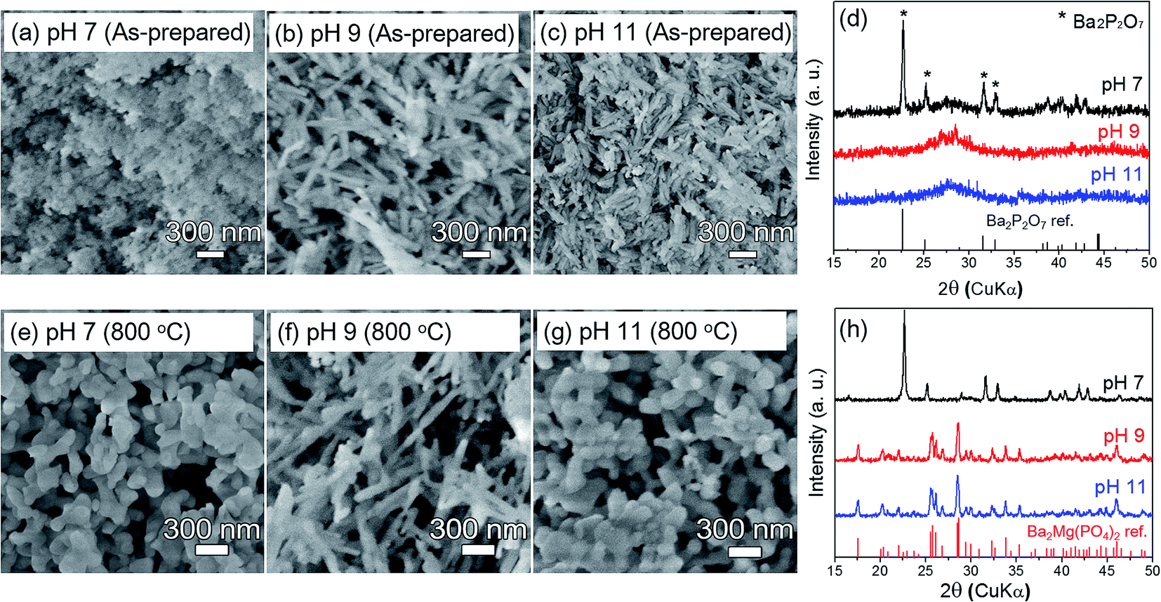 | ||
| Fig. 1 Effects of pH and post-annealing temperature on the morphology and crystal structure of the BMP:Eu phosphors. (a)–(c) SEM images and (d) corresponding XRD patterns of as-prepared BMP:Eu precursors, respectively. (e)–(g) SEM images and (h) corresponding XRD patterns of the BMP:Eu nanophosphors post-annealed at 800 °C under a flowing N2/H2 gas mixture, respectively. Phase-pure and nanorod-shaped Ba1.9Mg(PO4)2:0.05Eu2+ orthophosphate phosphor was synthesized at pH 9 and post-annealing temperature of 800 °C. | ||
Crystalline BMP:Eu phosphors were obtained and Eu3+ ions were reduced to Eu2+, by post-annealing the BMP:Eu precursors at various temperatures, under a reducing atmosphere. As the corresponding XRD patterns (Fig. S1†) show, BMP9 undergoes an amorphous-to-crystalline phase transformation at 700 °C. The reflection peaks of the BMP9 annealed at 700 °C and 800 °C were therefore indexed as the monoclinic Ba2Mg(PO4)2 reference structure (JCDPS no. 10-6521). This result indicates that phase-pure BMP was synthesized at these temperatures, and a slight shift of XRD peaks toward a high angle (smaller ionic radius of Eu2+ than Ba2+) implying successful incorporation of Eu2+ ions into Ba2+ sites. Fig. 1e–g shows the morphology of the BMP:Eu samples annealed at 800 °C. The BMP7, BMP9, and BMP11 samples annealed at 800 °C are denoted as BMP7_800, BMP9_800, and BMP11_800, respectively. BMP7_800 consisted of coarse agglomerated particles (Fig. 1e). In contrast, the initial rod-shaped morphology of the BMP9 is maintained, even after annealing at 800 °C (Fig. 1f). The morphology of BMP11 changed significantly, however, from nanorods to agglomerated particles after annealing at 800 °C (Fig. 1g). This change results from the partial melting/fusion of the small highly aggregated nanorods. Fig. 1h shows the XRD patterns of the BMP7_800, BMP9_800, and BMP11_800 samples. The pattern of BMP7_800 exhibited a close correspondence to that of Ba2P2O7, indicating that Mg did not precipitate in the low-pH (i.e., pH of 7) BMP7 precursor. In contrast, the reflection peaks of BMP9_800 and BMP11_800 were all well-matched to the reference BMP data. This indicates that stoichiometric Ba–Mg–P–O elements are initially co-precipitated in the high-pH (pH ≥ 9) amorphous precursors that consist of nanorods. A high-pH (pH = 9) reaction precursor and post-annealing at temperatures above 700 °C are therefore essential for attaining phase-pure and rod-shaped Ba2Mg(PO4)2:Eu2+ nanophosphors.
Fig. 2 summarizes the formation process of the BMP:Eu nanorod phosphor and shows corresponding microscopy images of the products. As the figure shows, amorphous nanoparticles are precipitated by adjusting the pH value to 9 and are then reacted at 90 °C for 6 h in a water bath. The SEM image of the intermediate product (90 °C/2 h), shows the simultaneously formed amorphous nanoparticles and nanorods. This observation reflects that the nanoparticles were self-assembled into nanorods during the reaction by an oriented attachment mechanism, which is enabled due to electrostatic forces from selectively adsorbed anions.34–36 The nanorods are subsequently crystallized and the Eu3+ activator is reduced/incorporated into the BMP host matrix during post-annealing at 800 °C, under a flowing reducing gas.
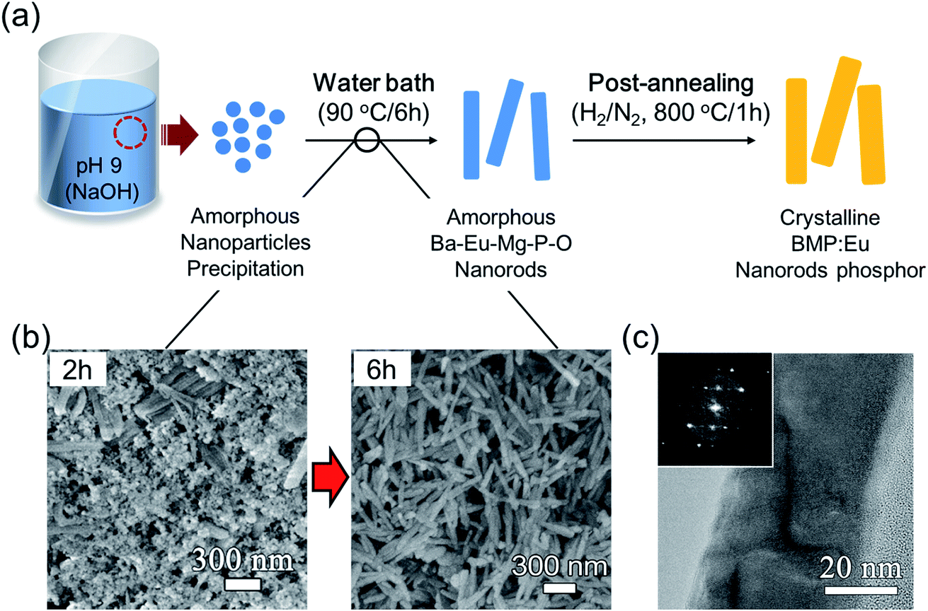 | ||
| Fig. 2 Synthesis process and formation mechanism of the BMP:Eu nanorod phosphor. (a) Schematic illustration of the synthesis process. (b) SEM and (c) TEM images of the products. The inset shows FFT pattern of the BMP:Eu nanorod. During the water bath of the reaction mixture, amorphous nanoparticles are firstly formed and they are self-assembled into nanorods after further reaction, which in turn transformed into single crystal nanorods by a post-annealing (H2/N2, 800 °C/2 h). | ||
Fig. 3a shows the crystal structure and atomic positions of the BMP matrix. According to the literature,27 BMP has monoclinic lattice symmetry that is described by the P21/n space group. Mg and P interact with octahedral and tetrahedral O intersitices, thereby forming a MgO6 octahedron and PO4 tetrahedron, respectively. Furthermore, Ba element is surrounded by two types of polyhedral: eight O atoms that form a bi-capped trigonal prism (Ba1) or seven O atoms that form a mono-capped trigonal prism (Ba2), which can influence the emission properties of the BMP:Eu phosphor.28 The crystallographic characteristics of the BMP9_800 (5 mol% of Eu2+) nanorod phosphor were investigated in detail via TEM analysis. As the resulting image in Fig. 3b shows, the BMP9_800 sample consists of ∼350 nm (length) × ∼60 nm (width) nanorods that are only mildly aggregated. The SAED patterns obtained from the rectangular region shown in Fig. 3b reveal the high crystallinity of the synthesized phosphor. These patterns also exhibit a close correspondence to those of the reference BMP structure (Fig. 2c). Fig. 3d and e show HR-TEM images of a single nanorod. These images reveal continuous lattice fringes with lattice spacings of 0.35 and 0.31 nm that correspond to the (113) and (122) planes, respectively, thereby confirming the single-crystalline nature of the nanorods.
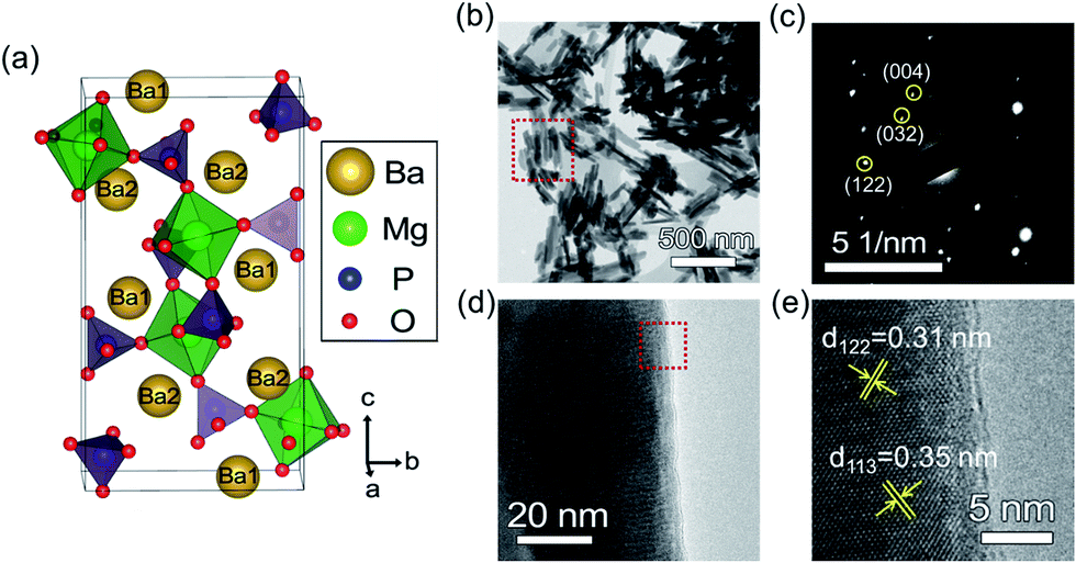 | ||
| Fig. 3 Crystal structure and TEM analysis of the BMP:Eu nanorod phosphor. (a) Crystal structure of the Ba2Mg(PO4)2 orthophosphate compound (monoclinic, P21/n, a = 5.294, b = 8.837, c = 16.143 Å, and β = 90.60° and two different Ba2+ sites in the unit cell). (b) Low-mag TEM image and (c) SAED pattern of the BMP9_800 nanorod phosphor. (d and e) High-resolution-TEM images of a single nanorod of the BMP9_800 nanorod phosphor. | ||
Fig. 4 shows the room-temperature excitation and emission spectra of the BMP9_800 (nanorods) and BMP11_800 (nanoparticles) nanophosphors. The spectra collected from the BMP9_800 sample exhibit similar tendencies to those of their BMP11_800 counterparts. The excitation spectra, which were collected over wavelengths ranging from 200–450 nm, exhibit three broad distinct bands, with peaks at 297, 334, and 390 nm. These peaks result from the 4f7 → 4f65d1 transition of the Eu2+ ions.28 The emission spectra of the BMP:Eu nanophosphors excited at 334 nm, were collected over wavelengths ranging from 400–750 nm. The maximum intensity of these spectra occurs at 580 nm, which is associated with an orange-yellow emission band that is slightly red-shifted compared to that (∼560 nm) of conventional YAG:Ce3+ phosphors. These spectra result from the f–d orbital energy transition of activated Eu2+ ions, indicating that Eu2+ ions substituted Ba2+ ions in the BMP host matrix.
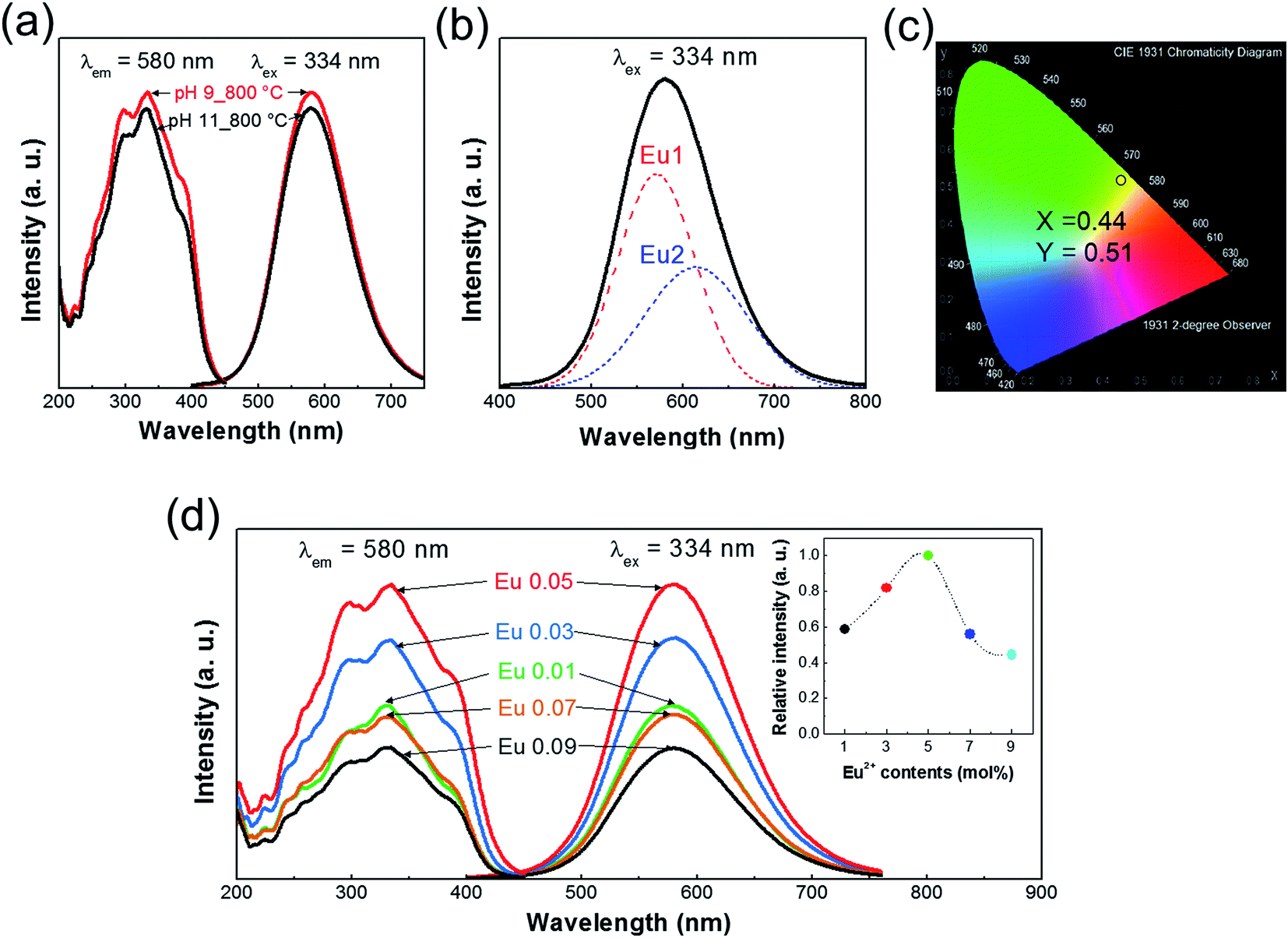 | ||
| Fig. 4 Photoluminescence properties of the BMP:Eu nanorod phosphor. (a) Photoluminescence excitation and emission spectra of the BMP9_800 (nanorod) and BMP11_800 (nanoparticles) phosphors. (b) Deconvolution of the emission spectrum of the BMP9_800 phosphor into two Gaussian peaks. (c) CIE chromaticity coordinate diagram of BMP9_800 nanorod phosphor. (d) Photoluminescence excitation and emission spectra of the BMP9_800 phosphor as a function of the Eu2+ concentration. The inset shows the emission intensity variation of the BMP9_800 phosphor as a function of the Eu2+ concentration. | ||
Owing to their crystallographic characteristics and effective average ionic radius, Eu2+ ions occupied Ba2+ sites rather than Mg2+ sites in the host matrix (REu2+: ∼1.25 Å, RBa2+: ∼1.42 Å, and RMg2+: ∼0.89 Å).37 Moreover, the asymmetric shape of the emission spectrum indicates that the Eu2+ ions occupy two or more emission centers in the matrix. The spectrum was deconvoluted into two Gaussian peaks (Fig. 4b) centered at 571 (Eu1) and 614 nm (Eu2), by using a Gaussian peak fitting method. As previously mentioned, there are two crystallographic Ba2+ sites and hence, the Eu2+ ions would occupy these two sites, thereby resulting in two emission bands.28
The photoluminescence (PL) properties of phosphors are affected by various factors such as the crystallinity of the host material and the concentration of the activator. In this work, the BMP9_800 sample exhibits a high crystallinity (high XRD peak intensity and smaller full width at half maximum, Fig. S1†), and hence higher PL emission intensity, than the BMP9_700 (Fig. S2†). The concentration (x) of Eu2+ ions was varied (x = 0.01, 0.03, 0.05, 0.07, and 0.09) in order to determine the effect of activator concentration on PL. After annealing at 800 °C, the XRD reflection peaks of the Ba2−2xMg(PO4)2:Eu2x2+ phosphors all exhibited close correspondence to the reference BMP data (Fig. S3†). Fig. 4c shows that the intensities of the PL excitation and emission spectra increased with increasing concentration of up to x = 0.05. However, owing to the concentration quenching effect, the intensities decreased with further increases in the concentration. Therefore, as in the case of BMP:Eu phosphors synthesized via a solid-state reaction (Fig. S4†), 5 mol% constitutes the optimum concentration of Eu2+ ions in the BMP:Eu phosphor. The critical distance (Rc) of Eu2+ ions at which concentration quenching occurs, is determined from:38
The morphological and size effects of the nanorod phosphor were determined by comparing the PL spectra and optical properties of the BMP nanophosphor (BMP9_800, 5 mol% of Eu2+) with those of a micro-sized bulk BMP:Eu phosphor. The bulk BMP:Eu phosphors (5 mol% of Eu2+) consisted of 1–5 μm-large particles and were prepared at 1200 °C, by using a solid-state reaction method (Fig. S5†). A comparison (Fig. 5a) of the PL excitation and emission spectra reveals that the spectra of the nanorod phosphor exhibit higher intensities than those of their bulk counterpart. This higher PL intensity of the nanorod BMP:Eu phosphor might be originated from our synthesis method, in which Eu2+ ions are homogeneously precipitated/incorporated into the amorphous nanorod precipitates during the water bath and they are further activated by the post-annealing step. More importantly, surface characteristics such as surface morphology and defects generally affects the PL intensity. For instance, rough surface morphology and high concentration of surface defects in nanophosphors result in a large amount of non-radiative surface recombination, lowering the PL intensity.39 As shown in the TEM images of both bulk and nanorod BMP:Eu phosphors (Fig. S6† and 5b), the bulk BMP:Eu phosphor showed thin and irregular surface amorphous layer, while the nanorod BMP:Eu phosphor showed thick and smooth amorphous layer (∼9 nm) at the surface, which may passivate the surface defects and reduce surface scattering, thereby leading to the higher PL intensity.40,41 Fig. 5c shows the UV-vis absorbance spectra of the nanorods and bulk BMP:Eu phosphors. Compared to the bulk phosphor, the nanorod phosphor exhibit higher light absorption in the UV region, especially at wavelengths ranging from 300–350 nm where the BMP phosphor can be most actively excited. The BMP:Eu nanorod phosphor also exhibited a higher excitation intensity at wavelengths ranging from 300–400 nm, as shown in the normalized PL excitation spectra (inset of Fig. 5c). This indicates that, compared to its bulk counterpart, the nanorod phosphor is more effective in absorbing and hence becomes more excited by the near-UV light. This enhanced light absorption and PL emission characteristics might be result from the smaller light scattering (compared to that associated with conventional synthesis methods) by the smooth and thick amorphous layer and homogeneous Eu2+ doping in the matrix i.e., the amorphous layer at the surface passivate the surface defects and suppress light scattering. Also the wet-chemical process may lead to a more homogeneous distribution of Eu2+ in the BMP matrix, than that achieved via conventional methods.
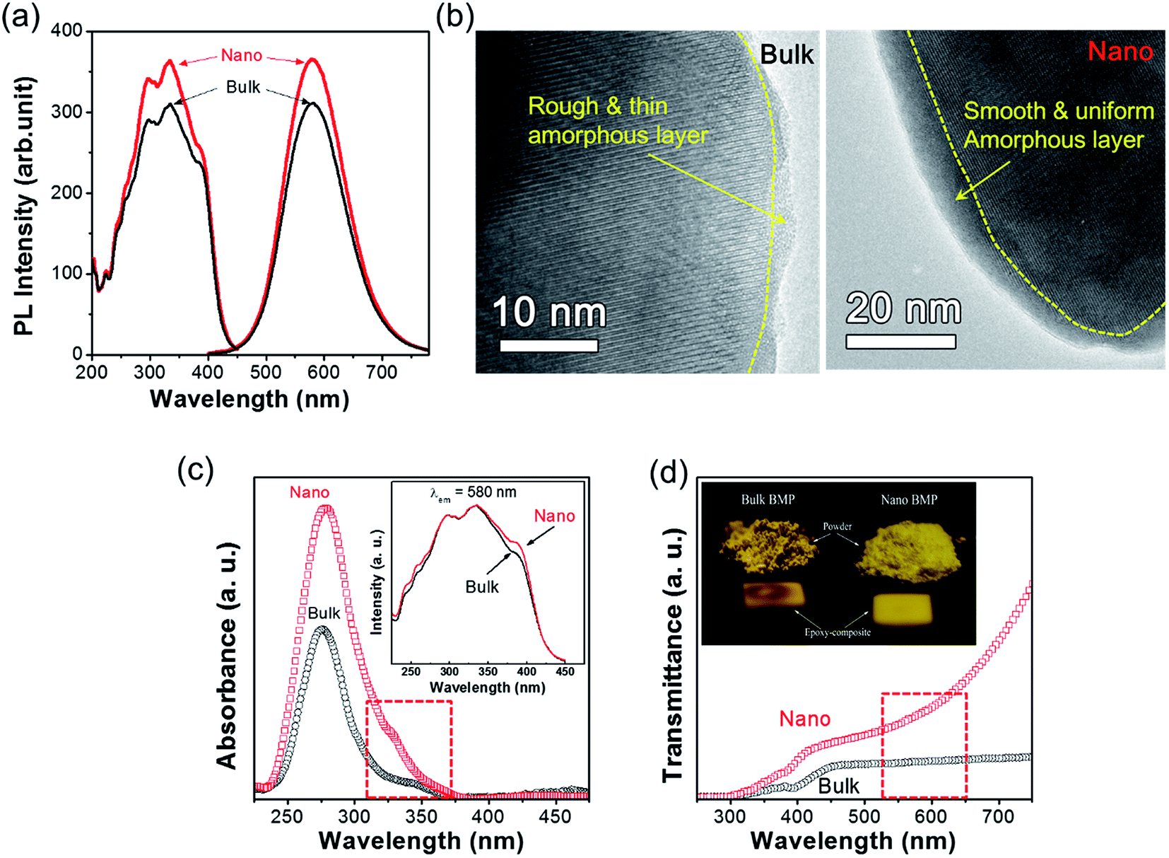 | ||
| Fig. 5 Comparison of PL and light absorption properties in between the nanorod and bulk BMP:Eu phosphors (5 mol% of Eu2+). (a) PL spectra, (b) TEM images, showing a difference in the surface characteristics. The nanorod BMP:Eu showed thick and smooth amorphous layer at the surface. (c) UV-vis absorbance spectra, the inset shows normalized excitation spectra of nano- and bulk-sized BMP phosphors. (d) Transmittance spectra of epoxy/BMP:Eu phosphor composite plates. The inset shows a photograph of the nanorod and bulk BMP:Eu phosphor powders and epoxy-composites under UVA (315–400 nm) irradiation. | ||
Lastly, we determined the light-transmittance characteristics, and hence suitability, of in-house-fabricated epoxy/BMP:Eu phosphor composite films for use in pc-LEDs. Fig. 5d shows the UV-vis transmittance spectra of tape-cast bulk and nano BMP:Eu phosphors. As the figure shows, the nanorod phosphor exhibits a higher visible-light transmittance than the bulk phosphor. This is especially true at near-580 nm wavelengths where the BMP phosphor emits the most intense light. The significantly higher intensity than that of bulk phosphor, is attributed to the smaller amount of light scattering in the phosphor-epoxy composite. This smaller amount of scattering results from the smaller particle size and more homogeneous distribution of particles in the epoxy (inset of Fig. 5c), compared to those in the bulk phosphor. These factors, lead, in turn, to a high emission efficiency.
Conclusion
We synthesized Ba2Mg(PO4)2:Eu2+ nanorod phosphor that produces strong yellow emission, by using a facile wet-chemical method followed by post-annealing in a reducing atmosphere. Amorphous and rod-shaped ∼350 nm (length) × ∼60 nm (width) BMP:Eu precursors were firstly obtained by controlling pH value in the reaction mixture. A crystalline BMP:Eu phosphor, with the same morphology as the precursor, was obtained by post-annealing the amorphous BMP:Eu precursor at 800 °C with a rapid heating rate of 10 °C min−1. In addition, emission and excitation spectra similar to those of bulk-sized BMP:Eu phosphors were obtained at the Eu2+ concentration that results in the maximum PL intensity. The BMP:Eu nanorod phosphor has several excellent properties, such as higher absorption of n-UV light and higher transmittance of long-wavelength light than those of the bulk-sized BMP:Eu phosphor. These results indicate that the BMP:Eu nanorod phosphor can be used as a yellow-emitting phosphor in-UV-based pc-LEDs.Acknowledgements
This research was supported by Basic Science Research Program through the National Research Foundation of Korea (NRF) funded by the Ministry of Science, ICT and Future Planning (NRF-2015R1C1A1A01053785).References
- S. Nakamura, Blue-green light-emitting diodes and violet laser diodes, MRS Bull., 1997, 22(02), 29–35 CrossRef CAS.
- N. Narendran and Y. Gu, Life of LED-based white light sources, J. Disp. Technol., 2005, 1(1), 167 CrossRef CAS.
- S. Pimputkar, J. S. Speck, S. P. DenBaars and S. Nakamura, Prospects for LED lighting, Nat. Photonics, 2009, 3(4), 180–182 CrossRef CAS.
- E. F. Schubert and J. K. Kim, Solid-state light sources getting smart, Science, 2005, 308(5726), 1274–1278 CrossRef CAS PubMed.
- M. Fan, M. Liang, D. Guo, F. Yang, L. Wang, G. Wang and J. Li, in Color filter-less technology of LED back light for LCD-TV, Photonics Asia 2007, International Society for Optics and Photonics, 2007, p. 68410G Search PubMed.
- S. Neeraj, N. Kijima and A. Cheetham, Novel red phosphors for solid-state lighting: the system NaM (WO4)2−x(MoO4)x:Eu3+ (M·Gd, Y, Bi), Chem. Phys. Lett., 2004, 387(1), 2–6 CrossRef CAS.
- P. Schlotter, R. Schmidt and J. Schneider, Luminescence conversion of blue light emitting diodes, Appl. Phys. A: Mater. Sci. Process., 1997, 64(4), 417–418 CrossRef CAS.
- G. Xia, S. Zhou, J. Zhang and J. Xu, Structural and optical properties of YAG:Ce3+ phosphors by sol–gel combustion method, J. Cryst. Growth, 2005, 279(3), 357–362 CrossRef CAS.
- S. C. Allen and A. J. Steckl, A nearly ideal phosphor-converted white light-emitting diode, Appl. Phys. Lett., 2008, 92(14), 143309 CrossRef.
- J. K. Park, K. J. Choi, K. N. Kim and C. H. Kim, Investigation of strontium silicate yellow phosphors for white light emitting diodes from a combinatorial chemistry, Appl. Phys. Lett., 2005, 87(3), 031108 CrossRef.
- P. Li, Z. Yang, Z. Wang, Q. Guo and X. Li, Preparation and luminescence characteristics of Sr3SiO5:Eu2+ phosphor for white LED, Chin. Sci. Bull., 2008, 53(7), 974–977 CAS.
- C.-H. Kuo, J.-K. Sheu, S.-J. Chang, Y.-K. Su, L.-W. Wu, J.-M. Tsai, C. Liu and R. Wu, n-UV + blue/green/red white light emitting diode lamps, Jpn. J. Appl. Phys., Part 1, 2003, 42(4S), 2284 CrossRef CAS.
- M. G. Craford, N. Holonyak and F. A. Kish, In pursuit of the ultimate lamp, Sci. Am., 2001, 284(2), 62–67 CrossRef CAS.
- E. Radkov, R. Bompiedi, A. M. Srivastava, A. A. Setlur and C. A. Becker, in White light with UV LEDs, Optical Science and Technology, SPIE's 48th Annual Meeting, International Society for Optics and Photonics, 2004, pp. 171–177 Search PubMed.
- J. McKittrick, M. Hannah, A. Piquette, J. Han, J. Choi, M. Anc, M. Galvez, H. Lugauer, J. Talbot and K. Mishra, Phosphor selection considerations for near-UV LED solid state lighting, ECS J. Solid State Sci. Technol., 2013, 2(2), R3119–R3131 CrossRef CAS.
- H. Menkara, R. Gilstrap, T. Morris, M. Minkara, B. Wagner and C. Summers, Development of nanophosphors for light emitting diodes, Opt. Express, 2011, 19(104), A972–A981 CrossRef CAS PubMed.
- K.-H. Lee, Y.-J. Bae and S.-H. Byeon, Nanostructures and photoluminescence properties of Gd2O3:Eu red-phosphor prepared via hydrothermal route, Bull. Korean Chem. Soc., 2008, 29(11), 2161–2168 CrossRef CAS.
- D. Haranath, H. Chander, P. Sharma and S. Singh, Enhanced luminescence of Y3Al5O12:Ce3+ nanophosphor for white light-emitting diodes, Appl. Phys. Lett., 2006, 89(173118), 3 Search PubMed.
- N. R. Taskar, R. N. Bhargava, J. Barone, V. Chhabra, V. Chabra, D. Dorman, A. Ekimov, S. Herko and B. Kulkarni, in Quantum-confined-atom-based nanophosphors for solid state lighting, Optical Science and Technology, SPIE's 48th Annual Meeting, International Society for Optics and Photonics, 2004, pp. 133–141 Search PubMed.
- D. Cooke, J. Lee, B. Bennett, J. Groves, L. Jacobsohn, E. McKigney, R. Muenchausen, M. Nastasi, K. Sickafus and M. Tang, Luminescent properties and reduced dimensional behavior of hydrothermally prepared Y2SiO5:Ce nanophosphors, Appl. Phys. Lett., 2006, 88(10), 103108 CrossRef.
- D. Jia, Y. Wang, X. Guo, K. Li, Y. Zou and W. Jia, Synthesis and characterization of YAG:Ce3+ LED nanophosphors, J. Electrochem. Soc., 2007, 154(1), J1–J4 CrossRef CAS.
- D. Jia, Sr4Al14O25:Eu2+ nanophosphor synthesized with salted sol–gel method, Electrochem. Solid-State Lett., 2006, 9(10), H93–H95 CrossRef CAS.
- K. Yamada, Y. Imai and K. Ishii, A Study of the Light Source Device Composed of Blue LEDs and YAG Phosphor by Using Optical Simulation, J. Inst. Eng. (India), Chem. Eng. Div., 2002, 86, 308–312 Search PubMed.
- C. F. Bohren and D. R. Huffman, Absorption and scattering of light by small particles, John Wiley & Sons, 2008 Search PubMed.
- Y. Xia and P. Yang, Guest editorial: chemistry and physics of nanowires, Adv. Mater., 2003, 15(5), 351–352 CrossRef CAS.
- Z. L. Wang, Nanobelts, Nanowires, and Nanodiskettes of Semiconducting Oxides From Materials to Nanodevices, Adv. Mater., 2003, 15(5), 4 Search PubMed.
- F. Lucas, G. Wallez, S. Jaulmes, A. Elfakir and M. Quarton, Dibarium magnesium phosphate, Acta Crystallogr., Sect. C: Cryst. Struct. Commun., 1997, 53(12), 1741–1743 CrossRef.
- Z. Wu, M. Gong, J. Shi, G. Wang and Q. Su, Dibarium magnesium diphosphate yellow phosphor applied in InGaN-based LEDs, Chem. Lett., 2007, 36(3), 410–411 CrossRef CAS.
- Z. Pan, Q. Hu, Y. Xu and L. Wang, Phase Identification and Dopant Site Occupancy of Yellow-Emitting Phosphor BaSrMg(PO4)2:Eu2+, J. Am. Ceram. Soc., 2015, 98(8), 2518–2522 CrossRef CAS.
- Q. Hu, Z. Pan, Y. Xu, L. Wang and L. Ning, Dopant Site Environment and Spectrum Blue Shift of Yellow-Emitting Solid Solution Phosphor Ba2−xSrxMg(PO4)2:Eu2+, J. Am. Ceram. Soc., 2015, 99(2), 645–650 CrossRef.
- G. Ju, Y. Hu, L. Chen, Y. Jin, Z. Yang and T. Wang, Photoluminescence properties of Ce3+ and Tb3+-activated Ba2Mg(PO4)2, Opt. Mater. Express, 2015, 5(1), 1–10 CrossRef CAS.
- C. Henrist, J.-P. Mathieu, C. Vogels, A. Rulmont and R. Cloots, Morphological study of magnesium hydroxide nanoparticles precipitated in dilute aqueous solution, J. Cryst. Growth, 2003, 249(1), 321–330 CrossRef CAS.
- F. Schrey, Effect of pH on the Chemical Preparation of Barium–Strontium Titanate, J. Am. Ceram. Soc., 1965, 48(8), 401–405 CrossRef CAS.
- Q. Zhang, S.-J. Liu and S.-H. Yu, Recent advances in oriented attachment growth and synthesis of functional materials: concept, evidence, mechanism, and future, J. Mater. Chem., 2009, 19(2), 191–207 RSC.
- I. S. Cho, D. W. Kim, S. Lee, C. H. Kwak, S. T. Bae, J. H. Noh, S. H. Yoon, H. S. Jung, D. W. Kim and K. S. Hong, Synthesis of Cu2PO4OH hierarchical superstructures with photocatalytic activity in visible light, Adv. Funct. Mater., 2008, 18(15), 2154–2162 CrossRef CAS.
- M. Tong, J. Zhao, Y. Liang, Y. Zhu, X. Wu, S. Liu, C. Yan and G. Li, Synthesis and characterization of micro/nano-structured BaHPO4/Ba3(PO4)2/Ba5(PO4)3 OH phases and their luminescence, RSC Adv., 2015, 5(35), 27517–27525 RSC.
- R. T. Shannon, Revised effective ionic radii and systematic studies of interatomic distances in halides and chalcogenides, Acta Crystallogr., Sect. A: Cryst. Phys., Diffr., Theor. Gen. Crystallogr., 1976, 32(5), 751–767 CrossRef.
- G. Blasse, Energy transfer in oxidic phosphors, Phys. Lett. A, 1968, 28(6), 444–445 CrossRef CAS.
- B. L. Abrams and P. H. Holloway, Role of the Surface in Luminescent Processes, Chem. Rev., 2004, 104(12), 5783–5802 CrossRef CAS PubMed.
- S. H. Sohn, J. H. Lee and S. M. Lee, Effects of the surface coating of BaMgAl10O17:Eu2+ phosphor with SiO2 nano-particles, J. Lumin., 2009, 129(5), 478–481 CrossRef CAS.
- C. Shang, H. Jiang, X. Shang, M. Li and L. Zhao, Investigation on the Luminescence Improvement of Nanosized La2O3/Eu3+ Phosphor under Charge-Transfer Excitation, J. Phys. Chem. C, 2011, 115(6), 2630–2635 CrossRef CAS.
Footnote |
| † Electronic supplementary information (ESI) available. See DOI: 10.1039/c6ra11156d |
| This journal is © The Royal Society of Chemistry 2016 |