Experimental and theoretical study on the reactivity of maghemite doped with Cu2+ in oxidation reactions: structural and thermodynamic properties towards a Fenton catalyst

Maíra dos Santos Piresa, Francisco G. E. Nogueirab, Juliana A. Torresa, Lívia C. T. Lacerdaa, Silviana Corrêaa, Márcio C. Pereirac and Teodorico C. Ramalho*ad
aDepartment of Chemistry, Federal University of Lavras, 37200-000 Lavras, MG, Brazil. E-mail: teo@dqi.ufla.br; Fax: +55 35 3829 1271; Tel: +55 35 3829 1271
bDepartment of Chemical Engineering, University of São Carlos, 13565-905 São Carlos, SP, Brazil
cInstitute of Science, Engineering and Technology, Federal University of the Jequitinhonha and Mucuri Valleys, Campus Mucuri, 39803-371 Teófilo Otoni, MG, Brazil
dCenter for Basic and Applied Research, University Hradec Kralove, Hradec Kralove, Czech Republic

Received 28th April 2016 , Accepted 12th August 2016

First published on 15th August 2016


Abstract

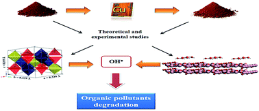
In this work, a polymeric method was used to prepare undoped and Cu-doped iron oxide catalysts for the H2O2 decomposition reaction. These catalysts were characterized by powder X-ray diffractometry (XRD), scanning electronic microscopy (SEM) coupled to an energy dispersive X-ray spectrometer (EDX), and H2-Temperature Programmed Reduction (H2-TPR). The SEM images show an inhomogeneous particle cluster in both samples, tending to decrease in size with Cu-doping. EDX mapping reveals a good dispersion of Cu2+ in the iron oxide. In addition, Rietveld refinement of the XRD patterns reveals that the samples are constituted of hematite and maghemite, but only maghemite has octahedral Fe3+ ions isomorphically replaced by 2 wt% Cu2+. Cu-doping produces an active catalyst for H2O2 decomposition. Tests using phenol show the strong inhibition of H2O2 decomposition by the Cu-doped catalysts, suggesting that H2O2 may be decomposed via a radical mechanism. Furthermore, phenol degradation kinetics confirm that the doping of maghemite with Cu2+ brings about a significant improvement in catalytic activity. Theoretical calculations reveal that Cu-doping in maghemite produces low electronic density sites, favoring the interactions between the surface oxygens of H2O2 and Cu2+, thus improving the catalytic activity. This strategy can be extended to other materials to design active heterogeneous catalysts for environmental purposes.


Introduction

The growth of agribusiness in recent years has led to a certain insecurity regarding the use of natural resources, among which water may be highlighted. This sector generates a substantial volume of wastewater containing many highly toxic organic compounds,1 which can cause severe damage to the environment and public health. In the aquatic medium, they may kill fish even at concentrations as low as 1 mg L−1. Furthermore, contaminants at sub-lethal dosages affect the nervous and circulatory systems, thus reducing the growth of blood cells in human beings.2,3

Among the technologies that may be applied to treat contaminated aqueous effluents, advanced oxidation processes (AOPs) are considered to be the most promising mainly because of the formation of less harmful compounds.4 AOPs are processes that generate hydroxyl radicals (HO˙), which are highly oxidative species, in amounts sufficient to induce the mineralization of organic matter to carbon dioxide, water, and inorganic ions from heteroatoms. These processes are classified as homogeneous and heterogeneous systems, in which hydroxyl radicals are generated with or without light radiation and may involve the use of ozone (ozonolysis), hydrogen peroxide (Fenton) or semiconductors (photocatalysis).5 Among these, heterogeneous Fenton-like systems are attractive, because they act at neutral pH and facilitate operational processes for the treatment of effluents, such as the cleaning of tanks and reactors.6 These systems are based on the activation of H2O2 by metal ions (e.g. Fe2+ and Fe3+ ions) in a solid structure, by electron transfer from the metal ions to H2O2 molecules, according to the modified Haber–Weiss radical mechanism.7,8

Different iron oxides, including hematite (α-Fe2O3), goethite (α-FeOOH), feroxyhyte (δ-FeOOH), maghemite (γ-Fe2O3) and magnetite (Fe3O4) have been described as excellent catalysts for heterogeneous Fenton-like reactions.9–13 The use of an iron oxide catalysts is advantageous due to the high availability of their raw material, since iron is one of the most abundant elements in the Earth’s crust, corresponding to 35% of the total mass of the planet.14 Therefore, iron oxide catalysts can be synthesized at a low cost. Moreover, iron oxides can accommodate several foreign ions in their structure, such as Al3+, Mn3+, Cr3+, V3+, Ni2+, Co2+, Zn2+ and Cu2+ which can modify their electronic, structural and catalytic properties.15–20

Several non-iron catalysts such as Al, Ru, Co, Ce, Mn, Cr and Cu in multiple redox states have been reported to directly decompose H2O2 into HO˙ through a conventional Fenton-like mechanism.21 Among these metals, the use of copper in Fenton-like reactions has several advantages such as: (i) it presents redox properties similar to iron, (ii) the Cu2+/H2O2 system works over a broader range, compared to the Fe3+/H2O2 system which works only at acidic pH, (iii) both Cu+ and Cu2+ ions react with H2O2 forming intermediate complexes that decompose forming highly active HO˙ radical22 and (iv) Cu2+ complexes with organic degradation intermediates are readily decomposed by HO˙ radicals, whereas the corresponding Fe3+ complexes are highly stable. It is important to highlight that the copper reactions are three times faster than the ferrous.23 On the other hand, the use of a Cu2+ catalyst requires much more H2O2 compared to Fe3+ based catalysts, to compensate for competitive scavenging by O2 in aerobic conditions.

An attractive approach to design active Fenton-like catalysts consists of combining the properties of Fe and Cu ions in the same crystal structure to generate synergic effects, thus improving their electronic, structural and catalytic properties. Previous studies have indicated that Cu is highly active in the heterogeneous Fenton reaction for degrading different organic compounds.24–28

Several heterogeneous solid catalysts based on Fe–Cu have been developed in recent years for the Fenton process, such as copper ferrite29–34 and composite materials containing ions of these metals (or small Cu–Fe oxide aggregates) deposited onto a porous support,35–39 among others. However, experimental and theoretical studies on the reactivity of iron oxides containing a small percentage of Cu in Fenton reactions have not yet been fully explored.

Thus, the objective of the present work was to evaluate, through experimental and theoretical studies, the effect of small amounts of Cu2+ doping on the catalytic properties of iron oxides prepared by a polymeric method for the H2O2 decomposition reaction.

Materials and methods

Synthesis of the catalysts

The catalysts were synthesized using a polymeric precursor method, which provides nanostructures with good homogeneity, besides being economically feasible.40 The synthesis of γ-Fe2O3 was carried out by dissolving 0.181 g of ferric nitrate (Fe(NO3)3·9H2O) in 25 mL of water under stirring for 20 min at 50 °C. To prepare an iron citrate complex, a solution of 0.1 mol L−1 of citric acid was added to the aqueous solution of the precursor iron, for which the citric acid/metallic cation ratio was kept at a proportion of 3[thin space (1/6-em)]:[thin space (1/6-em)]1. The resultant solution was maintained at 70 °C for 50 minutes. To obtain the polymerized resin, a previously prepared 0.2 mol L−1 ethylene glycol solution was mixed with the iron citrate solution in a citric acid/ethylene glycol volume ratio of 40[thin space (1/6-em)]:[thin space (1/6-em)]60, and kept at 110 °C for 4 h. The resin obtained after this process was ground and treated at 300 °C for 3 h to expand and break the polymer (puff). The pyrolyzed polymer “puff” was then calcined at 650 °C for 3 h to yield the undoped catalyst.

The Cu-doped catalyst was prepared by a method similar to the previous, but a second polymeric precursor, copper nitrate(II) (Cu(NO3)2·3H2O), was added in order to obtain the doped material at a 2 wt% Cu proportion (Fig. 1).


image file: c6ra11032k-f1.tif
Fig. 1 Simplified flow chart for the preparation of the Cu-doped catalyst.

Characterization of the catalysts

The crystalline phases of the catalysts were determined using a X’Pert Pro multi-purpose X-ray diffraction (MPD) system employing Cu Kα radiation (λ = 0.154 nm) operated at 40 mA and 45 kV. Silicon was used as an external standard. Rietveld structural refinement was performed using FullProf_Suite 2015 software. The behavior of materials under a reducing atmosphere was monitored by H2-Temperature-Programmed Reduction (TPR) using a ChemiSorb 2750 (Micromeritics, USA) with a TCD detector at a heating rate of 10 °C min−1. TPR ranged from 50 °C to 1000 °C in 10% H2 in Ar at a flow rate of 20 mL min−1. Morphologies were investigated using scanning electron microscopy (MEV-FEG FEI Magellan 400 L) with an electron beam operated at 5 kV. The elemental composition of the catalysts was determined using energy-dispersive X-ray (EDX) spectroscopy.

Catalytic tests

Decomposition of H2O2. The reaction of hydrogen peroxide decomposition was used to evaluate the catalytic behavior of the catalysts. The volumetric amount of O2 formed in a closed system containing 5 mL of distilled water, 2 mL of 50% (v/v) H2O2 and 30 mg of catalyst allowed us to measure the H2O2 decomposition rate. The system was set up at room temperature under magnetic stirring. H2O2 decomposition was monitored by O2 evolution according to the following eqn (1):
 
H2O2 → H2O + 0.5O2 (1)

The catalytic decomposition of H2O2 was also studied in the presence of 5 mL of phenol (50 mg L−1) as a radical scavenger.

The Fe and Cu content leached to the solution after the H2O2 decomposition reactions was analyzed using atomic absorption spectroscopy (AAS) (Spectra AA 55 Model, Varian).

Phenol degradation kinetics. Kinetic studies for phenol degradation were obtained for intervals of 5, 15, 30, 60, 90 and 120 minutes of reaction. Reactions were also performed using multiple and single additions of H2O2 for the Cu-doped catalyst. For multiple addition, aliquots of 0.1 mL of H2O2 30% (v/v) were added to the reaction medium at 30 minute intervals. The dosage of phenol was obtained using the procedures of the 4-aminoantipyrine colorimetric method. Initially 0.3 mL of 4-aminoantipyrine solution and 0.3 mL of ferricyanide solution were added to 1.8 mL of reaction medium containing 10 mg of catalyst, 0.1 mL of H2O2 30% (v/v) and 9.9 mL phenol (50 ppm). The absorbance reading was performed after an interval of 10 minutes at a wavelength of 510 nm.41
Computational studies. The ADF BAND package,42 a program that enables electronic structural methods involving periodic conditions, was used for all calculations. The theoretical method applied was DFT (Density Functional Theory) together with the functional PBE (Perdew, Burke and Ernzerhof),43 which is a generalized gradient approximation (GGA). The basis was Slater triple-zeta polarized type (TZP) for copper, iron and oxygen atoms.

The structures of maghemite and Cu-doped maghemite were constructed based on the crystallographic data obtained by Rietveld refinement (Table 1). The structure of Cu-doped maghemite was built through the isomorphic substitution of Fe atoms by Cu atoms in octahedral positions. The mechanism of H2O2 decomposition catalyzed by Cu-doped and undoped maghemite was investigated by inserting the peroxide molecule at the surface of the catalysts. The geometries of the reactants, possible intermediates and products involved in the decomposition reaction were then optimized.

Table 1 Crystallographic data of undoped and Cu-doped maghemite obtained by Rietveld refinement of XRD data
Sample Space group Lattice parameter/Å Atom Atomic coordinates
X Y Z
Undoped P43212 a = 8.3452, c = 8.3423 Fe1 0.744 0.996 0.120
Fe2 0.620 0.620 0.000
Fe3 0.364 0.867 0.984
Fe4 0.140 0.140 0.000
O1 0.615 0.869 0.986
O2 0.119 0.377 0.995
O3 0.137 0.861 0.007
O4 0.383 0.631 0.997
Cu-doped P43212 a = 8.3393, c = 8.3552 Fe1 0.744 0.996 0.120
Fe2 0.620 0.620 0.000
Fe3 0.364 0.867 0.984
Fe4 0.140 0.140 0.000
Cu1 0.620 0.620 0.000
Cu2 0.364 0.867 0.984
O1 0.615 0.869 0.986
O2 0.119 0.377 0.995
O3 0.137 0.861 0.007
O4 0.383 0.631 0.997


Results and discussion

Characterization of the catalysts

SEM images of the undoped and Cu-doped catalysts are shown in Fig. 2a, b, d and e. It is observed that both samples are made up of an agglomeration of inhomogeneous particles. Also, Cu doping (Fig. 2d and e) causes a reduction in grain size when compared to the undoped catalyst (Fig. 2a and b). EDX spectra of the samples (Fig. 2c and f) collected at four different points reveal that the undoped catalyst is formed only by Fe and O, whereas Cu, Fe, and O compose the Cu-doped catalyst. The Cu content was found to be 1.98 wt%, which is close to the 2 wt% nominal content used in the synthesis.
image file: c6ra11032k-f2.tif
Fig. 2 SEM images of the undoped (a and b) and Cu-doped catalysts (d and e) and EDX spectra of the undoped (c) and Cu-doped catalysts (f).

In order to verify the distribution of the elements Cu, Fe and O in the Cu-doped catalyst, EDX mapping was carried out in an area of 53 × 53 μm with a magnification of 7000× (Fig. 3). The images show that Fe, Cu and O atoms are well dispersed throughout the sample, suggesting that the polymeric method of synthesis proposed in this work allows a good dispersion of Cu in the iron oxide matrix (Fig. 3d).


image file: c6ra11032k-f3.tif
Fig. 3 EDX mapping images of the Cu-doped catalyst.

To better understand the effect of Cu doping on the structure of the synthesized iron oxides phases, powder XRD analysis was performed. Qualitative analysis of the XRD patterns of the undoped and Cu-doped catalysts indicates that maghemite (JCPDS 25-1402) and hematite (JCPDS 13-534) are the crystalline phases in both samples. The subsequent Rietveld refinement of the XRD data with Thompson–Cox–Hastings pseudo-Voigt axial divergence asymmetry peak fitting gave the structural parameters and refinement reliability factors, which are summarized in Table 2. Fig. 4 shows the refinement of the undoped and Cu-doped catalyst XRD patterns. The Rietveld refinement yielded a goodness of fit indicator, S, of approximately 1.4 for both samples, indicative of good quality refined models. The lattice parameters “a” and “c” of the hexagonal unit cell of hematite did not change after the Cu doping, indicating that Cu2+ ions did not replace Fe3+ ions in the hematite structure. On the other hand, the tetragonal unit cell of maghemite in the Cu-doped catalyst is strongly distorted, decreasing in the “a” direction and increasing in the “c” direction (Fig. 5). This suggests the substitution of Fe3+ by Cu2+ ions in the maghemite structure. The high spin Fe3+ ionic radius in an octahedral coordination is 65 pm while the ionic radius of Cu2+ is 73 pm. To keep the charge balance, 3 Cu2+ are required for each 2 Fe3+, and as a result of this replacement, a strong structural distortion in the maghemite structure is observed. Quantitative analysis of the XRD patterns shows that the undoped catalyst is formed of 41 wt% maghemite and 59 wt% hematite, whereas the maghemite content in the Cu-doped catalyst increases to 69.5 wt% and the hematite amount decreases to 30.5 wt%, suggesting that Cu2+ ions play a significant role in the stabilization of the maghemite structure under heating in an air atmosphere.

Table 2 Crystallographic phase, lattice parameters, and agreement factors for the refinements obtained from the Rietveld refinement of the undoped and Cu-doped maghemite
Sample Space group Lattice parameter/Å Phase Agreement factors Phase percentage/wt%
RF RB Rwp Rexp S χ2
Undoped P43212 a = 8.3452(9), c = 8.3423(9) γ-Fe2O3 5.04 5.26 9.15 6.61 1.38 1.91 40.8(3)
R[3 with combining macron]c a = 5.0330(1), c = 13.7509(3) α-Fe2O3 2.90 3.37 59.2(3)
Cu-doped P43212 a = 8.3393(5), c = 8.3552(8) γ-Fe2O3 4.82 5.19 8.38 5.94 1.41 1.99 69.5(3)
R[3 with combining macron]c a = 5.0332(1), c = 13.7504(5) α-Fe2O3 3.16 3.84 30.5(2)


image file: c6ra11032k-f4.tif
Fig. 4 Rietveld refinement of the powder XRD patterns of the undoped and Cu-doped catalyst.

image file: c6ra11032k-f5.tif
Fig. 5 Crystallographic structure of (a) undoped tetragonal maghemite and (b) Cu-doped tetragonal maghemite. Blue polyhedra = Fe3+ octahedral sites, red polyhedra = Fe3+ tetrahedral sites and yellow polyhedra = Cu2+ octahedral sites.

The TPR profiles of the undoped and Cu-doped catalysts are displayed in Fig. 6a. It can be seen that the undoped catalyst presents three main reduction peaks centered at 406, 652 and 801 °C. The first reduction stage that occurs at 406 °C is due to the chemical reduction of hematite and maghemite to magnetite (eqn (2)). The reduction peak centered at 652 °C is assigned to the reduction of magnetite into wüstite (eqn (3)), followed by the reduction of wüstite into metallic iron (eqn (4)).44,45

 
3Fe2O3 + H2 → 2Fe3O4 + H2O (2)
 
Fe3O4 + 6H2 → 3FeO + H2O (3)
 
FeO + H2 → Fe + H2O (4)


image file: c6ra11032k-f6.tif
Fig. 6 (a) Temperature-programmed reduction of the undoped and Cu-doped catalysts, and (b) deconvolution of the main reduction peaks of the Cu-doped catalyst.

In contrast, the Cu-doped catalyst exhibits a significant decrease in the reduction temperature of iron species. The overlapped peaks can be related to the phase transformations of CuO → Cu and Fe2O3 → Fe3O4. However, deconvolution of the TPR profile of the Cu-doped catalyst (Fig. 6b) reveals five reduction peaks. The small peak at approximately 180 °C is assigned to the reduction of Cu2+ → Cu+ and the peak centered at 290 °C is attributed to the complete reduction of Cu2+ → Cu+ → Cu0.46,47,48 The peak centered at 250 °C is due to the reduction of Fe2O3 → Fe3O4. It is important to note that the presence of copper in the catalyst decreases the temperature of reduction of Fe3O4 into FeO and FeO into Fe, from 650 °C to 540 °C and 800 °C to 600 °C, respectively. The lower reduction temperatures for the doped catalyst, compared with the undoped catalyst, may be attributed to the smaller particle size of iron oxides and the Cu doping of maghemite in the Cu-doped catalyst. Previous studies by Khan and Smirniotis49 have shown that the addition of Cu into iron oxides favours the reducibility of the Fe3+ to Fe2+ species. Moreover, doping with Cu increases the mobility of oxygen and hydroxyl groups in the iron oxide framework, thus making their reduction easier.49 In addition, according to Jin and Datye47 the promotional effect of Cu is attributed to its ability to dissociate H2 and provide a source of atomic H to assist in the reduction of Fe2O3.

To better understand the TPR profiles observed in Fig. 6, XRD analysis was performed in situ in an H2 reducing atmosphere (Fig. 7). Our data indicates that from 250 °C to 400 °C, the diffraction peaks of the Cu-doped catalyst are gradually shifted to smaller Bragg angles due to the partial reduction of Fe3+ ions in the maghemite and hematite structure, indicating the beginning of the formation of the magnetite phase. While each maghemite cell unit contains 21.33 iron atoms, all as Fe3+, and 32 oxygen atoms, in magnetite there are 24 iron atoms (16 Fe3+ and 8 Fe2+) and 32 oxygen atoms per unit cell. Thus, when a transition to this phase occurs, the 2.67 vacancies present in the maghemite cease to exist, since they are filled by more than 2.67 iron atoms.50 Knowing that the calculated diffraction pattern utilizes a unit cell as a basis to define the positions of the peaks, the change to magnetite phase justifies the shifting of the Bragg peaks to smaller angles. However, at temperatures between 500 °C and 600 °C a phase mixture of Fe3O4 and FeO is formed, as is evidenced by the small peak at a 2θ of 42.4° corresponding to the (200) plane of FeO, which suggests the reduction of Fe3O4 to produce FeO. However, the Fe3O4 and FeO peaks at a 2θ of about 36.9° are close to each other and therefore they do not clearly show the phase transition. It is important to note that the presence of a phase mixture at 600 °C (Fe3O4 and FeO) is probably due to the experimental conditions used in situ for the reduction process (5%H2 + He), where not all the magnetite may be reduced to form FeO.


image file: c6ra11032k-f7.tif
Fig. 7 XRD patterns of the Cu-doped catalyst obtained by an in situ H2 reducing atmosphere using a synchrotron source (λ = 1.5498 Å).

Catalytic tests of H2O2 decomposition

The H2O2 decomposition profiles are presented in Fig. 8. As can be seen from the figure, Cu doping significantly improves the O2 evolution from H2O2 compared with the undoped catalyst. This behavior may be related to the fact that Cu is responsible for the formation of very reactive catalytic sites in Fenton processes.26 This suggests that the Cu2+ ions play a fundamental role in the catalytic activity of the material since they react with H2O2 generating hydroperoxide radicals (HOO˙) and Cu+ species (eqn (5)). Then, the Cu+ ions can generate HO˙ radicals (eqn (6)).51
 
Cu2+ + H2O2 → Cu+ + HOO˙ + H+ (5)
 
Cu+ + H2O2 → Cu2+ + HO˙ + OH (6)

image file: c6ra11032k-f8.tif
Fig. 8 Decomposition of H2O2 in the presence of the catalysts (data: 25 °C, 5 mL H2O, 30 mg of catalyst, 2 mL H2O2 50% (v/v) and 5 mL of 50 mg L−1 phenol).

Experiments using phenol as a radical scavenger were performed to study the H2O2 decomposition mechanism in the presence of the Cu-doped catalyst. The results indicate a reduction in O2 evolution of approximately 50% after 50 minutes of reaction, suggesting that the mechanism of H2O2 decomposition on the Cu-doped catalyst takes place via the formation of radicals as intermediate species. Thus, once the radical is formed, it can react in a competitive pathway with the phenol, decreasing the O2 evolution.

The amounts of Fe and Cu leached from the materials were quantified using AAS. The results indicate that the iron content in solution was below the detection limit of AAS (detection limit for iron is 0.0062 μg mL−1) whereas the Cu content in solution was 1.7 ppm (detection limit for copper is 0.04 μg mL−1). These data suggest that H2O2 decomposition may occur on the surface of the catalysts rather than in solution.

Catalytic tests were conducted to degrade phenol in the presence of H2O2 (Fig. 9). The Cu-doped catalyst showed better performance in the degradation of the pollutant over time compared to the undoped catalyst. In the absence of copper the ability of maghemite to degrade phenol remained at values close to 16 and 17%. However, after the addition of this metal, about 32% degradation was initially obtained; this value increased to 45% with 30 minutes of reaction and remained constant to the end of the process. This suggests the rapid consumption of hydrogen peroxide by the catalyst, which limits the reaction rate over time. To better understand the catalytic behavior of the Cu-doped catalyst, multiple additions of hydrogen peroxide were made every 30 minutes to the reaction medium. As expected, the degradation of phenol increased expressively to 70% after 2 hours of reaction with stepwise addition of H2O2 (Fig. 9), confirming the fast activity of the Cu-doped catalyst towards the generated hydroxyl radicals (HO˙). Thus, the addition of copper makes the Cu-doped catalyst highly efficient and promising in the degradation of organic pollutants.


image file: c6ra11032k-f9.tif
Fig. 9 Effect of reaction time on the degradation of phenol (50 ppm) by the Cu-doped catalyst (image file: c6ra11032k-u1.tif), undoped catalyst (image file: c6ra11032k-u2.tif) and Cu-doped catalyst with multiple additions of H2O2 (image file: c6ra11032k-u3.tif).

Theoretical study of the H2O2 decomposition mechanism

The surface of maghemite and Cu-doped maghemite catalysts were modelled in the index plane (311), since the energy of this plane is 48.85 and 100.78 kcal mol−1 lower than the energy of the (220) and (440) planes, in the undoped catalyst, and 55.21 and 122.80 kcal mol−1 in the Cu-doped catalyst, respectively. The order of increasing energy values found, i.e. E311 < E220 < E440, is consistent with experimental studies of X-ray diffraction of this material (Fig. 4).

The improved catalytic activity of the Cu-doped catalyst may occur due to changes in the electronic properties of maghemite. The presence of more reactive catalytic sites after the insertion of Cu2+ atoms can be understood by analyzing the electronic densities map. The formation of more positive regions (lesser electronic density) close to Cu ions can be seen (Fig. 10), inferring that Cu is more susceptible to interact with a hydrogen peroxide molecule than the iron ions, which can favor the occurrence of Fenton processes.


image file: c6ra11032k-f10.tif
Fig. 10 Charge density of Fe, O, and Cu atoms in the (a) undoped maghemite and (b) Cu-doped maghemite. Red and green indicate regions of low and high electron densities, respectively.

To investigate the mechanism involved in the H2O2 decomposition via radical generation, a hydrogen peroxide molecule was placed on the surface of the catalysts (Fig. 11).


image file: c6ra11032k-f11.tif
Fig. 11 Illustration of the interaction between hydrogen peroxide and the surface of the (a) undoped catalyst and (b) Cu-doped catalyst. Atoms: red = oxygen, white = hydrogen, pink = iron and brown = copper.

Different reaction routes were investigated to obtain the HO˙ radicals by the most thermodynamically favorable pathway (Fig. 12). In all the analyzed cases, the interaction between the oxygen atoms of the hydrogen peroxide and the metal on the catalyst surface which produced the complex HO—M—OH and the peroxo groups M—O—O—M, M—H2O2 and M—OOH was considered.


image file: c6ra11032k-f12.tif
Fig. 12 Mechanisms of H2O2 decomposition on the catalyst surface. The energy values in each step are given in kcal mol−1 regarding the formation of an intermediate. ΔET = total energy involved in the process. M = Cu for the Cu-doped catalyst and M = Fe for the undoped catalyst.

A thermodynamic study of the reactions involved in the catalytic process of H2O2 decomposition allowed us to observe that in all the suggested routes, the first intermediate complexes formed become more stable in the presence of Cu2+ ions. This may be due to the formation of more positive regions around the Cu atoms on the catalyst surface (Fig. 10b), as previously discussed.

Based on theoretical calculations of the thermodynamic parameters of the H2O2 decomposition over the catalyst surfaces, we verified that the generation of HO˙ radicals occurs preferentially by Route 3 (Fig. 13), which is the route most influenced by Cu2+ ions, since the energy is reduced by about 25.02 kcal mol−1 in the presence of the dopant.


image file: c6ra11032k-f13.tif
Fig. 13 The most favorable route for the generation of HO˙ radicals from the H2O2 decomposition on the catalyst surface.

The favoring of this reaction pathway over the others may be initially explained by the fact that the peroxide O–O chemical bond is unstable and easily broken into reactive radicals via homolytic cleavage.52

Route 1 has the lowest probability of the generation of HO˙ radicals (Fig. 14). From this route, (–OH) groups are formed by bonds between the hydrogen atoms of hydrogen peroxide and the oxygen atoms of the catalyst, and are consequently less willing to leave the surface. Another factor which may explain the absence of this route is the formation of a three-membered ring, capable of strengthening the compound.


image file: c6ra11032k-f14.tif
Fig. 14 Route 1, less favorable for the generation of HO˙ radicals from the H2O2 decomposition on the catalyst surface.

Route 2 is likely limited in the first stage of the mechanism due to the formation of a cationic compound. This step is difficult to occur, since it requires a large amount of energy.

Hydroperoxide (HOO˙) radicals may be produced from Route 4. In this perspective, they may react with Fe3+ species generating Fe2+ ions, which are more reactive in Fenton processes (eqn (7)). In this route, the complex formed, as well as those responsible for the generation of (HO˙), present higher stability after Cu-doping.

 
Fe3+ + HOO˙ → Fe2+ + O2 + H+ (7)

Conclusions

Undoped and Cu-doped iron oxide catalysts were prepared by a simple polymeric method. The catalysts are made up of hematite and maghemite, but Rietveld refinement of the XRD data confirms that only maghemite was doped with Cu2+ ions. EDX mapping confirms the good dispersion of Cu on the maghemite surface, which is essential in obtaining an active catalyst for H2O2 decomposition. Catalytic tests of H2O2 decomposition suggest that Cu ions in the maghemite structure are the active site for O2 evolution, but in the presence of phenol the H2O2 molecule was strongly inhibited, suggesting a radical mechanism. Phenol degradation kinetics confirm that the doping of maghemite with Cu2+ ions brings about an improvement in the catalytic activity of the material. In the analyzed period, the Cu-doped catalyst shows excellent performance at all times, reaching 45% degradation. In addition, multiple addition of hydrogen peroxide at every 30 min of reaction considerably increases the degradation of phenol after 2 hours to 70%, confirming the fast activity of the Cu-doped catalyst towards the generated hydroxyl radicals (HO˙). Theoretical calculations indicate that Cu-doping in maghemite produces low electronic density sites, indicating that the Cu2+ ions are more likely to react with hydrogen peroxide, thus favoring the formation of HO˙ radicals. This simple strategy can be extended to other materials for the production of heterogeneous catalysts active in environmental recovery processes.

Conflicts of interest

The authors declare that there are no conflicts of interest regarding the publication of this paper.

Acknowledgements

The authors thank the Brazilian funding agencies FAPEMIG, CNPq, and FAPESP and the Brazilian Synchrotron Light Laboratory (LNLS) in Campinas, Brazil as well as the Federal University of Lavras. This work was also supported by Excellence project FIM.

References

  1. C. de Fraiture and D. Wichelns, Agr. Water Manag., 2010, 97, 502–511 CrossRef.
  2. G. M. Zhou and H. H. P. Fang, Bioresour. Technol., 1997, 61, 47–52 CrossRef CAS.
  3. R. Guerra, Chemosphere, 2001, 44, 1737–1747 CrossRef CAS.
  4. M. Gonçalves, M. C. Guerreiro, L. C. A. Oliveira and C. L. Da Rocha, Quim. Nova, 2008, 31, 1636–1640 CrossRef.
  5. R. F. P. Nogueira and W. F. Jardim, Quim. Nova, 1998, 21, 69–72 CrossRef CAS.
  6. R. F. Pupo Nogueira, A. G. Trovó, M. R. A. Da Silva, R. D. Villa and M. C. De Oliveira, Quim. Nova, 2007, 30, 400–408 CrossRef.
  7. D. L. Pardieck, E. J. Bouwer and A. T. Stone, J. Contam. Hydrol., 1992, 9, 221–242 CrossRef CAS.
  8. F. Haber and J. Weiss, Proc. R. Soc. London, Ser. A, 1934, 147, 332–351 CrossRef CAS.
  9. M. C. Pereira, L. C. A. Oliveira and E. Murad, Clay Miner., 2012, 47, 285–302 CrossRef CAS.
  10. M. C. Pereira, C. M. Tavares, J. D. Fabris, R. M. Lago, E. Murad and P. S. Criscuolo, Clay Miner., 2007, 42, 299–306 CrossRef CAS.
  11. A. L. Andrade, D. M. Souza, M. C. Pereira, J. D. Fabris and R. Z. Domingues, J. Nanosci. Nanotechnol., 2009, 9, 3695–3699 CrossRef CAS PubMed.
  12. M. C. Pereira, L. C. D. Cavalcante, F. Magalhães, J. D. Fabris, J. W. Stucki, L. C. A. Oliveira and E. Murad, Chem. Eng. J., 2011, 166, 962–969 CrossRef CAS.
  13. I. S. X. Pinto, P. H. V. V. Pacheco, J. V. Coelho, E. Lorençon, J. D. Ardisson, J. D. Fabris, P. P. de Souza, K. W. H. Krambrock, L. C. A. Oliveira and M. C. Pereira, Appl. Catal., B, 2012, 119–120, 175–182 CrossRef CAS.
  14. L. C. A. Oliveira, J. D. Fabris and M. C. Pereira, Quim. Nova, 2013, 36, 123–130 CrossRef CAS.
  15. E. Romero, R. Soto, P. Durán, J. Herguido and J. A. Peña, Int. J. Hydrogen Energy, 2012, 37, 6978–6984 CrossRef CAS.
  16. L. C. A. Oliveira, M. Gonçalves, D. Q. L. Oliveira, A. L. N. Guarieiro and M. C. Pereira, Quim. Nova, 2007, 30, 925–929 CrossRef CAS.
  17. L. C. R. Esteves, T. R. O. Oliveira, E. C. Souza Jr, C. A. Bomfeti, A. M. Gonçalves, L. C. A. Oliveira, F. Barbosa Jr, M. C. Pereira and J. L. Rodrigues, Talanta, 2015, 135, 75–80 CrossRef CAS PubMed.
  18. H. S. Oliveira, L. C. A. Oliveira, M. C. Pereira, J. D. Ardisson, P. P. Souza, P. O. Patrício and F. C. C. Moura, New J. Chem., 2015, 39, 3051–3058 RSC.
  19. T. D. S. Rocha, E. S. Nascimento, A. C. Da Silva, H. D. S. Oliveira, E. M. Garcia, L. C. A. Oliveira, D. S. Monteiro, M. Rodriguez and M. C. Pereira, RSC Adv., 2013, 3, 20308–20314 RSC.
  20. A. C. Silva, R. M. Cepera, M. C. Pereira, D. Q. Lima, J. D. Fabris and L. C. A. Oliveira, Appl. Catal., B., 2011, 107, 237–244 CrossRef CAS.
  21. A. D. Bokare and W. Choi, J. Hazard. Mater., 2014, 275, 121–135 CrossRef CAS PubMed.
  22. A. Aguiar, A. Ferraz, D. Contreras and J. Rodríguez, Quim. Nova, 2007, 30, 623–628 CrossRef CAS.
  23. S. Caudo, G. Centi, C. Genovese and S. Perathoner, Top. Catal., 2006, 40, 207–219 CrossRef CAS.
  24. J. L. Sotelo, G. Ovejero, F. Martínez, J. A. Melero and A. Milieni, Appl. Catal., B, 2004, 47, 281–294 CrossRef CAS.
  25. J. F. Perez-Benito, J. Inorg. Biochem., 2004, 98, 430–438 CrossRef CAS.
  26. M. N. Timofeeva, S. T. Khankhasaeva, E. P. Talsi, V. N. Panchenko, A. V. Golovin, E. T. Dashinamzhilova and S. V. Tsybulya, Appl. Catal., B, 2009, 90, 618–627 CrossRef CAS.
  27. S. Rahim Pouran, A. A. Abdul Raman and W. M. A. Wan Daud, J. Cleaner Prod., 2014, 64, 24–35 CrossRef CAS.
  28. X. Zhang, Y. Ding, H. Tang, X. Han, L. Zhu and N. Wang, Chem. Eng. J., 2014, 236, 251–262 CrossRef CAS.
  29. C.-C. Kuan, S.-Y. Chang and S. L. M. Schroeder, Ind. Eng. Chem. Res., 2015, 54, 8122–8129 CrossRef CAS.
  30. Y. Wang, H. Zhao, M. Li, J. Fan and G. Zhao, Appl. Catal., B, 2014, 147, 534–545 CrossRef CAS.
  31. X. Zhang, Y. Ding, H. Tang, X. Han, L. Zhu and N. Wang, Chem. Eng. J., 2014, 236, 251–262 CrossRef CAS.
  32. A. S. Albuquerque, M. V. C. Tolentino, J. D. Ardisson, F. C. C. Moura, R. de Mendonca and W. A. A. Macedo, Ceram. Int., 2012, 38, 2225–2231 CrossRef CAS.
  33. P. Baldrian, V. Merhautova, J. Gabriel, F. Nerud, P. Stopka, M. Hruby and M. J. Benes, Appl. Catal., B, 2006, 66, 258–264 CrossRef CAS.
  34. N. Ameta, J. Sharma, S. Sharma, S. Kumar and P. B. Punjabi, Indian J. Chem., Sect. A: Inorg., Bio-inorg., Phys., Theor. Anal. Chem., 2012, 51, 943–948 Search PubMed.
  35. R. Zhang, H. You and D. H. Wu, Desalin. Water Treat., 2016, 57, 12010–12018 CrossRef CAS.
  36. E. G. Garrido-Ramirez, J. F. Marco, N. Escalona and M. S. Ureta-Zanartu, Microporous Mesoporous Mater., 2016, 225, 303–311 CrossRef CAS.
  37. Y. Ling, M. Long, P. Hu, Y. Chen and J. Huang, J. Hazard. Mater., 2014, 264, 195–202 CrossRef CAS PubMed.
  38. M. Xia, M. Long, Y. Yang, C. Chen, W. Cai and B. Zhou, Appl. Catal., B, 2011, 110, 118–125 CrossRef CAS.
  39. Y. Dong, Z. Han, S. Dong, J. Wu and Z. Ding, Catal. Today, 2011, 175, 299–309 CrossRef CAS.
  40. H. Tu, X. Liu and Q. Yu, J. Power Sources, 2011, 196, 3109–3113 CrossRef CAS.
  41. Standard Methods for the Examination of Water and Wastewater, ed. L. S. Clesceri, A. R. Greenberg and R. R. Trussell, 1989, vol. 17. p. 4AA Search PubMed.
  42. G. Te Velde, F. M. Bickelhaupt, E. J. Baerends, C. Fonseca Guerra, S. J. A. Van Gisbergen, J. G. Snijders and T. Ziegler, J. Comput. Chem., 2001, 22, 931–967 CrossRef CAS.
  43. J. P. Perdew, K. Burke and M. Ernzerhof, Phys. Rev. Lett., 1996, 77, 3865–3868 CrossRef CAS PubMed.
  44. L. C. A. Oliveira, R. V. R. A. Rios, J. D. Fabris, V. Garg, K. Sapag and R. M. Lago, Carbon, 2002, 40, 2177–2183 CrossRef CAS.
  45. R. C. C. Costa, F. C. C. Moura, J. D. Ardisson, J. D. Fabris and R. M. Lago, Appl. Catal., B, 2008, 83, 131–139 CrossRef CAS.
  46. S. D. Peter, E. Garbowski, V. Perrichon, B. Pommier and M. Primet, Appl. Catal., A, 2001, 205, 147–158 CrossRef CAS.
  47. Y. Jin and A. K. Datye, J. Catal., 2000, 196, 8–17 CrossRef CAS.
  48. A. Pineau, N. Kanari and I. Gaballah, Thermochim. Acta, 2006, 447, 89–100 CrossRef CAS.
  49. A. Khan and P. G. Smirniotis, J. Mol. Catal. A: Chem., 2008, 280, 43–51 CrossRef CAS.
  50. R. M. Cornell and U. Schwertmann, The iron oxides: structures, properties, reactions, occurrences and uses, Wiley-VCH, Weinheim, 2003 Search PubMed.
  51. M. A. Fontecha-Cámaraa, C. Moreno-Castillab, M. V. López-Ramóna and M. A. Álvareza, Appl. Catal., B, 2016, 196, 207–2015 CrossRef.
  52. S. C. Middleburgh, K. P. D. Lagerlof and R. W. Grimes, J. Am. Ceram. Soc., 2013, 96, 308–311 CrossRef CAS.

This journal is © The Royal Society of Chemistry 2016
Click here to see how this site uses Cookies. View our privacy policy here.