Design and strategies for bile acid mediated therapy and imaging

Md Nurunnabi ab, Zehedina Khatun a, Vishnu Revuric, Md Nafiujjaman c, Seungbin Cha d, Sungpil Choe, Kang Moo Huh *a and Yong-kyu Lee *bc
aDepartment of Polymer Science & Engineering, Chungnam National University, Daejeon 305-764, Republic of Korea. E-mail: khuh@cnu.ac.kr
bDepartment of Chemical & Biological Engineering, Korea National University of Transportation, Chungju 380-702, Republic of Korea. E-mail: leeyk@ut.ac.kr
cDepartment of Green Bioengineering, Korea National University of Transportation, Chungju 380-702, Republic of Korea
dDepartment of Biomedical Chemistry, Konkuk University, 268 Chungwondaero, Chungju-si, Chungcheongbuk-do 27478, Republic of Korea
eKB Biomed Inc., Chungju 380-702, Republic of Korea

Received 28th April 2016 , Accepted 8th July 2016

First published on 29th July 2016


Abstract

Bioinspired materials have received substantial attention across biomedical, biological, and drug delivery research because of their high biocompatibility and lower toxicity compared with synthetic materials. Bile acids, well-established biomimetic biomolecules, have been reported with respect to their potential applications as carriers of drugs or imaging contrast agents and, most importantly, as oral absorption enhancers. This review introduced the potential mechanisms involved in the oral absorption of bile acids and their derivatives and further focused on the intelligent applications of bile acids or modified bile acids that respond to biological cues as potential oral absorption enhancers for peptides and macromolecular drugs. Our investigations via the modifications of bile acids with various linkers have demonstrated their effects on the degree of oral absorption. Furthermore, we summarized the reports regarding the development of bile acid formulations for the oral delivery of optical imaging contrast agents for GI tract imaging, as well as anticancer drug delivery. Our opinions regarding the utilization of bile acids for biological and biomedical applications provide clear and concise guidance to investigators with respect to the merits and demerits of bile acid use and the selection of appropriate bile acids based on the requirements for improved biomedical applications.
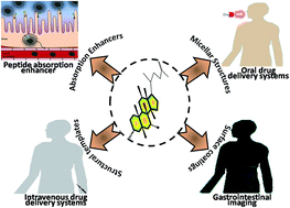

image file: c6ra10978k-p1.tif

Md Nurunnabi

Md Nurunnabi is a postdoctoral fellow at Chungnam National University with Prof. Kang Moo Huh, and simultaneously associating his graduate supervisor Prof. Yong-kyu Lee. He has received BS in Pharmacy, M. Eng in Chemical & Biological and PhD in Polymer Science & Engineering in 2007, 2010 and 2014, respectively. He was a visiting scholar at University of Utah and has experienced of research in industries for ∼3 years. His current research interests are; biomaterials for targeting drug/gene delivery and tissue engineering, developing of imaging agent for noninvasive diagnosis, and nanorobot for theragnosis.

image file: c6ra10978k-p2.tif

Zehedina Khatun

Zehedina Khatun is a postdoctoral fellow at Chungnam National University with Prof. Kang Moo Huh where she focuses on controlled drug delivery and photomedicine (PTT/PDT). Prior to join here, she was a short postdoc at Korea National University of Transportation with Prof. Yong-kyu Lee from where she graduated MS and PhD in Chemical & Biological Engineering and Green Bioengineering in 2011 and 2014, respectively. Her interests of research are oral therapeutics delivery, nanomedicine and polymeric drug delivery.

image file: c6ra10978k-p3.tif

Md Nafiujjaman

Md Nafiujjaman completed BS in Pharmacy from University of Development Alternative (UODA), Bangladesh in 2009 and Masters' in Chemical & Biological Engineering under Prof. Yong-kyu Lee from KNUT, South Korea in 2013. Currently, he is pursuing doctoral degree in Green Bio engineering department in the same lab. His research activity focus on the fabrication of hybrid nanomaterials for cancer imaging and therapy in Prof. Yong-kyu Lee's lab.

image file: c6ra10978k-p4.tif

Seungbin Cha

Seungbin Cha received her BS degree in Applied Chemistry from Konkuk University in 2016 and currently pursuing her masters in Biomedical Chemistry under the guidance of Prof. Young Jun Kim, at Konkuk University and Prof. Young-kyu Lee, at Korea University of Transportation. Currently she is working on using peptides as carriers for oral gene delivery.

image file: c6ra10978k-p5.tif

Kang Moo Huh

Kang Moo Huh is a full professor of Polymer Science & Engineering at Chungnam National University (CNU). He earned his BS in Polymer Science and Engineering from CNU, MS in Materials Science and Engineering from Gwangju Institute of Science and Technology and PhD in Materials Science from Japan Advanced Institute of Science and Technology in 2002. He completed his Post-doctoral training at the Biomedical Research Center of Korea Institute of Science and Technology and in the School of Pharmacy of Purdue University. He joined the faculty of Chungnam National University in 2004. Current research works involve design of new biofunctional polymers and polymeric nano-carriers, including polymer bioconjugates, polymer micelles, polymersomes, and polysaccharide based thermogels for efficient delivery of various drugs and cells. Dr Huh has published 110 papers and 9 book chapters and owns 20 domestic and international patents.

image file: c6ra10978k-p6.tif

Yong-kyu Lee

Yong-kyu Lee is full a professor of the Department Chemical & Biological and adjunct professor of Green Bioengineering at Korea National University of Transportation. He received MSc and PhD in Material Science and Engineering from Gwangju Institute of Science and Technology in 1999 and 2003, respectively. He has received postdoctoral training at Emory School of Medicine and Georgia Institute of Technology with Prof. Shuming Nie. He is the PI of Nano Biomedicine Laboratory at KNUT and founder of two start-up where he is leading various research projects on drug delivery, material chemistry, biomedical engineering, tissue engineering, and nanomedicine and coating technology. He is authored of more than 60 high impact journal articles and inventor of around 15 patents.


1. Introduction

Synthetic materials, such as polymers, inorganic nanocrystals and two-dimensional materials, have gained significant importance in the research and development of biological and biomedical applications.1–3 These materials possess scintillating properties with promising results; however, biocompatibility and biosafety issues impede their translation to preclinical studies.4–6 As a basic criterion, the materials selected as a carrier for drug delivery should meet the essential requirements of both biocompatibility and biodegradability in vitro and in vivo, as well as the facilitation of enhanced drug loading and controlled drug release properties. The bioinspired and biomimetic materials that are acquired or derived from biological systems have received substantial attention in drug delivery research because of their high biological compatibility and negligible toxicity profiles.

Oral drug delivery is well established as the easiest and most convenient strategy for therapeutic administration among all routes of administration, such as intravenous, intramuscular, subcutaneous, transdermal, and sublingual routes.7–12 According to the International Marketing Services IMS, the worth of the oral drug market is approximately US$ 30 billion worldwide, and orally delivered products accounted for 84% of the sales of the top 50 selling drugs worldwide.13,14 However, the advancements in oral drug delivery research are substantially affected by the poor bioavailability of orally delivered drugs. Moreover, the bioavailability of a drug molecule strongly depends on both its stability and absorption ability, which are related to the physicochemical and biological properties of individual molecules.15–17 The stability and absorption ability of individual molecules are further associated with the physiological states and parameters of the gastrointestinal tract. For example, many sophisticated molecules may be easily degraded in the acidic environment of an empty stomach and prevent them from reaching the blood circulation. Simultaneously, the existing gastrointestinal juices, such as enzymes and mucus, prevent interactions between drug molecules and intestinal membranes, which results in reduced gastrointestinal absorption and negligible therapeutic effects.18 Several strategies, such as the encapsulation of a drug/biomolecule in a micro/nano size micelle, the use of cationic nanocarriers to deliver the drugs across the intestinal membranes, the use of enteric coatings to prevent stomach acidic degradations and the conjugation of oral absorption enhancers to enhance the bioavailability, have been proposed to overcome these limitations.19,20 However, these carriers use the paracellular pathways or the rupture of tight junctions to deliver the drug molecules. Moreover, these uptake profiles are inefficient compared with receptor mediated active transport pathways (Fig. 1). Furthermore, these carriers presented significant toxicities during long-term administration.


image file: c6ra10978k-f1.tif
Fig. 1 Scheme represents the mechanism of ileal bile acid transporter mediated oral drug delivery. Bile acid–drug conjugates interact with bile acid transporters of the small intestine and facilitate drug absorption into the small intestine to distribute drugs throughout the body via the systemic circulation.

Bile acids are bioinspired and biomimetic small molecules that are secreted from the liver. The bile acids are derivatized to primary bile acids after conversion of water insoluble cholesterol to water soluble bile acids. The primary bile acids are further conjugated with taurine and glycine groups in order to increase their hydrophilicity. The taurine or glycine conjugated primary bile acids are secreted into the bile and released into the intestines. The intestinal bacteria further convert the primary bile acids to secondary bile acids and are responsible for the chemical diversity of the bile acids.21 Several comprehensive reviews detailed the biological significance,22 synthesis,23 physiochemical properties,24 metabolism25,26 and transport27 of bile acids in the biological systems and also emphasized the importance of bile acids and bile acid receptors in regulating the metabolic disorders.26,28 Moreover, the recent studies on the activation of diverse nuclear receptors and transmembrane G protein coupled receptors by the bile acids have gained significant attention in developing novel bile acid analogues for targeting these receptors that play a predominant role in carcinogenesis, inflammation, fibrosis and carbohydrate metabolism.21

Apart from their conventional roles in absorption and solubilisation of dietary lipids, bile acids have been used as potential oral absorption enhancers because of their ability to withstand the gastrointestinal impediments and aid in the active delivery of drug/biomolecules. The dedicated ileal bile acid receptors (IBAT) in the ileal regions of the small intestines aid in the receptor mediated endocytosis of bile acid conjugated drug/biomolecules (Fig. 1). The peculiar arrangement of rigid hydrophobic domains in the bile acids has gained significant attention in the development of novel drug delivery systems.29 The hydroxyl and carboxyl groups distributed within the bile acid molecules could be utilized for the covalent conjugation of polymers and drug molecules (Table 1). Several reviews highlighted the detergent properties of bile acids and detailed the development of bile acid mediated micellar systems,30 bile acid–drug/polymer conjugates,31 and bile acid conjugated liposomes or bilosomes32 for the delivery of drugs, vaccines and biomolecules. However, it is very important to understand the significance of bile acids conjugated nano drug delivery systems in targeting the receptors and transporters for oral delivery of drugs, imaging agents and biomolecules. Recently, our group thoroughly investigated the importance of bile acids in the oral delivery of drugs and biomolecules, as well as the oral delivery of bile acid conjugated biomolecules, drugs and optical contrast agents.

Table 1 Summary of bile acid mediated therapeutic delivery
  Bile acids Compounds Conjugation methods Drugs Therapeutic applications References
1 DOCA Heparin Amide/ester/covalent   Angiogenesis 63
2 DOCA PSDM Ester/amide   Cancer 64–66
3 DOCA Carboxymethyl curdlan Ester/amide Epirubicin Cancer 67
4 DOCA O-Carboxy-methylated chitosan Ester/amide Folic acid Cancer 68
5 DOCA Ethylenediamine Electrostatic interactions Ceftriaxone Cancer 69 and 70
6 DOCA DMSO Physical interaction Heparin Oral delivery 71 and 72
7 DOCA   Amide bond Low molecular weight heparin Deep venous thrombosis 73
8 Lithocholic acid RGD Amide bond Heparin Angiogenesis 74 and 75
9 TCA   Amide bond Heparin Angiogenesis 76 and 77
10 DOCA Heparin Amide bond Doxorubicin Cancer drug 78 and 79
11 DOCA DMSO Amide bond Heparin Deep vein thrombosis 73
12 DOCA Heparin Amide bond Doxorubicin Antitumor activity 80
13 DOCA Heparin Amide bond   Oral anticoagulant agent 81
14 Lithocholic acid Folic acid Amide bond/covalent Heparin Cancer 80
15 Cholic acid Alkyl chain spacer Ligand interaction Carboplatin Cancer 82
16 Cholic acid PEG Azide group Paclitaxel Cancer 46
17 Chenodeoxycholic acid Amino pyridine Linker   Oral bioavailability 47
18 Glycocholic acid   Combination Epirubicin Cancer 49


This review article details the recent trends in the development of receptor mediated active transport of targeted oral drug delivery systems. Here, we summarize the importance, structural significance of bile acids and pathways involved in the uptake of bile acid conjugated drug delivery systems. We have reviewed the recent advancements in the development of bile acid conjugated drug delivery systems and highlighted the interactions between bile acid conjugated drug delivery systems and receptors/transporters to facilitate active transport and enhance the bioavailability of orally delivered drugs, biomolecules and imaging contrast agents. We also summarize the use of bile acids in the design of bile acid mediated self-assembled nanoparticles, their drug loading, and retention properties, as well as the mechanisms involved in the stability, targeted delivery, and drug release patterns from these nanoparticles following oral administration. Furthermore, the roles of specific polymers in improving drug absorption through the GI mucosa by increasing the membrane permeability of the drug and/or carrier or inhibiting drug efflux transporters in the GI mucosa are discussed. We also proposed key aspects that must be investigated for the development of novel bile acid based biomaterials and drug delivery systems. Furthermore, we highlighted the use of bile acids as polymerization initiators and templates for the development of branched nanostructures. Finally, this review reports the use of bile acids in improving the oral delivery of optical imaging agents, such as Quantum Dots (QDs), PET trackers, and MR contrast agents, through the design of theranostic nanosystems that are directed towards non-invasive imaging and therapy.

2. Bile acids and mechanisms

Bile acids, amphiphilic steroidal molecules, are secreted from hepatocytes in the liver via the catabolism of cholesterol in the presence of enzymes and are stored in the gall bladder for further release into the intestinal lumen during meal ingestion. Two biosynthetic pathways, namely, the classical and alternative pathways, are primarily involved in the synthesis of bile acids (Fig. 2). In general, the hydroxylation, oxidation, steroidal ring modification, and side chain truncation of cholesterol are required to produce primary bile acids. Furthermore, enzymes such as cholesterol 7α-hydroxylase (CHX), BA-CoA synthase (BACS), and BA-CoA: amino acid N-acyltransferase (BAT) are primarily involved in amidation and modifications via the introduction of taurine and glycine to bile acids.33,75,76
image file: c6ra10978k-f2.tif
Fig. 2 (A) Basic structure and nomenclature of bile acids. Changes in the position of the side chains determined the activity of the bile acids. The scale of hydrophilicity is not only dependent on the number of hydroxyl groups (A-a) but also the position of the hydroxyl groups (A-b). (B) Enterohepatic circulation of bile acids. The bile acids gain access into the intestines via the ASBT receptors lined over the apical side of the ileal and colonic regions. I-BABP aids the basolateral transport of bile acids followed by the interactions with OST α/β, MRP3, and t-ASBT inside of the intestines, where bile acids enters the enterohepatic circulation through the portal vein. NTCP and OATP are involved in the transport of bile acids into the hepatocytes followed by interactions with the MRP2 receptor and storage in the gall bladder. Some bile acids are driven into the systemic circulation via activation of OATP, OST and MRP receptors. These bile acids are further coupled to the ASBT receptors in the kidney and are recirculated back to the gall bladder via activation of OST and MRP receptors in the kidney. A small number of bile acids are excreted into the forms of faces or urine. Reprinted with permission28 from the Nature Publishing Group.

In the 1990s, numerous studies were focused on investigating the application of bile acids as ligands in the activation of various nuclear homing receptors (NHR). Following activation by bile acids, the downstream signaling of the NHR may augment or retrograde the gene expressions and regulate the functions of cells. farnesoid X receptor (FXR), which is abundantly expressed in the tissues that are involved in the enterohepatic circulation of bile acids, is the most influenced NHR. Bile acids that activate FXRs are translocated into the nucleus, form heterodimers with retinoic receptor X (RXR), and bind to definite regions on DNA. They subsequently recruit transcription factors and polymerase machinery to initiate gene expression. It is noteworthy that only specific bile acids may bind to the FXR and their binding affinities are strictly structure specific. Rankings regarding the specificity from highest to lowest include: chenodeoxycholic acids (CDCA), deoxycholic acid (DOCA), lithocholic acid and cholic acid.21,28,36 It is interesting to note that highly hydrophilic bile acids, such as muricholic acids that are significantly expressed in the mouse, have antagonist effects on FXR activity. Moreover, the regulatory properties of FXR have also made them significant targeting receptors for bile acid homeostasis.

Other lithocholic acid derivatives may also bind to specific nuclear receptors, such as pregnane X receptors and vitamin D receptors, and function as agonists on gene expression. The FXR machinery is predominately stressed in this review because it is crucial in the synthesis and regulation of CHX proteins and the expression of specific proteins, such as ileal lipid binding proteins (iLBP) and organic solute transporter (OST) α,β proteins, which play predominant roles in guiding bile acids into the enterohepatic circulations.33

The mode by which bile acids cross the enterohepatic circulation is crucial because most bile acid mediated formulations are designed for oral delivery. It is noteworthy that more than 90% of the secreted bile acids are reabsorbed from the small intestines through bile acid transporters, reach the liver through the systemic circulation and are ultimately stored in the gallbladder. Apical sodium dependent bile acid transporter (ASBT) proteins expressed on the ileal portions of the small intestines also have specificity for the reabsorption of bile acids.37 Following interaction with ASBT, the bile acids are endocytosed into the cells. The iLBP further interact with the formed vesicles, prevent lysosomal lysis and aid in the transport of these bile acids to the basolateral portions of the cell.37 The OST α and OST β transporter proteins, as well as multidrug resistant associated proteins (MRP-3 in particular) aid in driving bile acids into the portal circulation.37,38 When they reach the liver, Na+ taurocholate transporting polypeptides (NTCP) and organic anion transport proteins (OATP) expressed in hepatocytes aid in the further absorption of bile acids. The accumulated bile acids are effluxed via OST α, OST β, and MRP-3, as well as MRP-4 into the systemic circulations to prevent cholestasis.27,28 These pathways are worth investigating because they may assist in the development of novel drug delivery strategies.

Recent studies have demonstrated that NTCP receptors are crucial for the entry of hepatitis B virus (HBV) into hepatocytes.39 With advents in drug discovery and molecular biology, novel drug delivery systems conjugated with bile acids may aid in potential treatments for HBV. We along with other research groups have demonstrated that orally delivered TCA conjugated nano-formulations were efficient in targeting hepatic cancers.

2.1 Bile acids and their structural significance

Structurally, bile salts are tetracyclic steroidal ring structures with three six-member and one five-member ring, as well as hydroxyl and acidic groups attached to the hydrophobic steroid rings.30 The activities of each bile salt differ based on the position and number of the hydroxyl groups attached to the bile acids, as well as the conjugation of amino acids, such as taurine or glycine (Fig. 2). In contrast to classic amphiphiles that have distinctive hydrophilic and hydrophobic regions, the hydrophilic and hydrophobic domains in bile acids are not separated.40 The hydrophilic groups in bile acids are arranged in the concave α-side of the steroidal rings, whereas the hydrophobic domains are aligned towards the convex β-sides.41 However, as a result of the rigid steroidal structures and complex arrangement of hydroxyl groups, portions of the hydrophilic domain may bury within the micelles, whereas specific hydrophobic domains may interact with water molecules.

Two key forces, namely, hydrogen bonding and hydrophobic interactions, aid in the stabilization of bile acid micelles. The positioning of hydroxyl and acidic groups not only determines the scale of hydrophobicity but also enables them to participate in intermolecular hydrogen bonding with the hydroxyl and carboxylate groups of neighboring bile acids. As a result of the rigid steroidal structural properties and the directional hydrogen bonding, bile acids aid in the formation of more robust and rigid micelles compared with other conventional amphiphiles. A thorough analysis of the structure function relationship of bile acids may be referred in recent reviews.30

The stability of the bile acid micelles depends on the selection of bile acids and solution conditions, such as the pH, temperature and ionic strength. The critical micellar concentrations of bile acids play a significant role in the development of supramolecular assemblies. Compared with other classic amphiphilic molecules, bile acids have broader transition regions that result in two micellar concentrations. The first transition is related to the formation of multimers or small aggregates, which are referred to as noncritical multimerization concentrations, and the second transition includes conventional micelles, namely, the critical micellar concentration (CMC). The CMC of the bile acids may be affected by the ionic strength and the pH of the solution. As the ionic strength of a solution increase, the electrostatic repulsion that results from the charged carboxyl groups decrease, which favors the aggregation and reduction of CMC. All bile acids remain ionized when the pH of the solution is greater than their pKa values. However, when the pH of the solution is close to the pKa values of the bile acids, equal numbers of protonated and deprotonated groups favor the micellar formation and reduce the CMC.40

Different bile acids possess different activities in the biological system. These activities are a result of the differences in the chemical compositions, positioning of hydroxyl groups and conjugation of glycine and taurine groups.40 The principle reason behind the conjugation of glycine or taurine groups to bile acids is to reduce the pKa values of the bile acids and enable them to be ionized at any pH. Theoretically, the pKa values of unconjugated bile acids are approximately 5.0, whereas the conjugation of glycine and taurine groups to the bile acids further reduces the pKa values to 3.9 and 1, respectively. Ionized bile salts are more water soluble compared with protonated bile salts; thus, the conjugation of glycine and taurine groups may prevent aggregations at a lower pH. Therefore, taurine conjugated bile acids are more ionic and soluble even at a gastric pH of approximately 1 and can be helpful for designing oral drug delivery systems.

2.2 Bile acids and their biological significance

Bile acids are digestive agents secreted by the oxidation of cholesterol in the liver. Taurine, glycine, sulfate or glucuronide groups are conjugated to bile acids and are stored in the gall bladder.36 They help break the ingested fat in the stomach and regulate the cholesterol levels in biological systems.23,42,43 Bile acids released from the gall bladder are reabsorbed in the intestines and are instituted by hepatocytes for re-excretion into bile.43,44 Although it is not specific for the type of disease, moderately high concentrations of bile acid represent a good sign of hepatobiliary function. However, extremely high concentrations of bile acids may lead to extrahepatic diseases (e.g., metabolic diseases, such as hyperadrenocorticism). Substantial amounts of bile acids are secreted into the intestine every day; however, only small portions of the total bile acids are lost from the body. This is because approximately 95% of the bile acids delivered from the duodenum are absorbed back into the blood at the ileum.45 The bile acids subsequently enter the blood vessels through the portal vein via the sinusoids of the liver. Bile acids are transported from the hepatocytes for re-secretion into the canaliculi. The net efficiency of this enterohepatic recirculation is so high that each bile salt molecule is reused approximately twenty fold and may be reused two or three times during a single digestive phase. Several principal bile acids include cholic acid, chenodeoxycholic acid, glycocholic acid, TCA, DOCA and lithocholic acid.
2.2.1 Cholic acid. Cholic acid is the major bile acid produced from cholesterol in the liver.34,35 In contrast to common platinum drugs, such as cisplatin, that exhibit drug resistance in different cancer cells, Paschke et al. have reported that the conjugation of cholic acid to the anti-cancer drug carboplatin resulted in overcoming the drug resistance in tumor cells. Their results demonstrated a reduction in the tumor size in platinum resistant tumor models. Similarly, Li et al. demonstrated significant anti-tumor efficiency in ovarian cancer xenograft models when they used two arm oligomeric cholic acid micelles with PEGylation and cisplatin.34,35,46

Chenodeoxycholic acid (CDOCA) is one of the main organic bile acids produced in the liver. Similar to other bile acids, CDOCA is produced as a result of cholesterol catabolism and is conjugated with taurine or glycine groups in the liver. It plays a significant role in liver homeostasis. It is ionized below its pKa values and remains in the GI tract until it reaches the reabsorption site, the ileum.20 Zheng et al. investigated the importance of the chemical space around the C-24 position of bile acids in ASBT mediated transport properties. Consequently, aminopyridine and aminophenol conjugates to the glutamyl-chenodeoxycholate were designed. Their results suggest that hydrophobic and steroidal features around the C24 position influenced the interaction between bile acids and ASBT, which resulted in functioning as potent ASBT inhibitors.47 Glycocholic acid is involved in the emulsification of fats and is present as a sodium salt in the bile of mammals. It is a conjugate of cholic acid with glycine.48 Lo et al. synthesized anticancer drug epirubicin glycocholic acid drug conjugates to treat multidrug resistant cancer cells.49 The conjugation of bile acids to epirubicin improved the intracellular accumulation and cytotoxicity of epirubicin, inhibited the pump and non-pump resistances in the multidrug resistant cancer cell models and improved their chemosensitivity and therapeutic activity.

2.2.2 Taurocholic acid (TCA). Taurocholic acid (TCA) is a hydrophilic bile acid that is derived from cholic acid. It is produced as a sodium salt in the bile of mammals. Miyake et al. reported that the combinatorial use of TCA and spermine (a polyamine involved in cellular metabolism) is essential for improving the oral absorption of poorly water soluble drugs, such as rebamipide.50 Lee et al. reported significant antiangiogenic effects of the LMWH–taurocholate conjugates by blocking the interaction between VEGF165 and its receptors. The competitive binding of TCA to the VEGF165 receptor led to intimidate VEGF165 mediated angiogenesis signaling.51
2.2.3 Deoxycholic acid (DOCA). Deoxycholic acid (DOCA) is produced from the salt of glycolic and TCA in the small intestine in the presence of bacterial enzymes. Less than half of the DOCAs are absorbed by the intestines, recirculated, and stored in the gall bladder. DOCA is typically extracted from bovine bile in the meat processing industry. Historically, DOCA has been used as an intermediate for the production of corticosteroids, which have anti-inflammatory indications. The use of DOCA as a biological detergent to lyse and solubilize cellular and membrane components has gained substantial significance.52 Studies from Kim et al. have suggested that the conjugation of DOCA to low molecular weight heparin (LMWH) improved the intestine mediated oral absorption and bioavailability of LMWH compared with the pristine LMWH, which had no apparent oral absorption.53 In another study, Zhao et al. demonstrated that PGA microspheres loaded with insulin–oligoethylenimine-hyperbranched DOCA complexes improved oral insulin delivery.54 A study conducted by Lee and his group demonstrated that DOCA conjugated heparin nanoparticles were orally absorbed through ileal bile acid transporters, which is very promising for the inhibition of tumor growth (Fig. 3).55
image file: c6ra10978k-f3.tif
Fig. 3 Scheme represents a potential role of bile acid conjugated with polymeric carriers that contain therapeutic drugs. A polymeric carrier system without linking with bile acids may not have a potential interaction with the intestinal cell membrane. The carrier conjugated with hydrophobic bile acids demonstrated less interaction with the intestinal cell membrane because of the formation of self-assembled micelles, in which most of the hydrophobic bile acids are located inside the core. However, hydrophilic bile acids exposed to the outside of a polymeric carrier facilitated the transport of the polymeric carrier into the cells via an interaction between the bile acids and their receptors on the intestinal cell membrane.
2.2.4 Lithocholic acid. Lithocholic acid is a bile acid that acts as a detergent to solubilize fats and improve their absorption. Lithocholic acid has the capacity to specifically target and kill cancer cells, as well as prevent tumor growth, which would make it a healthy and more efficient approach compared with conventional chemotherapeutic procedures. Dietary fibers bind to lithocholic acid and aid in its excretion in stool; thus, fibers may protect against colon cancer.

3. Functions of bile acid in oral drug delivery

Oral delivery is the most favorable yet regressive clinical advancements because of the impediments associated with the gastrointestinal barriers. The harsh acidic environment associated with enzymatic degradations in the stomach projects the first barrier for the oral absorption of therapeutic molecules. Moreover, the variable pH system at different intestinal segments, mucus secretions, and tight packing of the intestinal epithelia further hamper the transport of orally delivered drug molecules.

Oral delivery of chitosan-nanomaterials exhibited positive results as a result of their ability to unpack tight junctions and facilitate drug transport.56 However, debates persist regarding the efficacy and specificity of chitosan mediated oral drug delivery because of the difficulty in understanding the transport mechanism of chitosan-based nanoparticles. Evidence indicates that many of these particles disintegrated at tight junctions, whereas other results suggest successful transport of the nanoparticles to the targeted sites.57 However, the theoretical value of membrane disintegration is hardly few ten nanometers, and it is difficult for chitosan nanoparticles to cross the tight junctions. This issue enforces the use of larger drug concentrations to attain the therapeutic plasma drug concentrations. Moreover, studies have demonstrated that the stability of chitosan based nanoparticles is strongly related to their pH value.58 These studies indicate the necessity for specialized carriers that are specific and stable for the successful oral delivery of therapeutics.

The bile acid based micellar drug delivery systems used hydrophobic bile acids to form self-assemble micelles and firmly pack the hydrophobic drugs with a high payload efficacy. Furthermore, the stability of the bile acids in acidic pH, their adaptability to dynamic pH variations and the presence of selective receptors on the intestinal surface project them as efficient carriers for oral drug delivery. The conjugation of secondary (i.e., DOCA and lithocholic acid) and conjugated bile acids (glycine and taurine derivatives of secondary bile acids, i.e., taurocholic acid (TCA), tauro-deoxycholic acid, and glycolithocholic acid) have further improved the bioavailability of orally delivered nanoparticles.

Several studies have indicated the importance of apical sodium bile acid transporters (ASBT) specifically expressed in the ileal portions of the small intestines as crucial transmembrane proteins involved in the transport of heparin–bile acid conjugates. Al-Hilal et al. primarily investigated the importance of oligomeric DOCA conjugated heparin molecules, their affinity towards apical sodium-dependent bile acid transporters (ASBT), and the mechanism of the conjugate transport following oral delivery.59,60 Their findings suggested that the conjugation of the oligomeric form of DOCA (tetra DOCA) to low molecular weight heparin (LMWH) exhibited better binding affinity to ASBT receptors and oral absorption compared with mono DOCA (approximately 50-fold). The oral administration of 10 mg kg−1 LMWH–tetra DOCA resulted in an approximately 97% reduction in the thrombus formation and improved the anticoagulation activity in monkeys.55 Because DOCA is hydrophobic, they were minimally exposed to the native gastric environments. To improve their drug transport ability, Zehedina et al. formulated docetaxel with LMWH composed of primary bile acid or conjugated with TCA, a secondary, hydrophilic bile acid.61,62 The formulations exhibited a preferential uptake of docetaxel in the small intestines.

4. Strategies and methods of bioconjugation and formulation design

Table 1 indicates the different strategies for chemical conjugation of bile acids with carriers. To conjugate a bile acid with a carrier or other molecules, various strategies have been performed, such as esterification, covalent bonding and ionic interaction. Regarding oral delivery formulations, in most cases, the bile acid was conjugated through covalent bonding (amide bonding), which has better potentiality compared with other bonds in terms of the stability in an acidic environment. Other conjugation strategies, such as esterification or an ionic complex, have been widely considered for designing a formulation for the intravenous administration of a bile acid mediated formulation. The physical and chemical properties of each bile acid are different; however, they have potential as oral absorption enhances. For example, both DOCA and TCA have potential as oral absorption enhancers; however, they have different physical properties. DOCA is hydrophobic, whereas TCA is hydrophilic. Therefore, based on the requirement and design, the rationale for the selection of a bile acid should be varied.

Self-assemble micelle formation is easier when hydrophobic bile acids, such as DOCA are conjugated, where bile acids predominately locate in the core of the core/shell micelles. Moreover, hydrophilic bile acid has no potential impact on the formation of a particle; however, it is primarily exposed on the outer surface of a formulation when dissolved in an aqueous solution. Therefore, the bile acid selection and conjugation method should be compatible with the desired form of the formulation and the route of administration. A bile acid cluster prepared with the self-conjugation of DOCA has demonstrated an exceptional absorption efficacy.59,83 Tetra-DOCA conjugated heparin formed a vesicle as a result of the higher molecular weight of the hydrophobic part than the hydrophilic part. Comparatively higher hydrophobic properties of self-conjugated tetra-DOCA than mono-DOCA potentially play important roles in transforming the bile acid transformer in vitro and in vivo.

5. Bile acid mediated oral delivery of macromolecular drugs

Specific macromolecular therapeutics, such as heparin and insulin, have substantial potential as therapeutic agents; however, their degradability and poor absorption properties hinder their further applications in oral delivery.84,85 These drugs are not absorbed orally as a result of their high molecular weight, negatively charged surface, high water solubility and degradability in an acidic stomach environment.86,87 When orally administered, these drugs must be conjugated with an oral absorption enhancer to overcome these limitations.88 Studies have demonstrated that both physical mixing and chemical conjugation of heparin with bile acids enhanced their oral absorption.89 In another study, DOCA and TCA were independently conjugated to the heparin backbone to assess their oral therapeutic efficacy (Fig. 4). The hydrophobic nature of DOCA helped DOCA conjugated heparin to form stable micelles in an aqueous solution compared with hydrophilic TCA conjugated heparin molecules. However, the oral absorption of TCA conjugated heparin was increased compared with DOCA conjugated heparin.90
image file: c6ra10978k-f4.tif
Fig. 4 Absorption of orally administered LMWH–DOCA in the GI tract. (A) Comparison of LMWH–DOCA absorption in different species (n = 8–10, mean ± SEM for mice and rats, n = 2, mean ± SD for monkeys). (B and C) Effect of bile acid on the absorption of 100 mg kg−1 (12[thin space (1/6-em)]680 IU kg−1) LMWH–DOCA in mice (0.4 mL of human bile solution and 200 mg kg−1 free DOCA). Reprinted with permission from Elsevier.55

The mechanism involved in the uptake of bile acids in the ileum is an active, carrier-mediated, and Na+-dependent co-transport process. The active transport of bile acids across the ileal epithelial bilayer has been described in terms of the maximal transport velocity (Vmax) and the concentration at half-maximal flux (KT). The KT is apparently related to whether the bile acid is conjugated with the carrier. In general, conjugated bile acids have a two-fold higher KT value compared with their un-conjugated parent compounds. In rats, the Vmax values are independent of the conjugations; however, they appear to be related to the number of hydroxyl groups attached to the sterol nucleus, such as a trihydroxy > dihydroxy > monohydroxy order. However, no single hydroxyl group is essential for transport. Krag and Phillips identified similar structure–Vmax relationships in humans; however, no correlation was identified between the KT values and conjugation.91–93

For efficient transport, a bile acid molecule must possess a single negative charge, which should be located on the C-17 side chain of the sterol nucleus. Bile acids with two negative charges around the C-17 position provide minimal active transport; however, the addition of an extra negative charge in the form of sulfonation at the C-3 position does not preclude active transport.94 Lee et al. used a novel conjugation strategy for the synthesis of heparin–DOCA conjugates. They used the hydroxyl group at the C3 position rather than the carboxyl group for their conjugation with heparin.55 4-Nitrophenyl chloroformate (4-NPC) was initially conjugated to the C3-hydroxyl group and was further aminated using ethylenediamine (EDA). The aminated DOCA was further conjugated to the carboxyl groups of heparin using traditional EDC/NHS chemistry. Using this conjugation method, many DOCA molecules were bound to the heparin backbone. The maximum concentration of DOCA–heparin VIII conjugate was 3.3 ± 0.5 IU mL−1 at 10 mg kg−1 of an oral dose, and the calculated value was 3 times higher than the baseline with no adverse effect on the GI tissue.37

Insulin is typically subcutaneously (SC) administered to treat diabetes. Byun et al. reported that DOCA conjugated insulin could be administered orally to reduce increased blood glucose levels.95 His research team developed an oral drug carrier referred to as Nα-deoxycholyl-L-lysyl-methylester (DCK), which may physically associate with therapeutic proteins via ion–pair interactions without altering their native structure. The administration of 21, 30 and 42 U kg−1 (based on insulin activity) of an insulin/DCK formulation reduced the plasma glucose levels by up to 33% (median; range 30.6–70.2%), 78.5% (39.4–86.8%) and 75.2% (67–87.4%), respectively, compared with the baseline levels.96,97 Furthermore, the plasma insulin concentrations rapidly increased. Sakai et al. demonstrated that the combinatorial administration of dipotassium glycyrrhizinate and sodium deoxycholate exhibited an increased oral absorption profile compared with the administration of pristine dipotassium glycyrrhizinate. However, the combined administration of these two enhancers also did not exhibit significant toxic effects as observed in vitro.98

6. Bile acid mediated oral delivery of optical imaging agents

Noninvasive imaging and in vitro biomolecule detection is one of the most promising areas in modern biomedical fields because of their precedence in the diagnosis and early detection of diseases.99–104 Quantum dots (QDs), gold nanoparticles (AuNs) and superparamagnetic iron oxide nanoparticles (SPIONs) have been widely considered for noninvasive biomedical imaging.105–108 Recently, SPIONs have been approved by the U.S. Food and Drug Administration (FDA) for biological applications, whereas QDs were approved for phase one (phase I) clinical trials in humans following the successful completion of their safety trials in mice.109,110 To date, researchers have developed bile acid conjugated biopolymers, semiconductor gold nanoparticles, quantum dots, magnetic nanoparticles and other compounds for drug delivery and imaging.111–114

Most recently, we have developed a unique ileal bile acid transporter mediated strategy for the oral delivery of QDs.91 A number of significant advantages of oral delivery of QDs were highlighted, such as the ease of administration, lower toxicity, and ease for real-time monitoring of oral drug absorption, gastrointestinal tract imaging, and biodistribution studies. Zehedina et al. loaded hydrophobic 7 nm QDs into heparin–DOCA micelles and investigated the biodistribution of orally delivered QDs. The QD-loaded heparin–DOCA nanoparticles exhibited excellent stability in different pH conditions, and the release profile demonstrated a slow release after 5 days of oral administration (Fig. 5A). Confocal laser microscopic scanning images indicated that QD-loaded heparin–DOCA nanoparticles penetrated into the cell membrane and were located in the cell cytosol. Pharmacokinetic (PK) studies with real-time imaging modalities have also demonstrated the absorption, distribution, metabolism, and elimination profile of QD-loaded heparin–DOCA nanoparticles.93 Noninvasive imaging in nude mice demonstrated that the orally administered QD-loaded heparin–DOCA nanoparticles had accumulated in the liver, lung, heart, spleen, and kidney (Fig. 5B). We subsequently investigated the in vitro and in vivo toxicological profiles of the orally administered QD-loaded heparin–DOCA nanoparticles. After multiple administrations and dose variations of the QD loaded nanoparticles for 45 days, the results from a comet assay, serum biochemistry, and tissue histology indicated sub-optimal inflammation in the liver.4


image file: c6ra10978k-f5.tif
Fig. 5 Bile acid conjugated heparin nanoparticle for in vivo imaging. (A) Process of QD loading into heparin–bile acid conjugates. (B) Optical imaging agent absorbed in the small intestine via the ileal bile acid transporter, which was noninvasively imaged by an optical imaging system. Reprinted with permission from American Chemical Society.93

Jia et al. conjugated bile acid to 18F, a PET imaging agent, via click chemistry for the early detection of human diseases.115 This biomolecule exhibited high serum stability in vitro and good metabolic stability in vivo. The PET images of 18F labelled bile acid exhibited uniform distributions and a proper clearance time from the liver.115 Uggeri and his group demonstrated that gadolinium (GD) particles conjugated to bile acid may be used as hepatocyte-targeted MRI contrast agents. The GD conjugated bile acids exhibited enhanced stability, MR contrast properties, and elimination through the biliary route. The targeting properties strictly depended on the selection of the bile acid and the site of conjugation between the GD and bile acid.116,117

The imaging prospects of bile acids have also been used to determine specific disorders associated with impaired bile acid transport in biological systems, such as bile acid malabsorption (BAM).118 The conjugation of 19F to cholic acid helped to determine the bile acid enterohepatic circulations. Moreover, the designed MRI contrast agents imposed negligible toxicology effects in the small intestines or liver. Bruyn et al. developed a fluorescent bile acid probe, tauro-nor-THCA-24-DBD, and investigated the activity of the bile acid export pump (BSEP). Their studies suggest that the designed bile acid fluorescent probe may facilitate an understanding of the interference of drugs, such as cyclosporine, on the BSEP and NTCP activity in hepatocytes.119

7. Bile acid mediated oral delivery of peptides

The use of peptides to treat chronic diseases is substantially limited because of their poor intestinal absorption and loss of active sites as a result of acidic and enzymatic degradation in the stomach. A review article by Kramer et al. demonstrated that the conjugation of bile acids to peptides could improve the oral absorption profiles of small peptides. In their study, they designed peptides with chain lengths of 10 amino acids that were covalently conjugated to the 3-position of modified bile acids to yield peptidyl-3β-(ω-aminoalkoxy)-7α,12α-dihydroxy-5β-cholan-24-oic acids. These compounds interacted with the ileal Na+/bile acid cotransport system and aided the effective active transport of the peptides. Studies have also demonstrated the concentration-dependent inhibition of Na+-dependent [3H]taurocholate uptake into brush-border membrane vesicles from the rabbit ileum following oral administration of bile acid conjugated peptides.17,95–97 In contrast, the corresponding parent peptides did not inhibit taurocholate transport or photoaffinity labeling of the transport proteins. Moreover, the affinity of the peptide–bile acid conjugates to the ileal bile acid carrier system decreased with the increase in the chain length of the model peptides (Fig. 6). Their results suggested that the Na+/bile acid cotransport system is crucial for ileal uptake and an enhanced absorption of bile acid conjugated peptides.
image file: c6ra10978k-f6.tif
Fig. 6 A hypothetical approach to the oral delivery of peptide in the conjugation of bile acids. (A) The modified peptide through conjugation with bile acids increased the oral absorption compared with the non-modified peptide. (B) Oral absorption of a peptide depends on the molecular weight of the peptide. Interaction between bile acid–peptide conjugates and a bile acid transporter is inversely proportional with the molecular weight of the peptide molecules.

Kramer et al. also demonstrated that both bile acids and modified bile acids may represent potential oral absorption enhancers when linked to therapeutic molecules, such as drugs and peptides.120 Peptide–bile acid conjugates exhibited an enhancement of the overall intestinal absorption up to approximately 7% compared with the intestinal absorption of the naked peptide, which exhibited less than 0.5% of intestinal absorption.

The bile acid derived liposome/lipid nanoparticles or bilosomes have gained significant attention in the oral delivery of vaccines and surface antigens.30 The presence of bile salts prevented bilosomes and their therapeutic biomolecules from enzymatic and acidic degradations and prevented the premature release of surface antigens from the bilosomes.121 Several surface antigens, including tetanus toxoid,122 hepatitis B surface antigen,123 and influenza A,124 have successfully been orally delivered using bilosomes. Bilosomes exhibit superior activities in terms of the chemical and storage stabilities. Moreover, these bilosomes may withstand the gastrointestinal and enzymatic disruptions compared with liposomes and niosomes. It was interesting that the immune responses of bilosomes could vary. A review article by Shukla et al. details the use of different bilosomes in the oral delivery of various surface antigens and concludes that bilosomes may represent a new hope in oral vaccine delivery.32

8. Non-orally delivered bile acid conjugated drug delivery systems

The amphiphilic properties of bile acids aid the formation of self-assembled drug delivery systems. The steroid structures of bile acids may assist the hydrophobic interactions with the hydrophobic drugs and aid the formation of stable nanomicelles. Several bile acid mediated drug delivery strategies have been employed to treat various diseases. Different synthetic polymers, such as polyethylene glycol,125–129 Pluronic F127,130,131 lactic acids,128 PLGA,132,133 PCL,128 and polyethylene imine,130 have been conjugated to bile acids to fabricate core shell nanostructures. For example, Le Dévédec et al. fabricated a PEGylated, bile acid mediated, self-assembled drug delivery system to treat fungal diseases.125 The specificity of these fabricated nanoparticles was further enhanced by conjugating target specific ligands, such as folic acid, which could be actively delivered to the targeted site that expressed folic acid receptors.134 Biological polymers, such as chitosan,135,136 chondroitin sulfate A (CSA),137 heparin,96 and hyaluronic acid (HA),138 have also been conjugated with bile acids to generate micelle mediated drug delivery systems. Apart from rendering biocompatibility, biological polymer complexes may be targeted to key receptors, such as CD44, which are vividly expressed on the surface of cancer cells.129

Telodendrimer based nanomicelles are PEG-dendritic polymers that are self-assembled to load hydrophobic drugs and treat cancer.126,129,139–141 In general, the (Fmoc)lysine-Fmoc(OH) were attached via DIC–HoBT chemistry to the terminal end of the PEG to form cationic multigeneration dendrimers. The ends of these telodendrimers were conjugated with cholic acid to form nanomicelles. Several drugs with loaded micelles have been designed for cancer drug delivery. Shi et al. used molecular docking and molecular dynamic simulations to optimize telodendrimer based drug formulations.141 They proposed that the conjugation of hydrophobic moieties, such as cholic acids and rhein, onto the telodendrimeric backbones could enhance the drug loading efficiency and stability of hydrophobic drugs, such as doxorubicin, via pi–pi interactions, polycyclic–pi interactions, and hydrogen bonding. They also suggested that the generation of dendrimers has an effect on the stability and toxicity profiles of the nanomicelles. Compared with Doxil, the second and third generations of rhein conjugated telodendrimers loaded with doxorubicin exhibited an enhanced in vivo therapeutic efficacy. However, third generation telodendrimers are toxic because of their fast drug release profiles. Xiao et al. accounted for the effect of the surface charge on the bio distribution of telodendrimers.142 In vivo studies have demonstrated that positively charged nanoparticles induced enhanced hemolysis compared with negatively charged nanoparticles.129 They proposed that the positively charged nanoparticles would interact and fuse with the erythrocyte membrane, thereby leading to the induction of cell lysis by disrupting the cell membrane. Their results also demonstrated that highly charged nanoparticles (both positive and negative) were cleared by the reticulo endothelial system (RES) in the liver compared with the slightly charged telodendrimer nanoparticles. Enhanced clearance of highly charged nanoparticles may be a result of the active phagocytosis of macrophages in the liver. However, slightly negative-charged nanoparticles exhibited meaningful results as a result of the low opsonization rates by immune cells compared with the highly charged nanoparticles and also enhanced the circulation time of the nanoparticles, which thereby resulted in an enhanced tumor uptake compared with the liver.

A new second generation of telodendrimers was designed to develop dual stimuli responsive micelles to treat ovarian cancer. The stability of the micelles was further improved via the introduction of boronic acid and catechol groups onto the polymer backbone.140 The ability of boronic acid to react with the catechol groups to form boronic esters further generated covalently crosslinked nanomicelles. The ability of mannitol and pH sensitivity were employed to induce drug release. Paclitaxel was loaded into the micelle cores, in which pH and mannitol dependent drug release and increased toxicities were identified in ovarian cancer cells. A FRET system was further employed to assess the stability of micelles in vivo. Compared with the control groups with no boronic crosslinking, the boron conjugated systems exhibited enhanced stability. However, the release of drugs from the micelle cores requires intravenous supplementation of mannitol for the efficient therapy and is susceptible to release the drug in glucose concentrations.

Bile acids have also been harvested as initiators for ring-opening polymerization and the subsequent formation of star-shaped polymeric micelles.143,144 The hydroxyl groups served as polyhydroxy initiators and aided the formation of controlled star-shaped architectures. Star shaped polymers with cholic acids exhibited increased degradation profiles, biocompatibility, and cellular binding properties compared with the control star shaped polymers without cholic acid. Different star shaped polymers based on polycaprolactone, such as PLA and PLGA, have been designed to treat different cancers. For example, Tao et al. fabricated docetaxel loaded random polycaprolactone, polylactic acid based star shaped nanomicelles and were further surface engineered by polyethylene glycol to treat liver cancers.128 It was demonstrated that the PEG coating further enhanced the stability of these particles. Moreover, stronger interactions between the cores of the star shaped micelles (hydrophobic interactions) and a hydrophobic drug further improved the encapsulation efficiency. With a mean particle size of 95 nm and a docetaxel encapsulation efficiency of approximately 97%, these nanomaterials exhibited enhanced therapeutic efficiencies both in vitro and in vivo. A six fold reduction in the tumor volume was identified compared with the control. Moreover, compared with the clinically approved drug Taxotere, these formulations demonstrated reduced IC50 values and enhanced therapeutic efficiencies at the same dosage.

Sodium deoxycholates (DOCA) have also been used as a template to branch gold nanoparticles.29 The concentrations of DOCA regulated the structures of gold nanoparticles from nano-sized particles to dendrimers and fiber like branched structures. The unique structural arrangement of DOCA, the hydrophobic interactions between the polar faces of the steric groups and secondary forces, such as the ion–dipole, ion–ion, and hydrogen bonding interactions, facilitated the creation of central cavities that could host the guest gold atoms. The free spaces between the cholate anions were further accessed by the Au ions to assist in the growth of the gold nanoparticles to different structures, such as dendrimers or fibers. These dendritic gold nanomaterials were further conjugated with cyclic RGD peptides to improve the stability of the nanoparticles. The fabricated nanomaterials were used for photothermal therapy (PTT), in which the localized heating near the tumor areas was assessed. The tumor bearing mice were exposed to a laser with an 808 nm wavelength (1.4 W cm−2) for 5 min. The results indicated a substantial reduction in the tumor volume compared with the control groups. The DOCA branched gold nanostructures exhibited a photothermal transduction efficiency of 61%, which was two-fold higher than the conventional PTT agents. At the edges and tips of these branched gold nanostructures, increased enhancements in the local electric fields at the plasmon resonance were identified. The local enhancement in electron excitation and relaxation may aid efficient transduction of incident light energy to heat and produce efficient photothermal activity.

9. Bile acids for oral co-delivery of therapeutic and imaging agents

To date, researchers have developed modified polymers conjugated with various bile acids, semiconductive gold nanoparticles, quantum dots, magnetic nanoparticles, and many other compounds for the delivery of drugs and imaging agents.111–114 In a previous study, our group developed QD loaded nanomicelles and reported DOCA mediated GI imaging with orally delivered QDs.145,146 However, the presence of hydrophilic bile acids on the surface of the nanoparticle resulted in an enhanced oral absorption compared with the hydrophobic bile acids, such as DOCA, which reside in the cores of the nanoparticles. Consequently, we selected the hydrophilic bile acid TCA and designed advanced theranostic nanoformulations to co-deliver both therapeutics and contrast agents. In our study, TCA was conjugated to heparin–docetaxel compounds and further loaded with rhodamine B for the enhanced oral delivery of imaging and therapeutic agents (Fig. 7). The resulting self-assembled nanoparticles contained docetaxel in their cores and TCA on the surface of the nanoparticles. This bile acid conjugated system improved the oral absorption of nanoparticles through the stimulation of bile acid transporters in the small intestine. Rhodamine B was used as a function of the imaging agent to diagnose diseases. An animal model bearing KB cancer cells was developed to evaluate the in vivo imaging, therapeutic activity, and feasibility of oral absorption of the nano-formulations.
image file: c6ra10978k-f7.tif
Fig. 7 (A) Scheme indicates the formation of self-assembled nanoparticle micelles. The modified heparin with TCA (TCA) contains docetaxel (DTX) and rhodamine B in the core and hydrophilic TCA exposed on the outer surface of the nanoparticle. Non-invasive optical imaging of the accumulation of nanoparticles in the xenograft tumor model in mice with (B) KB and (D) MDA-MB231 cell lines. Anticancer activity of the nanoparticles in both xenograft tumor models in mice (C and E). Reprinted from ref. 61 with permission from Elsevier.

The results from in vivo imaging studies demonstrated optical imaging signals with stronger intensities from rhodamine B conjugated nanoparticles in the tumor sites and the kidney post 24 h of oral administration. The results demonstrated that the nanoparticles specifically targeted the KB cancer through the reticuloendothelial system RES.61,147 The designed nano-formulations had substantial potential in imaging cancer, investigation of the quantitative and qualitative pharmacokinetics of the formulations, and, most importantly, as a therapy to inhibit cancer growth. The results demonstrated synergistic anticancer effects of docetaxel with heparins, which may inhibit cancer growth through their antiangiogenic properties.61

10. Advantages of using bile acid modified nanoparticles in drug delivery and imaging

Bile acids are a class of amphiphilic cholesterol-derived natural biomolecules that are primarily synthesized in liver hepatocytes.28 As a result of their amphiphilic properties, they are widely considered for the development of micelle based drug delivery systems. The presence of steroidal structures further aided in the induction of hydrophobic interactions with drug molecules and aided in the resulting enhancement of the drug loading efficiency. Compared with polymeric nanocarriers, chitosan nanocarriers or other oral drug delivery systems, bile acid mediated drug delivery systems exhibited an enhanced intestinal uptake of drug molecules. This occurs because the conventional nanomaterials use paracellular or passive transcellular transport or tight junction disruption pathways for the transport of drug molecules across the intestinal membranes. However, these mechanisms may fail to achieve active therapeutic profiles of the drug molecules as a result of saturation in the transport phenomenon. In contrast, bile acid modified drug delivery systems use active ASBT receptors lined throughout the ileal regions of the small intestines for the uptake and transport of the biomolecules.21 This approach may facilitate achieving the desired therapeutic profiles of the orally delivered therapeutic molecules.

One key aspect in the oral delivery of biomolecules, such as peptides or DNA, is the ability to withstand the gastric and enzymatic degradations in the stomach. Bilosomes have been demonstrated to protect surface antigens from gastric degradations in the stomach and aid in the development of oral vaccines.32 Moreover, the oral delivery of biological polysaccharides, such as heparin, may be achieved by conjugating bile acids to the heparin backbone. Several other studies have demonstrated an enhanced oral availability of insulin by conjugating bile acids, such as DOCA, to polymeric nanocarriers.95

The peculiar structural properties may also be utilized to develop hyper branched structures and hydrogels.29,148 Moreover, the carboxylic tails in the bile acids may aid in the initiation of covalent interactions with other synthetic or natural polymers. Bile acids have an enhanced affinity towards hydrophobic drugs; thus, bile acid micelles may facilitate enhanced drug loading. The bile acid turnover rate is approximately 95%; thus, the bile acid conjugated imaging agents have exhibited promising effects in understanding the mechanisms and factors involved in hepatic disorders, such as BAM.119

11. Conclusions and future prospects

It is apparent that biomimetic or bioinspired materials have substantial potential in biomedical and bioengineering fields, such as drug delivery, gene delivery, tissue engineering, and non-invasive imaging. Despite advancements in inhalable, injectable, transdermal, nasal, and other routes of administration with advanced therapeutic delivery strategies, oral therapeutic delivery remains the favorable mode of drug administration.

Bile acids are one of the limited number of bioinspired small molecules that have been investigated by various research groups for oral therapeutic delivery research because they do not exert significant toxicity in biological systems and have targeted receptors in the ileal regions of the small intestines. The physiological properties of bile acids vary; however, their functions are similar. As the solubility profiles of bile acids depend on the hydrophilic and hydrophobic distributions in the bile acids, the proper selection of bile acids may assist in the design of micelles and liposomes. The most widely used bile acid, DOCA, has demonstrated promising results in the oral delivery of drugs or biomolecules, such as heparin; however, they are insoluble in water and may affect the integrity of the nanoparticles.138–142

Recently we have demonstrated that TCA has more potential than DOCA because TCA is hydrophilic and may preferentially locate on the outer surface compared with DOCA, which is located in the cores of micelles. We have suggested different bile acid mediated strategies for the oral delivery of drugs, which are not stable or absorbed through oral delivery as a result the high molecular weight, negatively charged surface, and high water solubility. These bile acid derived techniques are convenient and enhance the oral absorption because they are absorbed in the small intestine of the GI tract through bile acid transporters. Based on the research and findings over previous decades, bile acids, such as DOCA and TCA, may be considered for the enhancement of oral absorption of orally non-absorbed agents.95,149 This review highlighted that the conjugation of macromolecules, optical imaging agents, and peptides with bile acids may improve their oral absorption and bioavailability.

Diverse bile acid based drug delivery strategies have been proposed; however, their clinical translation remains in the infancy stage. A thorough analysis of the excretion, degradation and toxicological profiles of bile acid based drug delivery or imaging systems must be performed. Moreover, the unique structural properties of bile acids may be further exploited to design novel drug delivery systems, dendritic structures, biosensors or hydrogels for diverse biomedical applications. From the molecular mechanisms, it is evident that bile acids have substantial potential to target other organs, such as the kidney, and may target cells that are infected by hepatitis, in addition to their conventional use as oral absorption enhancers. Therefore, persistent research is required to delineate the molecular mechanisms of bile acids in biological systems, design novel strategies and develop new bile acid based drug delivery systems for various applications.

Acknowledgements

This work was supported by the Industrial Technology Innovation Program (10054687 & 10060059) funded by the Ministry of Trade, Industry & Energy (MOTIE, Korea), and grants (NRF-2015R1D1A1A09060567 & NRF-2015R1D1A1A09056741) through the National Research Foundation of Korea (NRF) funded by the Ministry of Education, Science and Technology.

Notes and references

  1. S. S. Sohn, V. Revuri, M. Nurunnabi, K. S. Kwak and Y. Lee, Macromol. Res., 2016, 24, 282–291 CrossRef CAS .
  2. I. S. Cho, M. O. Cho, Z. Li, M. Nurunnabi, S. Y. Park, S.-W. Kang and K. M. Huh, Carbohydr. Polym., 2016, 144, 59–67 CrossRef CAS PubMed .
  3. Z. Khatun, M. Nurunnabi, M. Nafiujjaman, G. R. Reeck, H. A. Khan, K. J. Cho and Y.-K. Lee, Nanoscale, 2015, 7, 10680–10689 RSC .
  4. Z. Khatun, M. Nurunnabi, D. Y. Lee, Y. Kim, Y. Byun, K. J. Cho and Y. Lee, Macromol. Res., 2015, 23, 686–695 CrossRef CAS .
  5. M. Nurunnabi, Z. Khatun, K. M. Huh, S. Y. Park, D. Y. Lee, K. J. Cho and Y. K. Lee, ACS Nano, 2013, 7, 6858–6867 CrossRef CAS PubMed .
  6. B. Semete, L. I. J. Booysen, L. Kalombo, J. D. Venter, L. Katata, B. Ramalapa, J. A. Verschoor and H. Swai, Toxicol. Appl. Pharmacol., 2010, 249, 158–165 CrossRef CAS PubMed .
  7. N. E. Richard Soltero, Innovations in Pharmacy and Pharmaceutical Technology, 2000, 12, 106–110 Search PubMed .
  8. K. Thanki, R. P. Gangwal, A. T. Sangamwar and S. Jain, J. Controlled Release, 2013, 170, 15–40 CrossRef CAS PubMed .
  9. M. R. Rekha and C. P. Sharma, Int. J. Pharm., 2013, 440, 48–62 CrossRef CAS PubMed .
  10. R. I. Mahato, A. S. Narang, L. Thoma and D. D. Miller, Crit. Rev. Ther. Drug Carrier Syst., 2003, 20, 153–214 CrossRef CAS PubMed .
  11. J. H. Hamman, G. M. Enslin and A. F. Kotzé, BioDrugs, 2005, 19, 165–177 CrossRef CAS PubMed .
  12. E. S. Khafagy and M. Morishita, Adv. Drug Delivery Rev., 2012, 64, 531–539 CrossRef CAS PubMed .
  13. J. G. Hatlebakk, P. O. Katz, L. Camacho-Lobato and D. O. Castell, Aliment. Pharmacol. Ther., 2000, 14, 1267–1272 CrossRef CAS PubMed .
  14. J. A. Lieberman, J. M. Kane and J. Alvir, Psychopharmacology, 1987, 91, 415–433 CrossRef CAS PubMed .
  15. R. Kumar, A. Sharma and P. K. Varadwaj, J. Nat. Sci., Biol. Med., 2011, 2, 168–173 CrossRef CAS PubMed .
  16. R. Kumar, A. Sharma and P. K. Varadwaj, J. Nat. Sci., Biol. Med., 2011, 2, 168–173 CrossRef CAS PubMed .
  17. S. Mitragotri and J. Lahann, Nat. Mater., 2009, 8, 15–23 CrossRef CAS PubMed .
  18. S. Ahmad, B. F. Johnston, S. P. Mackay, A. G. Schatzlein, P. Gellert, D. Sengupta and I. F. Uchegbu, J. R. Soc., Interface, 2010, 7(suppl. 4), S423–S433 CrossRef CAS PubMed .
  19. H. K. Makadia and S. J. Siegel, Polymers, 2011, 3, 1377–1397 CrossRef CAS PubMed .
  20. R. Trivedi and U. B. Kompella, Nanomedicine, 2010, 5, 485–505 CrossRef CAS PubMed .
  21. F. Kuipers, V. W. Bloks and A. K. Groen, Nat. Rev. Endocrinol., 2014, 10, 488–498 CrossRef CAS PubMed .
  22. W. C. Duane, J. Lipid Res., 2009, 50, 1507–1508 CrossRef CAS PubMed .
  23. J. Y. L. Chiang, J. Lipid Res., 2009, 50, 1955–1966 CrossRef CAS PubMed .
  24. W. Kramer, Biol. Chem., 2011, 392, 77–94 CrossRef CAS PubMed .
  25. P. Lefebvre, B. Cariou and F. Lien, Physiological, 2009, 147–191 CAS .
  26. E. Halilbasic, T. Claudel and M. Trauner, J. Hepatol., 2013, 58, 155–168 CrossRef CAS PubMed .
  27. P. A. Dawson, Handb. Exp. Pharmacol., 2011, 201, 169–203 CAS .
  28. C. Thomas, R. Pellicciari, M. Pruzanski, J. Auwerx and K. Schoonjans, Nat. Rev. Drug Discovery, 2008, 7, 678–693 CrossRef CAS PubMed .
  29. D.-H. Kim and A. C. Larson, Biomaterials, 2015, 56, 154–164 CrossRef CAS PubMed .
  30. C. Faustino, C. Serafim, P. Rijo and C. P. Reis, Expert Opin. Drug Delivery, 2016, 13, 1133–1148 CrossRef CAS PubMed .
  31. M. Stojančević, N. Pavlović, S. Goločorbin-Kon and M. Mikov, Front. Life Sci., 2013, 7(3–4), 112–122 CrossRef .
  32. A. Shukla, V. Mishra and P. Kesharwani, Drug Discovery Today, 2016, 21, 888–899 CrossRef CAS PubMed .
  33. F. G. Schaap, M. Trauner and P. L. M. Jansen, Nat. Rev. Gastroenterol. Hepatol., 2013, 11, 55–67 CrossRef PubMed .
  34. S. Mukhopadhyay and U. Maitra, Curr. Sci., 2004, 87, 1666–1683 CAS .
  35. A. F. Hofmann, Hepatology, 2009, 49, 1403–1418 CrossRef CAS PubMed .
  36. Y. Alnouti, Toxicol. Sci., 2009, 108, 225–246 CrossRef CAS PubMed .
  37. A. Balakrishnan, J. E. Polli, A. Sodium, D. Bile and A. Transporter, Mol. Pharm., 2006, 3, 223–230 CrossRef CAS PubMed .
  38. N. Ballatori, W. V. Christian, J. Y. Lee, P. A. Dawson, C. J. Soroka, J. L. Boyer, M. S. Madejczyk and N. Li, Hepatology, 2005, 42, 1270–1279 CrossRef CAS PubMed .
  39. K. Watashi, S. Urban, W. Li and T. Wakita, Int. J. Mol. Sci., 2014, 15, 2892–2905 CrossRef PubMed .
  40. D. Madenci and S. U. Egelhaaf, Curr. Opin. Colloid Interface Sci., 2010, 15, 109–115 CrossRef CAS .
  41. L. Galantini, M. C. di Gregorio, M. Gubitosi, L. Travaglini, J. V. Tato, A. Jover, F. Meijide, V. H. Soto Tellini and N. V. Pavel, Curr. Opin. Colloid Interface Sci., 2015, 20, 170–182 CrossRef CAS .
  42. A. F. Hofmann, Arch. Intern. Med., 2014, 159, 2647–2658 CrossRef .
  43. K. Lu, M.-H. Lee and S. B. Patel, Trends Endocrinol. Metab., 2001, 12, 314–320 CrossRef CAS PubMed .
  44. C. J. Packard and J. Shepherd, J. Lipid Res., 1982, 23, 1081–1098 CAS .
  45. S. Pattni and J. R. F. Walters, Br. Med. Bull., 2009, 92, 79–93 CrossRef PubMed .
  46. Y. Li, K. Xiao, J. Luo, J. Lee, S. Pan and K. S. Lam, J. Controlled Release, 2010, 144, 314–323 CrossRef CAS PubMed .
  47. X. Zheng, Y. Pan, C. Acharya, P. W. Swaan and J. E. Polli, Bioconjugate Chem., 2010, 21, 2038–2048 CrossRef CAS PubMed .
  48. D. A. Vessey, Biochem. J., 1978, 174, 621–626 CrossRef CAS PubMed .
  49. Y. L. Lo, C. T. Ho and F. L. Tsai, Eur. J. Pharm. Sci., 2008, 35, 52–67 CrossRef CAS PubMed .
  50. M. Miyake, T. Minami, H. Toguchi, M. Odomi, K. I. Ogawara, K. Higaki and T. Kimura, J. Controlled Release, 2006, 115, 130–133 CrossRef CAS PubMed .
  51. E. Lee, Y.-S. Kim, S. M. Bae, S. K. Kim, S. Jin, S. W. Chung, M. Lee, H. T. Moon, O.-C. Jeon, R. W. Park, I. S. Kim, Y. Byun and S. Y. Kim, Int. J. Cancer, 2009, 124, 2755–2765 CrossRef CAS PubMed .
  52. E. W. Pomare, K. W. Heaton and R. Infirmary, Gut, 1973, 14, 753–762 CrossRef CAS PubMed .
  53. S. H. Kim, E. H. Lee, B. Vaishali, S. Lee, Y. K. Lee, C. Y. Kim, H. T. Moon and Y. Byun, J. Controlled Release, 2005, 150, 32–42 CrossRef PubMed .
  54. L. Zhao, J. Ding, P. He, C. Xiao, Z. Tang, X. Zhuang and X. Chen, J. Controlled Release, 2011, 152, e184–e186 CrossRef CAS PubMed .
  55. Y. Lee, S. K. Kim, D. Y. Lee, S. Lee, C.-Y. Kim, H.-C. Shin, H. T. Moon and Y. Byun, J. Controlled Release, 2006, 111, 290–298 CrossRef CAS PubMed .
  56. K. Roy, H. Q. Mao, S. K. Huang and K. W. Leong, Nat. Med., 1999, 5, 387–391 CrossRef CAS PubMed .
  57. K. Bowman and K. W. Leong, Int. J. Nanomed., 2006, 1, 117–128 CrossRef CAS PubMed .
  58. R. Hejazi and M. Amiji, Chitosan-based gastrointestinal delivery systems, 2003, vol. 89 Search PubMed .
  59. T. A. Al-Hilal, J. W. J. Park, F. Alam, S. W. Chung, J. W. J. Park, K. Kim, I. C. Kwon, I.-S. S. Kim, S. Y. Kim and Y. Byun, J. Controlled Release, 2014, 175, 17–24 CrossRef CAS PubMed .
  60. T. A. Al-Hilal, S. W. Chung, F. Alam, J. Park, K. E. Lee, H. Jeon, K. Kim, I. C. Kwon, I. S. Kim, S. Y. Kim and Y. Byun, Sci. Rep., 2014, 4, 4163 Search PubMed .
  61. Z. Khatun, M. Nurunnabi, G. R. Reeck, K. J. Cho and Y.-K. Lee, J. Controlled Release, 2013, 170, 74–82 CrossRef CAS PubMed .
  62. Z. Khatun, M. Nurunnabi, K. J. Cho, Y. Byun, Y. H. Bae and Y. K. Lee, J. Controlled Release, 2014, 177, 64–73 CrossRef CAS PubMed .
  63. M. M. Kemp and R. J. Linhardt, Wiley Interdiscip. Rev.: Nanomed. Nanobiotechnol., 2010, 2, 77–87 CrossRef CAS PubMed .
  64. K. Na, K. H. Lee and Y. H. Bae, J. Controlled Release, 2004, 97, 513–525 CrossRef CAS PubMed .
  65. K. Na, E. S. Lee and Y. H. Bae, J. Controlled Release, 2003, 87, 3–13 CrossRef CAS PubMed .
  66. S. H. Hong, S. W. Hong and W. H. Jo, Macromol. Chem. Phys., 2010, 211, 1054–1060 CrossRef CAS .
  67. F. Gao, L. Li, H. Zhang, W. Yang, H. Chen, J. Zhou, Z. Zhou, Y. Wang, Y. Cai, X. Li, L. Liu and Q. Zhang, Int. J. Pharm., 2010, 392, 254–260 CrossRef CAS PubMed .
  68. F. Wang, D. Zhang, C. Duan, L. Jia, F. Feng, Y. Liu, Y. Wang, L. Hao and Q. Zhang, Carbohydr. Polym., 2011, 84, 1192–1200 CrossRef CAS .
  69. S. Lee, S. K. Kim, D. Y. Lee, S. Y. Chae and Y. Byun, Antimicrob. Agents Chemother., 2006, 50, 1869–1871 CrossRef CAS PubMed .
  70. O.-C. Jeon, S. R. Hwang, T. A. Al-Hilal, J. W. Park, H. T. Moon, S. Lee, J. H. Park and Y. Byun, Pharm. Res., 2013, 30, 959–967 CrossRef CAS PubMed .
  71. S. K. Kim, J. Huh, S. Y. Kim, Y. Byun, D. Y. Lee and H. T. Moon, Bioconjugate Chem., 2011, 22, 1451–1458 CrossRef CAS PubMed .
  72. F. Alam, T. A. Al-Hilal, S. W. Chung, D. Seo, F. Mahmud, H. S. Kim, S. Y. Kim and Y. Byun, Biomaterials, 2014, 35, 6543–6552 CrossRef CAS PubMed .
  73. S. K. Kim, D. Y. Lee, C. Y. Kim, H. T. Moon and Y. Byun, Thromb. Haemostasis, 2006, 96, 149–153 CAS .
  74. K. Park, Y.-S. Kim, G. Y. Lee, R.-W. Park, I.-S. Kim, S. Y. Kim and Y. Byun, Pharm. Res., 2008, 25, 2786–2798 CrossRef CAS PubMed .
  75. P. K. Dubey, V. Mishra, S. Jain, S. Mahor and S. P. Vyas, J. Drug Targeting, 2004, 12, 257–264 CrossRef CAS PubMed .
  76. S. K. Kim, D. Y. Lee, E. Lee, Y. K. Lee, C. Y. Kim, H. T. Moon and Y. Byun, J. Controlled Release, 2007, 120, 4–10 CrossRef CAS PubMed .
  77. J. W. Park, O. C. Jeon, S. K. Kim, T. A. Al-Hilal, K.-M. Lim, H. T. Moon, C. Y. Kim and Y. Byun, Thromb. Haemostasis, 2011, 105, 1060–1071 CrossRef CAS PubMed .
  78. D. Y. Lee, K. Park, S. K. Kim, R.-W. Park, I. C. Kwon, S. Y. Kim and Y. Byun, Clin. Cancer Res., 2008, 14, 2841–2849 CrossRef CAS PubMed .
  79. D. Y. Lee, S. K. Kim, Y. S. Kim, D. H. Son, J. H. Nam, I. S. Kim, R. W. Park, S. Y. Kim and Y. Byun, J. Controlled Release, 2007, 118, 310–317 CrossRef CAS PubMed .
  80. S. K. Kim, D. Y. Lee, C. Y. Kim, J. H. Nam, H. T. Moon and Y. Byun, J. Controlled Release, 2007, 123, 155–163 CrossRef CAS PubMed .
  81. K. Park, G. Y. Lee, Y.-S. Kim, M. Yu, R.-W. Park, I.-S. Kim, S. Y. Kim and Y. Byun, J. Controlled Release, 2006, 114, 300–306 CrossRef CAS PubMed .
  82. M. K. Yu, D. Y. Lee, Y. S. Kim, K. Park, S. A. Park, D. H. Son, G. Y. Lee, J. H. Nam, S. Y. Kim, I.-S. Kim, R. W. Park and Y. Byun, Pharm. Res., 2007, 24, 705–714 CrossRef CAS PubMed .
  83. T. A. Al-Hilal, S. W. Chung, F. Alam, J. Park, K. E. Lee, H. Jeon, K. Kim, I. C. Kwon, I.-S. Kim, S. Y. Kim and Y. Byun, Sci. Rep., 2014, 4, 4163 Search PubMed .
  84. R. Paschke, J. Kalbitz, C. Paetz, M. Luckner, T. Mueller, H. J. Schmoll, H. Mueller, E. Sorkau and E. Sinn, J. Inorg. Biochem., 2003, 94, 335–342 CrossRef CAS PubMed .
  85. Y. Ozsoy, S. Gungor and E. Cevher, Molecules, 2009, 14, 3754–3779 CrossRef CAS PubMed .
  86. N. A. Motlekar and B.-B. C. Youan, J. Controlled Release, 2006, 113, 91–101 CrossRef CAS PubMed .
  87. D. F. Veber, S. R. Johnson, H. Cheng, B. R. Smith, K. W. Ward and K. D. Kopple, J. Med. Chem., 2002, 45, 2615–2623 CrossRef CAS PubMed .
  88. M. N. Martinez and G. L. Amidon, J. Clin. Pharmacol., 2002, 42, 620–643 CrossRef CAS PubMed .
  89. J. Shaji and V. Patole, Indian J. Pharm. Sci., 1989, 70, 269–277 CrossRef PubMed .
  90. D. Y. Lee, J. Lee, S. Lee, S. K. Kim and Y. Byun, J. Controlled Release, 2007, 123, 39–45 CrossRef CAS PubMed .
  91. Z. Khatun, M. Nurunnabi, K. J. Cho and Y. Lee, Carbohydr. Polym., 2012, 90, 1461–1468 CrossRef CAS PubMed .
  92. D. Keppler, Methods Enzymol., 2005, 400, 531–542 CAS .
  93. Z. Khatun, M. Nurunnabi, K. J. Cho and Y. K. Lee, ACS Appl. Mater. Interfaces, 2012, 4, 3880–3887 CAS .
  94. A. Balakrishnan, S. A. Wring and J. E. Polli, Pharm. Res., 2006, 23, 1451–1459 CrossRef CAS PubMed .
  95. S. Lee, K. Kim, T. S. Kumar, J. Lee, K. K. Sang, Y. L. Dong, Y. K. Lee and Y. Byun, Bioconjugate Chem., 2005, 16, 615–620 CrossRef CAS PubMed .
  96. K. J. Cho, H. T. Moon, G.-E. Park, O. C. Jeon, Y. Byun and Y.-K. Lee, Bioconjugate Chem., 2008, 19, 1346–1351 CrossRef CAS PubMed .
  97. S. K. Kim, S. Lee, S. Jin, H. T. Moon, O. C. Jeon, D. Y. Lee and Y. Byun, Mol. Pharm., 2010, 7, 708–717 CrossRef CAS PubMed .
  98. M. Sakai, T. Imai, H. Ohtake, H. Azuma and M. Otagiri, J. Pharm. Pharmacol., 1999, 51, 27–33 CrossRef CAS PubMed .
  99. M. Heijblom, J. M. Klaase, F. M. van den Engh, T. G. van Leeuwen, W. Steenbergen and S. Manohar, Technol. Cancer Res. Treat., 2011, 10, 607–623 CAS .
  100. M. Nurunnabi, K. J. Cho, J. S. Choi, K. M. Huh and Y. Lee, Biomaterials, 2010, 31, 5436–5444 CrossRef CAS PubMed .
  101. D. G. Lee, M. Nurunnabi, J. Hwang, E. S. Chung, M. Rahmatullah, K. M. Huh, K. S. Kwak and Y. K. Lee, Adv. Mater. Res., 2012, 581, 185–188 CrossRef .
  102. M. Nurunnabi, Z. Khatun, G. R. Reeck, D. Y. Lee and Y. Lee, Chem. Commun., 2013, 49, 5079–5081 RSC .
  103. M. Nurunnabi, Z. Khatun, G. R. Reeck, D. Y. Lee and Y. Lee, ACS Appl. Mater. Interfaces, 2014, 6, 12413–12421 CAS .
  104. M. Rana, M. Balcioglu, N. Robertson and M. V Yigit, Analyst, 2014, 139, 714–720 RSC .
  105. K. El-Boubbou and X. Huang, Curr. Med. Chem., 2011, 18, 2060–2078 CrossRef CAS PubMed .
  106. K. M. L. Taylor-Pashow, J. Della Rocca, R. C. Huxford and W. Lin, Chem. Commun., 2010, 46, 5832–5849 RSC .
  107. J. Kim, Y. Piao and T. Hyeon, Chem. Soc. Rev., 2009, 38, 372–390 RSC .
  108. D.-E. Lee, H. Koo, I.-C. Sun, J. H. Ryu, K. Kim and I. C. Kwon, Chem. Soc. Rev., 2012, 41, 2656 RSC .
  109. S. C. Abeylath, S. Ganta, A. K. Iyer and M. Amiji, Acc. Chem. Res., 2011, 44, 1009–1017 CrossRef CAS PubMed .
  110. C. Xu, L. Mu, I. Roes, D. Miranda-Nieves, M. Nahrendorf, J. A. Ankrum, W. Zhao and J. M. Karp, Nanotechnology, 2011, 22, 494001 CrossRef PubMed .
  111. L. Fass, Mol. Oncol., 2008, 2, 115–152 CrossRef PubMed .
  112. O. Akin and H. Hricak, Radiol. Clin., 2007, 45, 207–222 CrossRef PubMed .
  113. X. Gao, Y. Cui, R. M. Levenson, L. W. K. Chung and S. Nie, Nat. Biotechnol., 2004, 22, 969–976 CrossRef CAS PubMed .
  114. R. Weissleder, Science, 2006, 312, 1168–1171 CrossRef CAS PubMed .
  115. L. Jia, D. Jiang, P. Hu, X. Li, H. Shi, D. Cheng and L. Zhang, Nucl. Med. Biol., 2014, 41, 495–500 CrossRef CAS PubMed .
  116. P. L. Anelli, L. Lattuada, V. Lorusso, G. Lux, A. Morisetti, P. Morosini, M. Serleti and F. Uggeri, J. Med. Chem., 2004, 47, 3629–3641 CrossRef CAS PubMed .
  117. P. L. Anelli, L. Calabi, C. de Haen, L. Lattuada, V. Lorusso, A. Maiocchi, P. Morosini and F. Uggeri, Acta Radiol., Suppl., 1997, 412, 125–133 CAS .
  118. W. F. Balistreri, Gastroerol Hepatol, 2007, 3, 343–345 CrossRef .
  119. T. De Bruyn, W. Sempels, J. Snoeys, N. Holmstock, S. Chatterjee, B. Stieger, P. Augustijns, J. Hofkens, H. Mizuno and P. Annaert, J. Pharm. Sci., 2014, 103, 1872–1881 CrossRef CAS PubMed .
  120. W. Kramer and H. Glombik, Curr. Med. Chem., 2006, 13, 997–1016 CrossRef CAS PubMed .
  121. J. S. Wilkhu, S. E. McNeil, D. E. Anderson and Y. Perrie, J. Drug Targeting, 2013, 21, 291–299 CrossRef CAS .
  122. S. Jain, H. Harde, A. Indulkar and A. K. Agrawal, Nanomedicine: Nanotechnology, Biology and Medicine, 2014, 10, 431–440 CrossRef CAS PubMed .
  123. A. Shukla, O. P. Katare, B. Singh and S. P. Vyas, Int. J. Pharm., 2010, 385, 47–52 CrossRef CAS PubMed .
  124. J. F. S. Mann, V. A. Ferro, A. B. Mullen, L. Tetley, M. Mullen, K. C. Carter, J. Alexander and W. H. Stimson, Vaccine, 2004, 22, 2425–2429 CrossRef CAS PubMed .
  125. F. Le Dévédec, S. Strandman, P. Hildgen, G. Leclair and X. X. Zhu, Mol. Pharm., 2013, 10, 3057–3066 CrossRef PubMed .
  126. K. Xiao, J. Luo, Y. Li, J. S. Lee, G. Fung and K. S. Lam, J. Controlled Release, 2011, 155, 272–281 CrossRef CAS PubMed .
  127. Y. Li, Y. Shen, S. Wang, D. Zhu, B. Du and J. Jiang, RSC Adv., 2015, 5, 30380–30388 RSC .
  128. W. Tao, X. Zeng, J. Zhang, H. Zhu, D. Chang, X. Zhang, Y. Gao, J. Tang, L. Huang and L. Mei, Biomater. Sci., 2014, 2, 1262 RSC .
  129. K. Xiao, Y. Li, J. Luo, J. S. Lee, W. Xiao, A. M. Gonik, R. G. Agarwal and K. S. Lam, Biomaterials, 2011, 32, 3435–3446 CrossRef CAS PubMed .
  130. M. W. Amjad, M. C. I. M. Amin, H. Katas and A. M. Butt, Nanoscale Res. Lett., 2012, 7, 687 CrossRef PubMed .
  131. W. Xu, X. Fan, Y. Zhao and L. Li, Colloids Surf., B, 2015, 128, 165–171 CrossRef CAS PubMed .
  132. X. Zeng, W. Tao, L. Mei, L. Huang, C. Tan and S. S. Feng, Biomaterials, 2013, 34, 6058–6067 CrossRef CAS PubMed .
  133. Y. Wu, Z. Wang, G. Liu, X. Zeng, X. Wang, Y. Gao, L. Jiang, X. Shi, W. Tao, L. Huang and L. Mei, J. Biomed. Nanotechnol., 2015, 11, 1247–1260 CrossRef CAS PubMed .
  134. M. W. Amjad, M. C. I. M. Amin, H. Katas, A. M. Butt, P. Kesharwani and A. K. Iyer, Mol. Pharm., 2015, 12, 4247–4258 CrossRef CAS PubMed .
  135. H. Du, X. Yang, X. Pang and G. Zhai, Carbohydr. Polym., 2014, 111, 753–761 CrossRef CAS PubMed .
  136. Z. Pan, Y. Gao, L. Heng, Y. Liu, G. Yao, Y. Wang and Y. Liu, Carbohydr. Polym., 2013, 94, 394–399 CrossRef CAS PubMed .
  137. J.-Y. Lee, S.-J. Chung, H.-J. Cho and D.-D. Kim, Eur. J. Pharm. Biopharm., 2015, 94, 532–541 CrossRef CAS PubMed .
  138. X. Dong and C. Liu, J. Nanomater., 2010, 2010, 1–9 CrossRef .
  139. V. Sreekanth, S. Bansal, R. K. Motiani, S. Kundu, S. K. Muppu, T. D. Majumdar, K. Panjamurthy, S. Sengupta and A. Bajaj, Bioconjugate Chem., 2013, 24, 1468–1484 CrossRef CAS PubMed .
  140. Y. Li, W. Xiao, K. Xiao, L. Berti, J. Luo, H. P. Tseng, G. Fung and K. S. Lam, Angew. Chem., Int. Ed., 2012, 51, 2864–2869 CrossRef CAS PubMed .
  141. C. Shi, D. Guo, K. Xiao, X. Wang, L. Wang and J. Luo, Nat. Commun., 2015, 6, 7449 CrossRef PubMed .
  142. W. Xiao, J. Luo, T. Jain, J. W. Riggs, H. P. Tseng, P. T. Henderson, S. R. Cherry, D. Rowland and K. S. Lam, Int. J. Nanomed., 2012, 7, 1587–1597 CrossRef CAS PubMed .
  143. G. Giguère and X. X. Zhu, Biomacromolecules, 2010, 11, 201–206 CrossRef PubMed .
  144. E. Doganci, M. Gorur, C. Uyanik and F. Yilmaz, J. Polym. Sci., Part A: Polym. Chem., 2014, 52, 3390–3399 CrossRef CAS .
  145. Z. Khatun, M. Nurunnabi, K. Cho and Y. Lee, ACS Appl. Mater. Interfaces, 2012, 4, 3880–3887 CAS .
  146. Z. Khatun, M. Nurunnabi, K. J. Cho and Y.-K. Lee, Carbohydr. Polym., 2012, 90, 1461–1468 CrossRef CAS PubMed .
  147. Z. Khatun, M. Nurunnabi, K. J. Cho and Y. K. Lee, J. Controlled Release, 2014, 177, 64–73 CrossRef CAS PubMed .
  148. A. Pal, H. Basit, S. Sen, V. K. Aswal and S. Bhattacharya, J. Mater. Chem., 2009, 19, 4325 RSC .
  149. H. S. Kim, Y. K. Lee, J. W. Kim, S. K. Baik, S. O. Kwon and H. I. Jang, Korean Journal of Gastroenterology, 2006, 48, 9–18 Search PubMed .

This journal is © The Royal Society of Chemistry 2016