Enhanced efficacy and a better pharmacokinetic profile of tamoxifen employing polymeric micelles

Harsh Yadava, Pramod Kumara, Vikas Sharmaa, Gajanand Sharmab, Kaisar Raza*a and O. P. Katareb
aDepartment of Pharmacy, School of Chemical Sciences and Pharmacy, Central University of Rajasthan, Bandar Sindri, Ajmer, Rajasthan 305817, India. E-mail: drkaisar@curaj.ac.in; razakaisar_pharma@yahoo.co.in
bDivision of Pharmaceutics, UGC-Centre of Advanced Studies, University Institute of Pharmaceutical Sciences, Panjab University, Chandigarh 160014, India

Received 27th April 2016 , Accepted 26th May 2016

First published on 27th May 2016


Abstract

The present work aims to develop tamoxifen-loaded polymeric micelles and explore their potential in topical delivery of the drug to breast cancer cells. The PLGA–PEG copolymer was chemically synthesized and the critical micelle concentration (CMC) of the polymer was determined, drug loaded micelles were developed by a modified solvent dialysis method. The developed system was characterized for micromeritics, surface charge, drug entrapment and morphology. Furthermore, nanocarriers were evaluated for drug-release-kinetics, erythrocyte-compatibility, in vitro cytotoxicity against MCF-7 breast cancer cells and a dermal pharmacokinetic profile. The developed system was of a spherical shape with size of 76.4 ± 2.1 nm and a neutral surface charge (−4.89 mV). The system was able to offer 60.86 ± 3.21% drug entrapment. Along with drug release controlling behaviour, the cytotoxic potential of tamoxifen against MCF-7 cell lines was substantially enhanced. Dermatokinetic studies revealed better drug availability to both the epidermis and dermis than that of the plain drug. The PLGA–PEG-based micellar system offered a safer and effective option for better delivery of tamoxifen to breast cancer with immense promise of delivery across skin.

1. Introduction

Despite a lot of advancement in diagnosis and therapy, cancer still remains a major public health concern across the globe and it is also a leading cause of mortality.1–4 Amongst the various types of cancers, breast cancer is the second most prominent cancer globally and the fifth major cause of cancer related deaths worldwide. In women, breast cancer is the most prominent diagnosed malignancy. Annually, more than a million women are diagnosed and around five hundred thousand women die because of breast cancer and related issues.5 Chemotherapy, the major treatment modality employed so far, is limited due to poor specificity and also dose-related side-effects due to obvious reasons; anticancer drugs not only affect cancer cells, but also kill normal cells, resulting in serious side effects. Other challenges associated with these drugs include poor aqueous solubility, low bioavailability and drug resistance, which limit their proper usage.3,4,6 For these reasons, it is of great significance either to investigate an effective chemotherapeutic agent or to resolve the problems associated with the time-tested agents, using novel formulation approaches to improve the survival rate of cancer patients as well as quality of life.7

Tamoxifen (TAM) belongs to a class of nonsteroidal triphenyl ethylene derivatives and is the first prototype selective estrogen receptor modulator. It was approved by the US-FDA for the treatment of metastatic breast cancer in 1977.5 Presently, it is used for the management of both advanced and early (estrogen receptor positive, ER+) breast cancers in pre-menopausal as well as post-menopausal women.8 The drug offers competition to estrogen for estrogen receptors in breast tissues, resulting in prevention of stimulatory effects of estrogen on tumor growth. Although the drug is reported to offer promising success rates, still poor specificity is the major obstacle that results in failure of the therapy.9,10 The drug also seems to increase the risks of endometrial cancer, liver cancer, pulmonary emboli and ocular side effects including retinopathy and venous thrombosis.11 These all side effects have been reported to be concentration and dose-dependent.12 Moreover, it also suffers from solubility related problems and undergoes extensive metabolism, when given by oral route.13 As a consequence, there is a need to design a drug delivery system targeted to breast cancer for better therapy. Use of proper materials and methods for drug targeting offer promise to diminish these concerns by sustaining the concentration of drug in the target organs and decreasing the same in non-target tissues.14

Chemotherapeutic drug delivery systems based on biodegradable nanocarriers are becoming an important nanomedicine platform for applications in cancer management. Polymeric micelles have emerged as promising delivery vehicles for anticancer drugs.15 In segregated attempts, various groups have employed variety of copolymers for delivery of numerous anticancer agents and reported better drug delivery, safety and efficacy.16–19 Such biodegradable polymers, with amphiphatic properties are being employed, which get self-assembled into polymeric micelles in aqueous solutions.20 It has been reported that in tumor cells, due to leaky vasculature and ineffective lymphatic vessels, there is increase of drug accumulation by small-sized drug-loaded polymeric micelles.21 Poly-co-glycolic acid (PLGA) is one the most striking polymeric candidates with advantages like biocompatibility, biodegradability and variety in erosion times.22 It is approved by various federal agencies and is being continuously used in the field of drug delivery and tissue engineering from the past few decades.2,20 Though, a promising polymer, still it fails on the fronts of high lipophilicity and lower drug loading capabilities. The other polymer, i.e., methoxy polyethylene glycol (mPEG) is also biodegradable and relatively nontoxic, and is known to provide stealth effect, owing to its hydrophilic nature.23 The conjugation of both biocompatible polymers offers promises of a copolymer with both hydrophilic and hydrophobic properties along with substantial drug loading capabilities. Henceforth, it was envisioned to develop a PLGA–PEG copolymer-based micellar system to load TAM and deliver it to cancer cells in an effective and safer way.

2. Materials and methods

2.1 Materials

Poly(lactic-co-glycolic acid) (PLGA; 75[thin space (1/6-em)]:[thin space (1/6-em)]25; MW ∼90[thin space (1/6-em)]000; RESOMER® RG 752S) was supplied as gift sample from M/s Evonik Nutrition & Care GmbH (Darmstadt, Germany). Methoxy polyethylene glycol monomethyl ether (mPEG; Mw 5000), toluene, pyrene, dimethyl formamide (DMF) and stannous octoate were purchased from M/s Sigma-Aldrich chemicals Pvt. Ltd (Bangalore, India). Methylene chloride (DCM) was purchased from M/s Central Drug House (P) Ltd. (New Delhi, India). Dimethylsulfoxide (DMSO) was procured from M/s Molychem (Mumbai, India). MCF-7 cell lines were purchased from European Collection of Cell Cultures (ECACC), a Culture Collection of Public Health, England. Dialysis membrane was obtained from M/s Himedia Chemicals (Mumbai, India). Distilled water was employed throughout the studies.

2.2 Synthesis and purification of PLGA–PEG copolymer

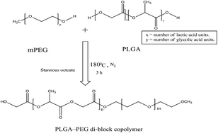
PLGA (500 mg) and mPEG (100 mg) were weighed and dissolved in a freshly distilled toluene (7.5 mL). The solution was poured in 250 mL round bottom flask with continuous stirring, using water-cooled condenser, and the inert environment was maintained using nitrogen gas. Stannous octoate (1 mg) was slowly added drop wise to the round bottom flask, which was being heated in an oil bath at 114 °C, with continuous stirring at 250 rpm for 8 h. The solvent was evaporated under reduced pressure and the residue was dissolved in 10 mL of DCM, then the solution was filtered, and the filtrate was added to stirred water (50 mL) at 60 °C. After the evaporation of DCM from the emulsion, the solid product was isolated from the aqueous phase by the process of decantation, as shown in Fig. 1.24
image file: c6ra10874a-f1.tif
Fig. 1 Schematic representation of PLGA–PEG di-block copolymer.

2.3 Characterization of PLGA–PEG copolymer

The structural investigation of the synthesized compound was done by Fourier Transform Infrared Spectroscopy (FT-IR). For FT-IR spectrum, the samples were mixed thoroughly with potassium bromide and punched to a tablet employing hydraulic press. The FT-IR data were recorded with FT-IR Spectrometer at a wave number range of 4000 cm−1 to 400 cm−1. The structure of PLGA–PEG copolymer was also confirmed by 1H-NMR spectrum. The NMR spectrum was recorded in deuterated chloroform (CDCl3) on Avance II 400 NMR Spectrometer (M/S Bruker Bio Spin Corporation, Billerica, USA).

2.4 Determination of critical micelle concentration (CMC)

The CMC value of the copolymer was determined using pyrene as an extrinsic probe. Briefly, pyrene (4 mg) pre-dissolved in DCM (10 mL), was added to a test tube and the solvent was evaporated. Deionized water (5 mL) was added and different amounts of PLGA–PEG copolymer dissolved in acetone (5 mL solution) was added to the aqueous phase. The solution was incubated at room temperature with mild stirring for complete evaporation of the organic solvent to induce the formation of micelles. The fluorescence intensity of the solution was measured at room temperature using a spectrofluorometer (LS 55 Fluorescence Spectrometer 230V, M/s Liantrisant, Talbot Green UK). The concentration of pyrene was fixed at 40.45 μg mL−1 and the final concentration of PLGA–PEG copolymer was varied from 1 μg mL−1 to 100 μg mL−1. Excitation spectra were obtained at 335 nm and 332 nm, which were determined from the scanning excitation spectrum of each sample from 300 to 600 nm, fixing the emission wavelength at 374 nm. The CMC was reported as the concentration, corresponding to the sharp increase in the ratio of intensity at 335 nm to intensity at 332 nm.25

2.5 Preparation of drug-loaded PLGA–PEG based polymeric micelles

TAM-loaded PLGA–PEG based polymeric micelles were prepared by modified solvent-dialysis method. In brief, 10 mg of the copolymer and 1 mg of TAM were dissolved in 1 mL of DMSO/DMF (3[thin space (1/6-em)]:[thin space (1/6-em)]1 v/v). Deionized water (10 mL) was slowly added, to the system with continuous stirring for 15 minutes. The organic solvent was removed by dialysis against deionized water for 12 h. Finally, the TAM-loaded polymeric micelles were filtered through a syringe filter (0.45 mm pore size, Millipore, USA) to remove unloaded TAM and filtrate. The obtained micellar dispersion was subjected to further characterization studies.26

2.6 Determination of entrapment efficiency and drug loading

Freeze dried polymeric micelles were packed in the dialysis membrane, which were hermetically sealed from both sides. Packed dialysis bag was dipped in the 25.0 mL of methanol for 2 hours. Dialysis bag was opened after 2 hours and analysed for entrapped drug using RP-HPLC. In brief, the RP-HPLC conditions were as: liquid chromatography was conducted on Shimadzu RP-HPLC (LC-2010 C HT, M/s Schimadzu Co., Ltd., Chiyoda-ku, Tokyo, Japan) equipped with an quaternary pump, a UV-visible detector and a HIBAR 250-4.6 mm, Lichrospher60 RP-select B (5 μm) column (M/s. Merck KGaA, Germany) at room temperature. The mobile phase for TAM determination was consisted of a mixture of acetonitrile (ACN) and potassium phosphate (50 mM, pH 3.0) (45.55% v/v), the flow rate was 2.0 mL min−1 and the detection wavelength was set at 254 nm. The injection volume was fixed as 5 μL.27

2.7 Characterization of TAM-loaded PLGA–PEG polymeric micelles

2.7.1 Particle size distribution, ζ-potential and morphological characterization. Particle size of the developed micellar system was determined using Malvern Zetasizer (M/s Malvern Instruments Co., Worcestershire, UK). The average value of three measurements for the sample was reported as the final result. For zeta-potential measurements, the same equipment was employed. For morphological studies, transmission electron microscopic (TEM) analysis was performed, using Hitachi H-7500 TEM, installed at Central Instrumentation Laboratory (CIL), Panjab University, Chandigarh, India, and microphotographs were clicked at suitable magnifications. For TEM, 1 mg of sample was dispersed in 1 mL of saline and the solutions were sonicated for 10 minutes and 2 drops were deposited on copper grids. Finally, the grid was air dried, before the initiation of the TEM experiment.

2.8 In vitro drug release studies

TAM-loaded micellar dispersion equivalent to 1 mg of TAM was sealed in dialysis bag, with molecular weight cut-off of 12[thin space (1/6-em)]000–14[thin space (1/6-em)]000 Da. The sealed bag was suspended in 20 mL solution of ethanol[thin space (1/6-em)]:[thin space (1/6-em)]PBS 5.6 pH (1[thin space (1/6-em)]:[thin space (1/6-em)]9 v/v). The system was stirred at 100 rpm at 37 °C. At predetermined time intervals, of 0, 0.25, 0.5, 0.75, 1, 1.5, 2, 4 and 6 hours, samples of 1 mL each, were withdrawn and replaced with equivalent volume of fresh diffusion medium. The amount of TAM in the release medium was quantified by UV-visible spectrophotometer at a wavelength of 301 nm (UVS-2700, Spectro UV-vis Dual beam, Labomed Inc., Los Angeles, USA).

2.9 In vitro cytotoxicity assays

Human breast cancer MCF-7 cells were used for this process. Briefly, 6 × 10³ cells were seeded in 96 well tissue-culture plates. Different concentrations of test samples (equivalent to 1 μg mL−1 and 10 μg mL−1 of TAM) were added to cells, and the control cells were treated with placebo samples. Plates were incubated at 37 °C for 48 h using CO2 incubator. To the incubated plates, MTT solution (5 mg mL−1), 20 μL, was added and kept for gentle stirring. Plates were incubated for 4 h and then centrifuged at 400 × g for 15 min. The resulting MTT formazan crystals were dissolved in DMSO, which were collected by discarding supernatant. The optical density was recorded at 570 nm, and 620 nm as reference wavelength with the help of microplate reader. The cells were visualized under an Olympus 1 × 70 inverted microscope for morphological changes.2,28 The microphotographs were clicked one out of three similar experiments (30×)2.

2.10 Ex vivo dermatokinetic studies

For dermatokinetic studies, excised skin of Laca mice was used. The skin was mounted on the Franz diffusion cell (M/s PermeGear, Inc., PA, USA).29 Formulation (equivalent to 1 mg of TAM) and plain drug (1 mg) were charged in the donor compartment on the skin. Plain TAM was dispersed in 1 mL of PBS 5.6 and was poured on to the surface of skin in the donor compartment by means of a syringe. Analogously, the TAM-loaded micellar dispersion, equivalent to 1 mg of TAM, was applied on to the skin in the donor compartment. The sink was composed of 30.0 mL of 9[thin space (1/6-em)]:[thin space (1/6-em)]1 v/v solution of PBS 5.6 and ethanol. The setup was maintained at 35 ± 2 °C and was continuously stirred. At specified time intervals, the skin was removed from the Franz cell. The epidermis and dermis were harvested, after removed of adhered drug/formulation. Each layer was cut into pieces and digested in method for 12 hours for complete drug extraction. After filtration, the protein was precipitated by addition of ether and samples were diluted with ACN. The resultant mixture was filtered and drug estimation was performed by HPLC. One compartment open body model approach was employed for dermatokinetic treatment to fetch [AUC]6 h0, first order permeation rate constant (Kp), first order elimination rate constant (Ke) and maximum dermal/epidermal concentration (Cmax).29,30

3. Results and discussion

3.1 Characterization of PLGA–PEG co-polymer

The FT-IR spectrum obtained was consistent with the structure of the expected copolymer, as shown in Fig. 2. It showed that absorption band at 3659.5 cm−1 was ascribable to terminal hydroxy groups. The band observed at 2943.7 cm−1 was due to C–H stretch of CH3 and the one at 1762.3 cm−1 could be assigned to C[double bond, length as m-dash]O stretch. Absorption at 1186.2 cm−1 and 1098.9 cm−1 were due to C–O stretch. The 1H-NMR spectrum of PLGA–PEG co-polymer is shown as Fig. 3. The prominent peak at 3.651 ppm corresponding to the methylene groups of mPEG was conspicuous. The overlapping doublets at 1.55 ppm were attributed to the methyl groups of the D- and L-lactide repeat units, the multiplets at 5.2 and 4.8 ppm corresponded to the lactide CH and glycolide CH2, respectively, and peak at 7.270 ppm corresponded to CDCl3. Both the spectra confirmed the successful conjugation of PLGA and PEG.
image file: c6ra10874a-f2.tif
Fig. 2 FT-IR spectrum of PLGA–PEG copolymer.

image file: c6ra10874a-f3.tif
Fig. 3 1H-NMR spectrum of PLGA–PEG co-polymer.

3.2 Critical micelles concentration (CMC) study

The synthesized polymer was assessed for its CMC using fluorescence spectrophotometry. Pyrene was employed as the fluorescent probe. Self-assembled copolymers are known to form micelles, when their concentration reaches CMC. Beyond CMC, pyrene gets solubilized into the hydrophobic micellar core, resulting in a sharp increase in fluorescence intensity. The CMC value of PLGA–PEG copolymer was reported by extrapolating the fluorescence intensity ratio (I335/I332) versus polymer concentration. The observed CMC value was 9.4 μg mL−1, as shown in Fig. 4. The findings are in close agreement with the reported values of CMC of similar polymers.2
image file: c6ra10874a-f4.tif
Fig. 4 Plot of the fluorescence intensity ratio I335/I332 from pyrene excitation spectrum as a function of the PLGA–PEG co-polymer concentration.

3.3 Particle size distribution, ζ-potential and morphological characterization

The average size of blank co-polymeric micelle was observed to be 68.2 ± 2.8 nm, but drug entrapped co-polymeric micelle were observed to be in size range of 76.4 ± 2.1 nm. The possible reason for increased in particle size may be the incorporation of lipophilic drug, TAM.2 The zeta potential values of the plain and drug-loaded micelles were observed to be −4.49 mV and −4.89 mV. The PDI values for both the both the systems were below 0.3. Similar findings have been reported by various researchers employing analogous carriers.2,26,31

3.4 Entrapment efficiency and drug loading

The observed entrapment efficiency and drug loading of TAM in the developed micelles was 60.86% ± 3.21% and 6.02% ± 0.12, respectively. This value indicated the appreciable drug loading capabilities of designed polymer. Substantial drug loading assured the adequate encapsulation of drug in the designed nanocarriers.

3.5 Transmission electron microscope (TEM)

The TEM image at 40[thin space (1/6-em)]000× magnification confirmed the formation of spherical and turgid micelles, as shown in Fig. 5. The image is devoid of any sign of aggregation, indicating the non-adherent nature of the micelles with each other.
image file: c6ra10874a-f5.tif
Fig. 5 TEM photograph of TAM-loaded PLGA–PEG co-polymeric micelles.

3.6 In vitro drug release

The in vitro drug release profile of the TAM-loaded PLGA–PEG copolymeric micelles is shown in Fig. 6. It can be seen from the figure that the drug release profile followed a biphasic pattern, with a small initial burst release, followed by a zero-order release. The initial fast release can be attributed to release of un-entrapped TAM and the molecules located near the outer surface of micelles; however, the zero order controlled release may be due to the inner located TAM molecules.32
image file: c6ra10874a-f6.tif
Fig. 6 In vitro release profile of TAM from PLGA–PEG TAM-loaded micelles.

3.7 In vitro cell cytotoxicity study

The cell viability on MCF-7 was assessed by the MTT assay. Naïve PLGA–PEG copolymeric micelles did not show significant toxicity towards the cells both at the conc. of 1 μg mL−1 and 10 μg mL−1, indicating that PLGA–PEG di-block co-polymeric micelles were nontoxic to tumour cells. However, plain TAM exhibited anticancer activity with approx. 70% growth inhibition on MCF-7 cell lines at the studied concentrations. On the other hand, there was marked increase in the cytotoxicity of TAM, after the loading with PLGA–PEG co-polymeric micelles at studied concentrations, as drawn in Fig. 7 and 8. This result suggested that the enhanced cytotoxicity in MCF-7 cells might be due to increased concentration of TAM.4
image file: c6ra10874a-f7.tif
Fig. 7 Bar diagram depicting the cancer cell cytotoxicity on MCF-7 cell lines offered by various treatments.

image file: c6ra10874a-f8.tif
Fig. 8 Microphotographs indicating the effect of various treatments on the morphological symmetry of MCF-7 cells.

3.8 Dermatokinetic studies

The results of dermatokinetic studies have been shown in Table 1, Fig. 9 and 10. The bioavailability of drug in epidermis was enhanced by approx. 3.47 times vis-à-vis plain drug gel. The skin layer permeation constants were of higher magnitudes in the skins treated with TAM-loaded micelles to that of plain drug gel. This indicated better permeation of drug, when PLGA–PEG-based micelles were employed. The drug elimination was also decreased to that of plain drug. The results of dermatokinetic unequivocally demonstrated the better delivery potential of PLGA–PEG-based micelles for topical delivery of TAM.
Table 1 Various dermatokinetic parameters of TAM topical formulations in epidermis and dermis of Laca mice (n = 3)
Dermatokinetic parameter TAM-gel TAM-micelles
Epidermis Dermis Epidermis Dermis
AUC0–6 h (μg cm−2 h) 119.92 89.63 549.91 311.26
Kp (h−1) 0.91 4.05 2.67 5.10
Ke (h−1) 0.301 0.378 0.138 0.246
Cmax (μg cm−2) 46.03 41.99 189.65 140.51


image file: c6ra10874a-f9.tif
Fig. 9 Graph showing the amount of drug present in the dermis and epidermis of Laca mice at various time points.

image file: c6ra10874a-f10.tif
Fig. 10 Graph showing the amount of drug TAM gel present in the dermis and epidermis of Laca mice at various time points.

4. Conclusions

The current studies demonstrated the merits of PLGA–PEG-based polymeric micelles in delivery of TAM to breast cancer cells, enhancement of haemo-compatibility and better dermatokinetic distribution. The concerns of poor tissue availability, faster drug clearance, compromised efficacy and biocompatibility have been reasonably taken care. The approach haves a path for the development of safer and effective topical products of TAM for the management of initial breast cancers. In conclusion, the approach and methodology can be rationally explored for other similar drugs.

Conflict of interests

None conflict of interest has been declared by the authors.

Acknowledgements

The kind support to the corresponding author in the form of UGC startup project from UGC, New Delhi is highly acknowledged. Generous gift samples of PLGA from M/s Evonik Industries AG, Kirchenalle, Germany is also acknowledged. The authors acknowledge the unconditional support from Dr Shashi Bhushan and Mr Santosh Kumar Guru from IIIM, Jammu, India for the conduct of MTT cytotoxicity assay.

References

  1. C. De Martel, J. Ferlay, S. Franceschi, J. Vignat, F. Bray, D. Forman and M. Plummer, Lancet Oncol., 2012, 13, 607–615 CrossRef PubMed.
  2. K. Raza, N. Kumar, C. Misra, L. Kaushik, S. K. Guru, P. Kumar, R. Malik, S. Bhushan and O. Katare, Int. J. Biol. Macromol., 2016, 88, 206–212 CrossRef CAS PubMed.
  3. P. Kumar, K. Raza, R. Malik, S. Arora and O. P. Katare, Curr. Pharm. Des., 2016, 22, 1 CrossRef.
  4. K. Raza, N. Thotakura, P. Kumar, M. Joshi, S. Bhushan, A. Bhatia, V. Kumar, R. Malik, G. Sharma and S. K. Guru, Int. J. Pharm., 2015, 495, 551–559 CrossRef CAS PubMed.
  5. A. Jemal, R. Siegel, E. Ward, Y. Hao, J. Xu, T. Murray and M. J. Thun, Ca-Cancer J. Clin., 2008, 58, 71–96 CrossRef PubMed.
  6. K. Raza, M. Kumar, P. Kumar, R. Malik, G. Sharma, M. Kaur and O. Katare, BioMed Res. Int., 2014, 2014, 406731 Search PubMed.
  7. P. M. Ravdin, S. Green, T. M. Dorr, W. L. McGuire, C. Fabian, R. P. Pugh, R. D. Carter, S. E. Rivkin, J. R. Borst and R. J. Belt, J. Clin. Oncol., 1992, 10, 1284–1291 CAS.
  8. V. Assikis, P. Neven, V. Jordan and I. Vergote, Eur. J. Cancer, 1996, 32, 1464–1476 CrossRef.
  9. A. J. Wood and C. K. Osborne, N. Engl. J. Med., 1998, 339, 1609–1618 CrossRef PubMed.
  10. F. Hu, K. Neoh and E. Kang, Biomaterials, 2006, 27, 5725–5733 CrossRef CAS PubMed.
  11. G. De Lima, G. Facina, J. Shida, M. Chein, P. Tanaka, R. Dardes, V. Jordan and L. Gebrim, Eur. J. Cancer, 2003, 39, 891–898 CrossRef CAS PubMed.
  12. F. Peyrade, M. Frenay, M. C. Etienne, F. Ruch, C. Guillemare, E. Francois, M. Namer, J. M. Ferrero and G. Milano, Clin. Pharmacol. Ther., 1996, 59, 401–410 CrossRef CAS PubMed.
  13. S.-C. Shin and J.-S. Choi, Anti-Cancer Drugs, 2009, 20, 584–588 CrossRef CAS PubMed.
  14. P. Voltairas, D. Fotiadis and L. Michalis, J. Biomech., 2002, 35, 813–821 CrossRef CAS PubMed.
  15. R. Plummer, R. Wilson, H. Calvert, A. Boddy, M. Griffin, J. Sludden, M. Tilby, M. Eatock, D. Pearson and C. Ottley, Br. J. Cancer, 2011, 104, 593–598 CrossRef CAS PubMed.
  16. G. Cavallaro, L. Maniscalco, M. Licciardi and G. Giammona, Macromol. Biosci., 2004, 4, 1028–1038 CrossRef CAS PubMed.
  17. M. Licciardi, G. Giammona, J. Du, S. P. Armes, Y. Tang and A. L. Lewis, Polymer, 2006, 47, 2946–2955 CrossRef CAS.
  18. M. Licciardi, E. F. Craparo, G. Giammona, S. P. Armes, Y. Tang and A. L. Lewis, Macromol. Biosci., 2008, 8, 615–626 CrossRef CAS PubMed.
  19. M. Licciardi, G. Cavallaro, M. Di Stefano, G. Pitarresi, C. Fiorica and G. Giammona, Int. J. Pharm., 2010, 396, 219–228 CrossRef CAS PubMed.
  20. M. Ma, F. Li, X.-h. Liu, Z.-f. Yuan, F.-j. Chen and R.-x. Zhuo, J. Mater. Sci.: Mater. Med., 2010, 21, 2817–2825 CrossRef CAS PubMed.
  21. S. Croy and G. Kwon, Curr. Pharm. Des., 2006, 12, 4669–4684 CrossRef CAS PubMed.
  22. H. K. Makadia and S. J. Siegel, Polymers, 2011, 3, 1377–1397 CrossRef CAS PubMed.
  23. M. Ashjari, S. Khoee and A. R. Mahdavian, Polym. Int., 2012, 61, 850–859 CrossRef CAS.
  24. Y.-P. Li, Y.-Y. Pei, X.-Y. Zhang, Z.-H. Gu, Z.-H. Zhou, W.-F. Yuan, J.-J. Zhou, J.-H. Zhu and X.-J. Gao, J. Controlled Release, 2001, 71, 203–211 CrossRef CAS PubMed.
  25. H. S. Yoo and T. G. Park, J. Controlled Release, 2001, 70, 63–70 CrossRef CAS PubMed.
  26. J. Huang, H. Zhang, Y. Yu, Y. Chen, D. Wang, G. Zhang, G. Zhou, J. Liu, Z. Sun and D. Sun, Biomaterials, 2014, 35, 550–566 CrossRef CAS PubMed.
  27. K.-H. Lee, B. A. Ward, Z. Desta, D. A. Flockhart and D. R. Jones, J. Chromatogr. B: Anal. Technol. Biomed. Life Sci., 2003, 791, 245–253 CrossRef CAS.
  28. K. Raza, D. Kumar, C. Kiran, M. Kumar, S. K. Guru, P. Kumar, S. Arora, G. Sharma, S. Bhushan and O. Katare, Mol. Pharm., 2016 DOI:10.1021/acs.molpharmaceut.6b00183.
  29. K. Raza, B. Singh, S. Singla, S. Wadhwa, B. Garg, S. Chhibber and O. P. Katare, Mol. Pharm., 2013, 10, 1958–1963 CrossRef CAS PubMed.
  30. G. Sharma, G. Dhankar, K. Thakur, K. Raza and O. Katare, AAPS PharmSciTech, 2015, 1–11 Search PubMed.
  31. M. Licciardi, C. Scialabba, C. Sardo, G. Cavallaro and G. Giammona, J. Mater. Chem. B, 2014, 2, 4262–4271 RSC.
  32. A. Bhatia, B. Singh, S. Wadhwa, K. Raza and O. P. Katare, Pharm. Dev. Technol., 2014, 19, 160–163 CrossRef CAS PubMed.

This journal is © The Royal Society of Chemistry 2016