One-pot synthesis of sulfur-doped graphene quantum dots as a novel fluorescent probe for highly selective and sensitive detection of lead(II)

Shiyue Bian a, Chao Shena, Hong Huab, Lin Zhoua, Hailin Zhua, Fengna Xia, Jiyang Liu*a and Xiaoping Dong*a
aDepartment of Chemistry, Zhejiang Sci-Tech University, Hangzhou, 310018, China. E-mail: liujy@zstu.edu.cn; xpdong@zstu.edu.cn
bComprehensive Technology Center, Nantong Entry-Exit Inspection and Quarantine Bureau of the P. R. C., Nantong, Jiangshu 226004, China

Received 27th April 2016 , Accepted 6th July 2016

First published on 8th July 2016


Abstract

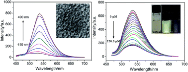
A novel one-pot synthesis of sulfur-doped graphene quantum dots (S-GQDs) was proposed based on water-phase molecular fusion with 1,3,6-trinitropyrene, Na2S, and NaOH in a hydrothermal process. A 75% yield was obtained and mass production of S-GQDs with high crystallinity was possible. The prepared S-GQDs gave a stable yellow-green emission within a wide pH range of 2.0–11.0 and exhibited excitation-independent photoluminescence behaviors. As investigated by atomic force microscopy (AFM), the synthesized S-GQDs possessed monolayer-graphene thickness. As illustrated by transmission electron microscopy (TEM), the synthesized S-GQDs exhibited high crystallinity and uniform size (∼3 nm). Successful doping of S atoms in graphene quantum dot lattices was proven by X-ray photoelectron spectroscopy (XPS) characterization. Compared with GQDs, the S-GQDs had drastically changed surface chemistry and showed a selective and sensitive response to Pb2+. Ions such as Na+, K+, Cu2+, Ca2+, Mg2+, Zn2+, Fe3+, Ni2+, Co2+, Cd2+ have no effect on the fluorescence of S-GQDs. Based on the fluorescence quenching of S-GQDs by Pb2+ in water, a facile and direct fluorescence sensor for Pb2+ detection was developed. Under the optimized conditions, the linear response ranged from 0.1 to 140.0 μM with a detection limit of 0.03 μM.


1. Introduction

Nowadays, poisonous transition metal ions in water and soils have aroused worldwide attention due to the adverse effects on ecosystems caused by their non-biodegradability and toxicity.1,2 The lead(II) ion (Pb2+) is one of the most poisonous substances to animals and humans, which damages the central nervous system and causes brain and blood disorders. To prevent lead poisoning, developing easy, fast and sensitive methods for Pb2+ detection is of great significance. In spite of traditional methods including graphite furnace atomic absorption spectroscopy (AAS) and inductively coupled plasma mass spectroscopy (ICP-MS), fluorescent sensors have witnessed immense progress in recent years because they do not need highly expensive and complex instruments, and well-trained personnel.3,4

Recently, fluorescent carbon nanomaterials including carbon quantum dots (CQDs) and graphene quantum dots (GQDs) have been attractive in bioimaging,5 matrix/catalysis for deposition of surface-clean metal nanoparticles,6,7 and sensing detection,5,8–10 and etc. Resulting from the pronounced quantum confinement and edge effects, zero-dimensional (0D) graphene quantum dots (GQDs) exhibited extraordinary optical and electrical characteristics. Compared with semiconductor quantum dots (e.g. CdS, CdSe, CdTe), GQDs possess low cost, low toxicity and stable photoluminescence properties. Consequently, fluorescent chemosensors with GQDs as fluorescent probe show promising potential for the detection of metal ions owing to their intrinsic sensitivity, selectivity and capacity for rapid, real-time monitoring.11–14,16 As recently demonstrated, the invasion of heteroatoms into the perfect hexagonal carbon sheet of GQDs serves as useful tool to bring new optical properties and selectivity. The structural, physicochemical and electronic properties of GQDs, such as charge transport, Fermi level, bandgap, localized electronic state and spin density, can be drastically altered by heteroatom doping.15–17 Amongst various heteroatoms (oxygen, boron, nitrogen, phosphor, sulfur, etc.), sulfur atom (S) doping is distinctive because the mismatch of the outermost orbitals of S and C induces an on-uniform spin density distribution, which consequently endows S-doped materials with unique properties and potential for many applications. Unlike B, N and P, S atom is larger than C atom and the C–S bond length (1.78 Å) is 25% longer than that of the C–C bond. Also, the difference of electronegativity between S (2.58) and C (2.55) appears to be too small to offer significant polarization (charge transfer) in C–S composites. Consequently, chemical doping of S atoms into the framework of GQDs appears to be very difficult. Until now, only few reports cover GQDs or CQDs with sulfur atoms doping. In term of S-CQDs, Kwon et al. synthesized S-CQDs by pyrolyzing glucose with thioglycolic acid being sulfur source.18 Xu et al. reported the preparation of S-CQDs with sodium citrate and sodium thiosulfate by a hydrothermal method.19 For S-doped or co-doped GQDs, Li et al. prepared S-GQDs by electrolysis of graphite in sodium p-toluenesulfonate medium.20 Li et al. prepared S-GQDs by a hydrothermal method using fructose and sulphuric acid as specific source materials.21 Qu et al. synthesized S, N co-doped graphene quantum dots (S, N-GQDs) using hydrothermal synthesis route with citric acid as the C source and urea or thiourea as N and S sources.22 Thus, developing a facile, low-cost and high yield method for the preparation of S-GQDs with high crystal quality is highly desired. Moreover, it is also necessary to further explore novel properties and promising applications of S-GQDs.

In this paper, a facile approach was developed to prepare the S-doped GQDs (S-GQDs) by using water-phase molecular fusion with 1,3,6-trinitropyrene, Na2S, and NaOH in one-step hydrothermal process. The method is simple, effective and mass production of the S-GQDs with high crystallinity is possible. Excitation-independent yellow-green fluorescence of the S-GQDs could be sensitively quenched in the presence of Pb2+ ions, whereas it was non-sensitive to other metal ions, such as Na+, K+, Cu2+, Ca2+, Mg2+, Zn2+, Fe3+, Ni2+, Co2+, Cd2+. Such selectivity was not observed for the control sulfur-free GQDs. Thus, sensitive detection of Pb2+ ion by sulphur doped GQDs was realized. The proposed preparation strategy and the main characteristic features were described and discussed in detail.

2. Experimental section

2.1 Materials and reagents

Pyrene, Na2S, NaOH, Na2CO3, NaHCO3, NaH2PO4 and Na2HPO4 were obtained from Aladdin Chemistry Co. Ltd. (China). Aqueous solutions of Na+, K+, Cu2+, Ca2+, Mg2+, Zn2+, Fe3+, Ni2+, and Co2+ were prepared from their chloride salts. Aqueous solutions of Cd2+ and Pb2+ were prepared from their nitrate salts. All chemicals used in our work were of analytical grade and used without further treatment. All aqueous solutions were prepared with ultrapure water (18.2 MΩ cm, Milli-Q, Millipore).

2.2 Instrumentation

Transmission electron microscopic (TEM) photograph was taken on a JEM-2100 transmission electron microscope (JEOL Ltd., Japan) at operating voltage of 200 kV. Atomic force microscopic (AFM) images were obtained by Nanascopy IVA system for height characterization (Digital Instruments Inc, USA). Elemental analysis of the S-GQDs was performed by X-ray photoelectron spectroscopy (XPS) with PHI5300 electron spectrometer using 250 W, 14 kV, Mg Kα radiation (PE Ltd., USA). UV-vis absorption and fluorescence spectra were recorded on UV-2450 spectrophotometry (Shimadzu Corporation, Japan) and RF-5301PC spectrofluorometer (Shimadzu Corporation, Japan).

2.3 Preparation of S-GQDs

Using pyrene as the initial substrate, 1,3,6-trinitropyrene was synthesized according to the literature.23 Then, 1,3,6-trinitropyrene (2.0 mg mL−1) were dispersed in NaOH (0.2 M) solution containing Na2S (0.1 M) to form trinary mixture (1,3,6-trinitropyrene, NaOH, Na2S) by ultrasonic treatment for 1 h. The obtained suspension was transferred into a 50 mL Teflon-lined autoclave and heated at 200 °C for 10 h. After being cooled to room temperature, a brown-black solution containing no solid precipitation was obtained and dialysed in a dialysis bag (retained molecular weight 1000 Da) for 2 days to remove ions and small organic molecules (e.g. sodium salt and unfused molecules). After being further dialyzed in a dialysis bag with retained molecular weight of 3500 Da for 24 h to remove larger impurities, the obtained S-GQDs were freeze dried for further investigation. When the trinary source materials for S-GQDs were changed to binary 1,3,6-trinitropyrene + NaOH or 1,3,6-trinitropyrene + Na2S under otherwise identical conditions, the obtained GQDs was named as S-free or S-control samples for comparison.

2.4 Measurement of quantum yield (QY)

QY was determined by using rhodamine 6G as the standard sample and was calculated according to the following equation:
image file: c6ra10836a-t1.tif
where Yμ is the quantum yield of unknown samples, Ys is the fluorescence quantum yield of standard substance, Fu and Fs respectively stand for the integral fluorescence intensity of the test sample and the standard dilute solution, Aμ and As respectively stand for the maximum absorbance value of the test sample and the standard dilute solution.

2.5 Detection of Pb2+ using S-GQDs as fluorescence probe

The detection of Pb2+ was carried out in phosphate buffer solution (PBS, 3.0 mM, pH 7.0). In a typical process, 0.95 μL of S-GQDs solution (1.0 mg mL−1) were dispersed in PBS (6.0 mL) followed by the addition of different amount of Pb2+ and then the solution was diluted to 10 mL with PBS. Final concentration of Pb2+ ranged from 0.1 to 220.0 μM. The resulting solution was shaken well and incubated for 5 min at room temperature before the fluorescence emission spectrum (excited at 460 nm) was recorded. The detection measurements were performed in triplicate. The relative fluorescence intensity (F0F)/F0 versus Pb2+ concentration were used for calibration. Here, F0 and F are the fluorescence intensities of S-GQDs in the absence and presence of Pb2+, respectively.

3. Results and discussion

3.1 Excitation-independent fluorescence properties of the S-GQDs

The obtained S-GQDs solution was pale-yellow and transparent, as illustrated in the inset of Fig. 1 (the left photograph). It emitted yellow-green fluorescence upon being irradiated by a 365 nm lamp (the right photograph, inset in Fig. 1). With rhodamine 6G as a reference, and based on equation, the quantum yield of the obtained S-GQDs was calculated to be 11.6% at 460 nm excitation wavelength. To further explore the optical properties of the S-GQDs, fluorescence spectroscopy was investigated. When the excitation wavelength changed from 410 nm to 490 nm, the emission all centred at 535 nm. The maximum excitation and emission wavelength at 490 nm and 535 nm was revealed, respectively. These results confirmed an excitation-independent fluorescence of S-GQDs. The single-emission fluorescence with excitation-independence indicated high uniformity of S-GQDs. For comparison, the S-free sample and the S-control sample were also prepared by replacing the ternary source materials for S-GQDs (1,3,6-trinitropyrene + NaOH + Na2S) to binary 1,3,6-trinitropyrene + NaOH (S-free) or 1,3,6-trinitropyrene + Na2S (S-control), respectively. When excited at 365 nm irradiation, almost no fluorescence was observed for S-control sample (Fig. S1 of the ESI). However, S-free sample emitted yellow-green fluorescence that was ascribed to the formation of OH-GQDs according to the literature (Fig. S1 of the ESI).23 These results may explain the addition of NaOH play a significant role in the formation of the S-GQDs.9 A 75% yield was calculated using the dry weight of the obtained S-GQDs as compared with the initial 1,3,6-trinitropyrene. In addition, mass production of the S-GQDs is possible by using Teflon-lined autoclave with large volume.
image file: c6ra10836a-f1.tif
Fig. 1 Fluorescence excitation and emission spectra of the S-GQDs. Fluorescence excitation spectrum was obtained at an emission wavelength of 480 nm. Fluorescence emission spectra were obtained at excitation wavelength ranged from 410 to 490 nm. The wavelength interval between two adjacent spectra was 10 nm. The inset were the photographs of the S-GQD aqueous solution under visible light (left) and UV light of 365 nm (right).

3.2 Structure characterization of the S-GQDs

Fig. 2 showed the TEM image of the prepared S-GQDs. Obviously, the obtained S-GQDs were well-dispersed and relatively uniform in size (about 3 nm). The high resolution TEM (HRTEM) image (inset of Fig. 2A) clearly depicted the good crystallinity of the S-GQDs. The lattice parameter was 0.23 nm, which was similar to (1120) lattice fringes of graphene.20 These phenomena indicated the water-phase molecular fusion with 1,3,6-trinitropyrene, Na2S, and NaOH in hydrothermal process resulted in high quality S-GQDs with almost perfect single crystals. That might be ascribed to the special structure of the initial substrate. Briefly, 1,3,6-trinitropyrene consists of four peri-fused benzene rings with a unique carbon skeleton similar to the primitive cell of graphene.23 Due to three positively charged sites of NO2 groups, enhanced reaction activity of 1,3,6-trinitropyrene has been demonstrated for the preparation of OH-GQDs (1,3,6-trinitropyrene + NaOH as source material) and amine-functionalized GQDs (1,3,6-trinitropyrene + ammonia or 1,3,6-trinitropyrene + hydrazine hydrate as source material) using hydrothermal process.23 Inspired by its specific structure and reaction activity, 1,3,6-trinitropyrene was also used in our work to form the lattice of graphene quantum dots. Na2S was used as the S source to achieve S-doping in molecular fusion of 1,3,6-trinitropyrene in alkaline NaOH medium. XPS measurements were applied to investigate the composition of the obtained S-GQDs (Fig. 3). The XPS survey spectrum of the S-GQDs (Fig. 3A) clearly displayed the presence of C, O, and S with atomic percentages of 61.7%, 37.0% and 1.3%, respectively. Those results confirmed that the successful S-doping via the one-step hydrothermal process. The C1s spectrum of S-GQDs (Fig. 3B) could be fitted into three peaks, which were assigned to C–O at 286.8 eV, C[double bond, length as m-dash]C at 282.6 eV and C–S at 285.4 eV, respectively.20 The O1s spectrum of S-GQDs (Fig. 3C) stated that there was a peak at 531.2 eV, which corresponded to C–OH or C–O–C.23 Thus, the obtained S-GQDs were rich in oxygen-containing groups on the surfaces, suggesting good water-dispersibility. The S2p spectrum of S-GQDs (Fig. 3D) could be fitted into three peaks, which were assigned to –C–SOx–C (x = 2, 3, and 4) sulphone bridges at 167.7 eV, 168.5 eV and 169.3 eV, respectively.24,25 Thus, S atoms were indeed covalently bonded to the framework of GQDs. In brief, 1,3,6-trinitropyrene has a mother nucleus structure similar to that of graphene. The nitro groups have a strong electrophilicity, and the addition reaction can occur with electron-rich groups. Sulfur ions in Na2S and hydroxide ions in NaOH reacted with nitro sites pyrene ring in the hydrothermal reaction to achieve the synthesis of S-doped GQDs.
image file: c6ra10836a-f2.tif
Fig. 2 (A) TEM and (B) HRTEM images of the as-prepared S-GQDs. The number indicated in (B) was the lattice parameter of S-GQDs.

image file: c6ra10836a-f3.tif
Fig. 3 (A) XPS survey spectrum, high-resolution (B) C1s spectrum, (C) O1s spectrum and (D) S2p spectrum of the S-GQDs.

AFM image in Fig. 4 showed the topographic morphology of S-GQDs. As illustrated in the inset, the heights of S-GQDs were similar and between 0.7 and 0.8 nm. The average height was 0.75 nm, indicating that the fabricated S-GQDs consisted of single graphene layer. Compared with the theoretical height of plain graphene, the increase of topographic height might be ascribed to the presence of rich oxygen-containing and sulfur-containing groups.


image file: c6ra10836a-f4.tif
Fig. 4 AFM images of the S-GQDs. Inset was the height profile along the blue line.

3.3 Fluorescence quenching of S-GQDs by Pb2+

To achieve a fluorescence sensor for metal ion, the effect of pH on the fluorescence of S-GQDs was firstly investigated. The pH value ranged from 2.0 to 11.0 (Fig. 5A). Results showed that the fluorescence intensity of S-GQDs increased dramatically with the increase of pH value in acid medium. When pH was above 7.0, the fluorescence intensity became stable (Fig. S2). The phenomena might attribute to the change of surface charge density resulting from the ionization of oxygen-containing groups.
image file: c6ra10836a-f5.tif
Fig. 5 (A) The effect of pH value on the fluorescence intensity of the S-GQDs. (B) The change ratio of fluorescence intensity (F/F0) in the presence of various metal ions (140.0 μM). (C) The effect of pH value on the response of S-GQDs to Pb2+ (140.0 μM). (D) Time-dependent fluorescence response of S-GQDs aqueous solutions upon the addition of Pb2+ (95.0 μM).

To assess the selectivity for metal ions, the fluorescence of S-GQDs upon mixing with different metal ions were investigated. The chosen ions were mainly related to environment including Na+, K+, Cu2+, Ca2+, Mg2+, Zn2+, Fe3+, Ni2+, Co2+, Cd2+ and Pb2+. The concentration of each metal ion was all controlled at 140.0 μM. When excited under UV light (365 nm), the photographs of S-GQDs aqueous solution mixed with different ions illustrated the selectivity (Fig. S3 of the ESI). Compared to other metal ions, Pb2+ significantly quenched the fluorescence of S-GQDs, indicating efficient selectivity of S-GQDs for Pb2+ over other cations. The change of fluorescence intensity was quantitatively investigated. Fig. 5B showed the difference of fluorescence intensity ratio (F/F0) of the S-GQDs solution in the absence and presence of various metal ions. Notably, only Pb2+ ions gave apparent quenching effect on the fluorescence of S-GQDs, and the effect of other tested metal ions can be neglected. The high selectivity of S-GQDs for Pb2+ could be due to the fact that Pb2+ ion might have a higher binding affinity with S and O functional groups of S-GQDs than other transition-metal ions. For comparison, fluorescence detection of Pb2+ using sulfur-free GQDs (prepared without Na2S) was studied (Fig. S4 of the ESI). However, the fluorescence intensity was almost un-changed at the presence of 140.0 μM Pb2+ at UV irradiation (365 nm). Therefore, the S dopant in the GQDs structure promoted the selectivity of GQDs towards Pb2+.

The effects of detection pH and the kinetic behavior on the reaction were further studied to optimize the detection conditions for Pb2+. To avoid the hydrolysis and precipitation of Pb2+, weakly acidic and neutral solution was investigated. As illustrated in Fig. 5C, the fluorescence quenching was pH-dependent. Obviously, Pb2+ showed the best quenching efficiency at pH 7.0. Thus, the neutral PBS (pH 7.0) buffer was chosen for further investigation. The kinetic behavior of reactions between the S-GQDs and Pb2+ was explored by plotting the values (F0F)/F0 as a function of time. It was evident that the quenching equilibrium reached in less than 5 min (Fig. 5D), suggesting the fast interaction between S-GQDs and Pb2+. Thus, 5 min was chosen as reaction time in the following experiments.

3.4 Fluorescence detection of Pb2+ using S-GQDs

Under the optimized conditions, a quantitative detection analysis of Pb2+ was carried out. As presented in Fig. 6, the fluorescence intensity of the S-GQDs decreased dramatically when Pb2+ concentration increased from 0.1 to 220.0 μM. There were two good linear correlation existed in the values of (F0F)/F0 as the Pb2+ concentration from 0.1 to 1.0 μM and 1.0 to 140.0 μM (inset of Fig. 6), giving calibration curves with linear regression equations of (F0F)/F0 = 0.0048C + 0.0538, and (F0F)/F0 = 0.0613C + 0.0019, respectively. Where F0 and F correspond to the fluorescence intensity of the S-GQDs in the absence and presence of Pb2+ ions, C is the concentration of Pb2+ ions. The detection limit was calculated to be 0.03 μM at a signal-to-noise ratio of 3, which was lower than most of the previous reported assays for Pb2+ detection.1–4,11,26–33 Table 1 listed the comparison of various fluorescent probes for Pb2+ detection, the detection limit (DL) obtained with the present method was lower than those achieved by organic fluorescent probes including rhodamine derivative,1 phenazine derivative,27 naphthalene derivative,4 pyridine derivative,11 thrombin aptamer and pyrene probe system,30 CPA-8-HQL functionalized SBA-15[thin space (1/6-em)]33 and thrombin aptamer and pyrene probe, but higher than those attained from organic fluorescent probes of BODIPY,28 DNA based system including G-quadruplex and graphene oxide composite,3 G-rich DNA and perylene derivative,2 graphene oxide and gold nanoparticles29 and semiconductor quantum dots-based system including ZnS@SiO2 QDs,34 core–shell CdSe/CdS QDs,35 3-mercaptopropionic acid functionalized CdTe QDs,36 CuInS2 QDs37 and DNA functionalized CdS QDs.38
image file: c6ra10836a-f6.tif
Fig. 6 (A) Fluorescence spectra of S-GQDs upon addition of different concentration of Pb2+ (from top to bottom: 0, 0.1, 0.3, 0.5, 0.8, 1.0, 5.0, 10.0, 20.0, 30.0, 40.0, 50.0, 60.0, 70.0, 80.0, 90.0, 100.0, 110.0, 120.0, 130.0, 140.0, 150.0, 160.0, 170.0, 180.0, 190.0, 200.0, 210.0 and 220.0 μM, respectively). (B) The curve of the fluorescence quenching values (F0F)/F0 versus Pb2+ concentration. Inset was the linear calibration plot for Pb2+ detection.
Table 1 Comparison of different fluorescent probes for Pb2+ detectiona
Fluorescent probe DL (μM) Linear range (μM) Ref.
a CPA-8-HQL, 5-(4-carboxy-phenylazo)-8-hydroxyquinoline; QDs, quantum dots.
Rhodamine derivative prepared by rhodamine and tris(2-aminoethyl)amine 100 1
Phenazine derivative prepared by phenazine, o-phenylene diamine and ninhydrin 1.3 1–63.1 27
Naphthalene derivative prepared by 2-aminophenol, 2-[2-(2-formylnaphthoxy)ethoxy]-naphthaldehyde 0.5 4
BODIPY prepared by 1,2-diaminobenzene, ethyl bromoacetate and 2,4-dimethylpyrrole 0.0134 0.05–1.25 28
Pyridine derivative prepared by 2,6-diaminopyridine and 2-hydroxy-3-isopropyl-6-methyl-benzaldehyde 0.49 11
G-Quadruplex and graphene oxide composite 0.003 0.005–0.3 3
Thrombin aptamer and pyrene probe 0.8 30
G-Rich DNA and perylene derivative 0.005 0.01–1 2
Graphene oxide and gold nanoparticles 0.0001 0.002–0.23 29
CPA-8-HQL functionalized SBA-15 0.49 33
ZnS@SiO2 QDs prepared by zinc acetate dihydrate and tetraethyl orthosilicate 0.001–260 34
Core–shell CdSe/CdS QDs 0.02 0.05–6 35
3-Mercaptopropionic acid functionalized CdTe QDs 0.00093 0.01–1 36
CuInS2 QDs 0.06 0.10–38.00 37
DNA functionalized CdS QDs 0.01 0.02–2 38
S-Doped GQDs 0.03 0.1–140.0 Present work


4. Conclusion

The successful preparation of S-doped GQDs demonstrated the efficiency of water-phase molecular fusion with 1,3,6-trinitropyrene, Na2S, and NaOH in hydrothermal process. The advantages of the proposed method and the prepared S-GQDs lie in four aspects. (1) Successful doping of S atom in GQDs was achieved in an easy and cheap strategy with the possibility of mass production. (2) The obtained S-doped GQDs exhibited stable and excitation-independent photoluminescence behaviors, monolayer-graphene structure and uniform size. (3) The S-GQDs showed a selective and sensitive response to Pb2+. (4) Based on the fluorescence quenching of S-GQDs by Pb2+, fluorescence sensor for the detection of Pb2+ was developed with fast response, wide linear range and low detection limit. The ease of the one-step hydrothermal process and the promising performance of the prepared S-GQDs make this methodological study and application attractive in bioimaging and sensing.

Acknowledgements

The authors gratefully acknowledge the financial support from the National Natural Science Foundation of China (No. 21305127), the Zhejiang Provincial Natural Science Foundation of China (Y15B050022, LY14E030012), the Science Foundation of Zhejiang Sci-Tech University (13062173-Y) and 521 talent project of ZSTU.

References

  1. H. Ju, M. H. Lee, J. Kim, J. S. Kim and J. Kim, Talanta, 2011, 83, 1359–1363 CrossRef CAS PubMed.
  2. X. H. Zhao, L. Gong, Y. Wu, X. B. Zhang and J. Xie, Talanta, 2016, 149, 98–102 CrossRef CAS PubMed.
  3. Y. Bai, L. Zhao, Z. Chen, H. Wang and F. Feng, Anal. Methods, 2014, 6, 8120–8123 RSC.
  4. R. Azadbakht, H. Vaisi, H. Mohamadvand and J. Khanabadi, Spectrochim. Acta, Part A, 2015, 145, 575–579 CrossRef CAS PubMed.
  5. J. Ju, R. Zhang, S. He and W. Chen, RSC Adv., 2014, 4, 52583–52589 RSC.
  6. J. Ju, R. Zhang and W. Chen, Sens. Actuators, B, 2016, 228, 66–73 CrossRef CAS.
  7. J. Ju and W. Chen, Anal. Chem., 2015, 87, 1903–1910 CrossRef CAS PubMed.
  8. R. Zhang and W. Chen, Biosens. Bioelectron., 2014, 55, 83–90 CrossRef CAS PubMed.
  9. X. Gao, Y. Lu, R. Zhang, S. He, J. Ju, M. Liu, L. Lia and W. Chen, J. Mater. Chem. C, 2015, 3, 2302–2309 RSC.
  10. J. Ju and W. Chen, Curr. Org. Chem., 2015, 19, 1150–1162 CrossRef CAS.
  11. K. C. Tayade, A. S. Kuwar, U. A. Fegade, H. Sharma, N. Singh, U. D. Patil and S. B. Attarde, J. Fluoresc., 2014, 24, 19–26 CrossRef CAS PubMed.
  12. M. S. Hosseini and A. Pirouz, Luminescence, 2014, 29, 798–804 CrossRef CAS.
  13. J. Deng, Q. Lu, H. Li, Y. Zhang and S. Yao, RSC Adv., 2015, 5, 29704–29707 RSC.
  14. F. Wang, Z. Gu, W. Lei, W. Wang, X. Xia and Q. Hao, Sens. Actuators, B, 2014, 190, 516–522 CrossRef CAS.
  15. T. V. Tam, N. B. Trung, H. R. Kim, J. S. Chung and W. M. Choi, Sens. Actuators, B, 2014, 202, 568–573 CrossRef CAS.
  16. F. Cai, X. Liu, S. Liu, H. Liu and Y. Huang, RSC Adv., 2014, 4, 52016–52022 RSC.
  17. J. Ju and W. Chen, Biosens. Bioelectron., 2014, 58, 219–225 CrossRef CAS PubMed.
  18. W. Kwon, J. Lim, J. Lee, T. Park and S. W. Rhee, J. Mater. Chem. C, 2013, 1, 2002–2008 RSC.
  19. Q. Xu, P. Pu, J. Zhao, C. Dong, C. Gao, Y. Chen, J. Chen, Y. Liu and H. Zhou, J. Mater. Chem. A, 2015, 3, 542–546 CAS.
  20. S. Li, Y. Li, J. Cao, J. Zhu, L. Fan and X. Li, Anal. Chem., 2014, 86, 10201–10207 CrossRef CAS PubMed.
  21. X. Li, S. P. Lau, L. Tang, R. Ji and P. Yang, Nanoscale, 2014, 6, 5323–5328 RSC.
  22. D. Qu, M. Zheng, P. Du, Y. Zhou, L. Zhang, D. Li, H. Tan, Z. Zhao, Z. Xie and Z. Sun, Nanoscale, 2013, 5, 12272–12277 RSC.
  23. L. Wang, Y. Wang, T. Xu, D. Pan, L. Sun and M. Wu, Nat. Commun., 2014, 5, 5357 CrossRef CAS PubMed.
  24. Z. Yang, Z. Yao, G. Li, G. Fang, H. Nie, Z. Liu, X. M. Zhou, X. A. Chen and S. M. Huang, ACS Nano, 2012, 6, 205–211 CrossRef CAS PubMed.
  25. D. Sun, R. Ban, P. H. Zhang, G. H. Wu, J. R. Zhang and J. J. Zhu, Carbon, 2013, 64, 424–434 CrossRef CAS.
  26. N. Singla, A. Tripathi, M. Rana, S. K. Goswami, A. Pathak and P. Chowdhury, J. Lumin., 2015, 165, 46–55 CrossRef CAS.
  27. K. Aggarwal and J. M. Khurana, J. Lumin., 2015, 167, 146–155 CrossRef CAS.
  28. J. Liu, K. Wu, S. Li, T. Song, Y. Han and X. Li, Dalton Trans., 2013, 42, 3854–3859 RSC.
  29. X. Shi, W. Gu, W. Peng, B. Li, N. Chen, K. Zhao and Y. Xian, ACS Appl. Mater. Interfaces, 2014, 6, 2568–2575 CAS.
  30. Y. Yang, S. Wang, X. Hu and X. Yang, Chin. Sci. Bull., 2014, 59, 502–508 CrossRef CAS.
  31. J. Y. Kwon, Y. J. Jang, Y. J. Lee, K. M. Kim, M. S. Seo, W. Nam and J. Yoon, J. Am. Chem. Soc., 2005, 127, 10107–10111 CrossRef CAS PubMed.
  32. Z. Q. Hu, C. Lin, X. M. Wang, L. Ding, C. L. Cui, S. F. Liu and H. Y. Lu, Chem. Commun., 2010, 46, 3765–3767 RSC.
  33. L. Zhao, D. Sui and Y. Wang, RSC Adv., 2015, 5, 16611–16617 RSC.
  34. H. Qu, L. Cao, G. Su, W. Liu, R. Gao, C. Xia and J. Qin, J. Nanopart. Res., 2014, 16, 1–12 CrossRef CAS.
  35. Q. Zhao, X. Rong, H. Ma and G. Tao, Talanta, 2013, 114, 110–116 CrossRef CAS PubMed.
  36. B. Du, C. Liu, Y. Cao, L. Gao and X. Zhao, Spectrosc. Spectral Anal., 2013, 33, 5 Search PubMed.
  37. X. Yan, H. Li, Y. Yan and X. Su, Food Chem., 2015, 173, 179–184 CrossRef CAS PubMed.
  38. S. Liu, W. Na, S. Pang and X. Su, Biosens. Bioelectron., 2014, 5, 17–21 CrossRef PubMed.

Footnotes

Electronic supplementary information (ESI) available. See DOI: 10.1039/c6ra10836a
These authors contributed equally to this work

This journal is © The Royal Society of Chemistry 2016
Click here to see how this site uses Cookies. View our privacy policy here.