G. S. Ouriquea,
J. F. Viannaa,
J. X. Lima Netoa,
J. I. N. Oliveiraa,
P. W. Maurizb,
M. S. Vasconcelosc,
E. W. S. Caetanod,
V. N. Freiree,
E. L. Albuquerquea and
U. L. Fulco*a
aDepartamento de Biofísica e Farmacologia, Universidade Federal do Rio Grande do Norte, 59072-970, Natal, RN, Brazil. E-mail: umbertofulco@gmail.com; Fax: +55-84-32153791; Tel: +55-84-32153793
bDepartamento de Física, Instituto Federal de Educação, Ciência e Tecnologia do Maranhão, 65030-005, São Luís, MA, Brazil
cEscola de Ciência e Tecnologia, Universidade Federal do Rio Grande do Norte, 59072-970, Natal, RN, Brazil
dInstituto Federal de Educação, Ciência e Tecnologia do Ceará, 60040-531, Fortaleza, CE, Brazil
eDepartamento de Física, Universidade Federal do Ceará, 60455-760, Fortaleza, CE, Brazil
First published on 7th June 2016
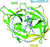
We present an in silico study of the interaction energy between NS2B–NS3, a serine protease of the dengue virus (DENV), and the inhibitor benzoyl-norleucine-Lys-Arg-Arg-aldehyde (Bz-nKRR-H), a crucial step in the design and development of dengue's antiviral drugs, using quantum chemistry calculations based on the density functional theory (DFT) at the generalized gradient approximation (GGA). The interaction energies between the inhibitor Bz-nKRR-H and each amino acid belonging to the binding site was calculated through the molecular fragmentation with conjugate caps (MFCC) approach employing a dispersion corrected exchange-correlation functional. Besides the interaction energy, we also calculated the distances, types of molecular interactions, and the atomic groups involved in the process. Our results show that the interaction energy of the system reached convergence at 15.0 Å, with the central residues identified in this interaction radius, as well as their attraction/repulsion energies, all of them being important inputs to improve the effectiveness of antiviral drugs to avoid the dissemination of the dengue virus.
Currently, there are four distinct serotypes (DENV-1 to 4) consolidated by the scientific community and subclassified into several different phylogenetic clusters or genotypes.5 Recently it was announced the discovery of a fifth serotype, DENV-5, found only in specific regions like the Malaysian and Indonesian jungles, usually following the sylvatic cycle and not the human one.6 It is known that the first four serotypes are genetically similar and share approximately 65% of their genomes.7 Despite this, although infection with one serotype confers lifelong immunity to that serotype, it does yield only short-term protection against a secondary infection with a heterologous serotype. Subsequent infection with a different type increases the risk of severe complications like the life-threatening dengue hemorrhagic fever, a rare case characterized by high fever, damage to lymph and blood vessels, bleeding from the nose and gums, enlargement of the liver, and failure of the circulatory system. The symptoms may progress to the dengue shock syndrome (DSS) with massive bleeding, shock, and ultimately death, a phenomenon often attributed to the antibody-dependent enhancement (ADE).8,9
Dengue viruses are small particles (50 nm in diameter) containing a single-stranded, positive-sense RNA of approximately 11 Kb. The genomic RNA encodes a polyprotein precursor that is processed proteolytically into 10 proteins: three structural proteins (C, capsid; prM, precursor membrane; and E, envelope) and seven non-structural ones (NS1, NS2A, NS2B, NS3, NS4A, NS4B and NS5).10,11 The coding of these ten viral proteins is arranged as |C|prM|E|NS1|NS2A|NS2B|NS3|NS4A|NS4B|NS5|.12 The structural proteins form the viral particle while the nonstructural ones participate in the replication of the RNA genome, virion assembly, and attenuation of the host antiviral response. However, according to recent studies, it has been found that NS3 protein is one of the most significant non-structural protein involved in the DENV infection. This region is named as NS3 protease (NS3 for short), and its activity depends on a cofactor named as NS2B, which is crucial in the virus replication process. They collectively forms the active serine protease complex known as NS2B–NS3, which has a function to cleave the precursor polyprotein at the NS2A–NS2B, NS2B–NS3, NS3–NS4A and NS4B–NS5 junctions.13 Thus, the interaction between NS2B and NS3 is essential for the composite function of active viral protease. The NS3 (69 kDa) is a serine protease that has a typical catalytic triad formed by the residues His51, Asp75 and Ser135.4,6 The NS3 is a multifunctional protein that not only promotes cleavage of genomic polyprotein (cleaving C, NS2A/NS2B, NS2B/NS3, NS3/NS4A and NS4B/NS5 proteins), but also has autoproteolytic function, helicase, ATPase and GTPase activity.6,13 It is precisely the autoproteolytic cleavage at the NS2B/NS3 site that results in the formation of an enzymatically active non-covalent complex between NS2B and NS3.4 Thereby, is predicted that the NS3 protease requires a portion of 40 residues of the NS2B protein to assume the activity conformation, where the NS2B will act as a cofactor.4,7 These observations suggest that competitive drugs targeted to the serine protease of the dengue virus NS2B–NS3 complex represent promising candidates for therapeutic agents that could inhibit the maturation of DENV proteins and thereby inhibit viral replication.11,14
The biochemical and structural properties of dengue protease have been recently extensively reviewed.15 Currently, four strategies have been pursued to identify inhibitors of DENV by targeting both viral and host proteins, namely: HTS (high-throughput screening) using virus replication assays; HTS using viral enzyme assays; structure-based in silico docking; and rational design repurposing hepatitis C virus inhibitors for DENV. Although a number of inhibitor candidates for dengue were investigated so far, like the pancreatic trypsin inhibitor (aprotinin) and Ribavirin inhibitor (to cite just a few), the tetrapeptide aldehyde Bz-nKRRH requires direct interactions with the subunit NS2B, essential for the protease catalytic activity, being the best-studied targets for the development of anti-infective therapeutics against dengue virus (see Fig. 1 for its structural illustration). Fig. 2 depicts the Bz-nKRR-H molecule with its main chemical regions. The electron density projected onto an electrostatic potential isosurface is also shown for completeness.
![]() | ||
Fig. 1 Structural representation of the serine protease of the dengue virus NS2B–NS3/inhibitor Bz-nKRR-H complex (PDB ID: 3U1I).26 The Protease NS3 is highlighted in green, while the NS2B cofactor in yellow. The binding pocket sphere with radius (r) is also shown in this picture as a blue dotted-line circle around the Bz-nKRR-H ligand. |
As mentioned before, the dengue protease is responsible for the post-translational cleavage of the viral polyprotein, being essential for viral replication. In the past, other viral proteases, such as those of HCV and HIV, have been successfully established as targets with therapeutic relevance. Recently Yin et al.16 examined some tetrapeptides as protease inhibitors and have suggested the tetrapeptide inhibitor benzoyl-norleucine-Lys-Arg-Arg-aldehyde (Bz-nKRR-H), with a Ki (equilibrium inhibition constant) of 5.8 to 7.0 μM as a potent inhibitor of DENV-2. Besides, Parida and co-workers investigate non structural NS3 inhibitors by means of a protein–protein interacting sites of the dengue virus.17,18
On the experimental side, a rapid assay for detecting and typing dengue viruses was already developed by using reverse transcriptase-polymerase chain-reaction.19 Although the contemporary worldwide distribution of the risk of dengue virus infection and its public health burden are still poorly known, data on clinical diagnosis and pathophysiology of vascular permeability and coagulopathy, parenteral treatment of the dengue, viral factors and new laboratory tests were recently reviewed.20 However, the resurgence of dengue in tropical and subtropical areas of the world, and its spread and establishment in new habitats and environments21 was always a big challenge, like the dengue severity associated with age and a new lineage of dengue virus-type 2 during an outbreak in Rio de Janeiro, Brazil.22 Immunological concerns in dengue virus vaccine development, leaving dengue viruses susceptible to antibody-dependent enhancement (ADE), was successfully investigated.23 Furthermore, technical feasibility of engineering dengue viruses to display targets of protective antibodies are being investigated aiming the development of new dengue vaccines and diagnostic assays (ref. 24 and the reference there in).
The purpose of this work is to describe the interaction energies between the serine protease of the dengue virus NS2B–NS3 and the peptide inhibitor Bz-nKRR-H (receptor–ligand complex) bounded to the site of greatest activity, whose structural representation is depicted in Fig. 1. Our theoretical/computational method makes use of a quantum molecular biochemistry calculations, particularly ab initio techniques based on the density functional theory (DFT) approach, together with the molecular fractionation with conjugate caps (MFCC) method (for a review of these techniques see ref. 25).
Our results allow us to provide a detailed energy profile of the interactions between the inhibitor and its residues in the binding site near the receptor. These data are essential to identify those residues that contribute more to the interactive process between receptor and ligand. Thus, adjustments could be made to improve the effectiveness in the design of new viral drugs leading to a quantitative description of the interaction mechanism of the NS2B–NS3/Bz-nKRR-H complex of DENV. Our ultimate aim is to shed light on the design and development of antiviral drugs to avoid the dissemination of this disease.
The total/individual interaction energy between the inhibitor Bz-nKRR-H molecule (ligand) and the serine protease NS2B–NS3 was calculated by using an MFCC-based scheme within a DFT framework. MFCC has been widely used to calculate protein-ligand binding energy,28–31 once it turns possible the investigation of a vast number of amino acid residues in a protein with small computational cost and no loss in accuracy.32,33 The fractionation scheme was initially aimed to provide an efficient linear-scaling ab initio calculation of protein-ligand interaction energies by dividing a protein molecule into amino acid fragments properly capped.34,35 A significant difference between the MFCC method and other fragmentation methods that employ capping of fractionated bonds is the nature of the caps used. Instead of simple hydrogen caps, the caps in the MFCC approach are portions of the neighboring sections of the molecule, providing an efficient method for representation of the local environment during individual fragment calculations.
Labelling the ligand molecule as L, and the residue interacting with L as Ri (i denotes the index of the i-th amino acid residue), the individual interaction energy between L and each Ri, E(L–Ri), at DFT level is calculated according to:
EI(L–Ri) = E(L + C1−iRiCi+1) − E(C1−iRiCi+1) − E(L + C1−iCi+1) + E(C1−iCi+1), | (1) |
All energetic calculations were made in the DMol3 program package,36,37 using the generalized gradient approximation (GGA)38 within the parameterizations developed by Perdew–Burke–Ernzerhof (PBE),39 together with the Grimme's long-range dispersion correction.40 It improves the description of the non-covalent interactions, providing a better compromise between the cost of first principles evaluation of the dispersion terms (non-covalent interactions). The orbital cutoff, which is a parameter used to control the quality of the numerical basis set and the numerical integrations performed during the computations, was set to 3.0 Å. Besides being suitable for the treatment of this system, this cutoff value reduce the computation time with little impact on the accuracy of the results.41 The total energy variation, which specifies the self-consistent field (SCF) convergence threshold, was selected to be 10−5 Ha, ensuring a well converged electronic structure for the system.
A convergence study of the interaction energy as a function of the ligand binding pocket radius was performed inputting a limit to the number of amino acid residues to be analyzed, without missing important interactions.42 Convergence here means the stabilization of the total binding energy by a variation less than 10% within sequential pocket radius r. The total binding energy is determined by adding up all residue-ligand energies within a pocket radius, and summing consecutive binding pocket energies. We investigated the variation of the total interaction energy considering the contributions of all amino acid residues within a sphere of radius r from 2.0 to 15.0 Å, with origin in the ligand, capping the dangling bonds of each amino acid residue (Ri) with the following three amino acids before (Ci−1) and after (Ci+1) them.
The representation of molecular environment is a necessary step for the theoretical study of molecular properties. Recently, models of implicit solvation have been used in MFCC scheme by Liu et al.43 and Dantas et al.44 to calculate binding affinity in protein–ligand complexes, by assuming different values of the dielectric constant ε to take into account the effect of the protein and solvent environment in the evaluation of the electrostatic energies. Here, we have employed the MFCC approach together with the COSMO (Conductor-like Screening Model) continuum solvation model and dielectric constant ε = 1, 2, 10 and 40 to increase the similarity with the protein environment and estimate energy effects, such as the electrostatic polarization promoted by the solvent.
We analyzed the binding pocket of the receptor employing the MFCC scheme to obtain the individual contribution of each amino acid residue, ranking the most relevant interactions between residues and ligands. The total binding energy was obtained by adding up the individual contributions. To evaluate the binding interactions through the fragment-based quantum mechanics method, it is important to take into account each significant attractive and repulsive amino acid residue which can influence this mechanism. Therefore, to avoid the adoption of an arbitrary binding site size, we performed a search for an optimal limit for which no significant variation in the total interaction energy could be observed after its increase. The result is depicted in Fig. 3, which shows the profile of the interaction energy (measured in kcal mol−1) as a function of r (varied from 2.0 to 15.0 Å).
According to Fig. 3, even for a large radius of 15.0 Å (meaning 131 amino acids), the interaction energy does not achieve a stabilization when the calculations have been made either in vacuum or for a small value of the dielectric constant (ε = 1 or 2). The convergence was broken for the pocket radius r equal to 5.5, 6.5, 7.5, 8.0, 9.5 and 11.0 Å, respectively, mainly due to the attractive (repulsive) interactions of Met49 for r = 5.5 Å, Asp79 for r = 6.5 Å, Asp80 for r = 7.5 Å, and Met75 for r = 9.5 Å (Lys87 for r = 8.0 Å, Lys26 for r = 11.0 Å, and Lys104 for r = 11.0 Å). The total binding energy considering the dielectric function ε = 10 and 40 has shown two destabilization pocket radius, namely at r = 5.5 and 9.5 Å, that are caused mainly by the two methionine residues Met49 and Met75. Beyond the pocket radius r = 9.5 Å, these total binding energies are shown within the convergence criteria. Furthermore, our results have shown that inside r, an increase in the dielectric constant is related to a decrease of the total binding energy, following the order: −313.11 kcal mol−1 (ε = 1) > −242.07 kcal mol−1 (ε = 2) > −129.06 kcal mol−1 (ε = 10) > −112.58 kcal mol−1 (ε = 40). Thus, destabilization of the total curves is mainly linked to the attractive or repulsive interaction energies between the inhibitor Bz-nKRR-H and the charged amino acid residues of the serine protease NS2B–NS3, which are overestimated when low dielectric constant values are used in the MFCC scheme.44,46
Once the MFCC scheme provides for us a possibility to inspect the binding energy of each amino acid individually, we can use these results to describe the amino acids with highest interactions and estimate the regions of the ligand that most contribute to its docking and stabilization. They are summarized in Table S1, shown in the ESI,† as well as in Fig. 4 which depicts a BIRD (an acronym of the keywords binding site, interaction energy and residues domain) panel with the highest binding energies found by our calculations.
Considering the dielectric constant ε = 1 (vacuum calculations), one can see from Table S1† that charged amino acids are the most relevant energetically, following the order: −111.19 kcal mol−1 (Asp81-NS2B) > −110.09 kcal mol−1 (Asp79-NS2B) > −84.31 kcal mol−1 (Asp80-NS2B) > −66.90 kcal mol−1 (Asp75-NS3) > −66.89 kcal mol−1 (Asp129-NS3) > −55.69 kcal mol−1 (Asp88-NS2B) > −43.20 kcal mol−1 (Glu101-NS3) > −41.14 kcal mol−1 (Glu54-NS2B) > −30.46 kcal mol−1 (Met84-NS2B) > −27.09 kcal mol−1 (Asn152-NS3) > −17.98 (Val155-NS3) > −17.08 kcal mol−1 (Met75-NS2B) > −14.67 kcal mol−1 (Met49-NS3) > −14.19 kcal mol−1 (Gly151-NS3) > 29.97 kcal mol−1 (Lys26-NS3) > 36.33 kcal mol−1 (Lys157-NS3) > 41.68 kcal mol−1 (Lys104-NS3) > 48.46 kcal mol−1 (Lys131-NS3) > 49.18 kcal mol−1 (Lys87-NS2B) > 50.34 kcal mol−1 (Lys74-NS3) > 64.18 kcal mol−1 (Arg54-NS3) > 79.82 kcal mol−1 (Arg85-NS2B). Under the effect of a small dielectric constant value (ε = 2), there are a decrease in each binding energies implying some small differences concerning the amino acids relevance sequence, such as Met84 and Asn152 which were the 9th and 10th most important residues for the vacuum calculation, becoming now the 7th and 8th ones.
For higher values of the dielectric constant (ε = 10 and 40), the importance of charged residues decreases, once ε influences mainly individual energies of these amino acids.47,48 As we can see from ε = 40 results, the order of importance of some amino acids was changed as follow: Met84 (−23.44 kcal mol−1) > Asn152 (−16.47 kcal mol−1) > Met75 (−15.47 kcal mol−1) > Met49 (−14.84 kcal mol−1) > Tyr161-NS3 (−8.18 kcal mol−1) > Asp129 (−7.68 kcal mol−1) > Gly151 (−6.15 kcal mol−1) > Gly153-NS3 (−4.53 kcal mol−1) > Asp81 (−4.40 kcal mol−1) > Asp79 (−3.64 kcal mol−1) > Val155 (−3.17 kcal mol−1) > Asp75 (−3.08 kcal mol−1) > Trp83-NS3 (−3.02 kcal mol−1) > Phe130-NS3 (−2.92 kcal mol−1) > Val36-NS3 (−2.81 kcal mol−1) > Asp80 (−2.00 kcal mol−1) > Pro132-NS3 (−1.89 kcal mol−1) > Lys74 (1.34 kcal mol−1) > Trp50 (1.57 kcal mol−1) > Lys157 (1.66 kcal mol−1) > His51 (2.49 kcal mol−1) > Arg54 (2.52 kcal mol−1) > Arg85 (4.98 kcal mol−1).
Comparing our energetic results with the map of the binding network in Bz-nKRR-H/protease, complex provided by Noble and co-workers,26 it is easy to see that the results obtained from ε = 40 are closer to the crystallographic binding scenario than any other ε-calculations showed here. In a recent study based on a MFCC scheme within the DFT framework, simulations performed for an averaged dielectric constant ε = 40 improve the results, not only showing better agreement with experimental data, but also giving a better convergence of the total interaction energy with the binding pocket size.44 A similar achievement was also found by Vicatos et al.46 regarding the best-fitted value of the dielectric constant ε for charge–charge interactions and for self-energies.
Fig. 5 depicts some of the main amino acid residues and respective intermolecular interactions with the inhibitor Bz-nKRR-H. In order to give a better spatial visualization, we show the hydrogen bonds (h-bonds) from three different outlooks. Fig. 5a presents the inhibitor binding with Gly153 (1.87 Å), Arg85 (2.04 Å), Met84 (2.30 Å) and Val155 (2.47 Å). Fig. 5b displays interactions with Asn152 (2.15 Å), His51 (2.86 Å), Asp81 (3.39 Å), Asp75 (3.59 Å) and Arg54 (3.73 Å). Finally, Fig. 5c give us a picture showing the interactions between Bz-nKRR-H with Gly151 (1.89 Å), Tyr161 (1.97 Å), Phe130 (2.09 Å), Asp129 (2.25 Å) and Val36 (4.49 Å).
The electron density distribution in the binding cleft of the serine protease NS2B–NS3/inhibitor Bz-nKRR-H complex can be observed in Fig. 6, which highlight the negative charge concentrations at carbonyl oxygen atom of all amino acid residues, as well as the anionic carboxylate (hydroxyl group) of Asp81 and Asp129 (Asn152). The approximation of these residues with the guanidinium groups of the inhibitor explains the strength of its attractive interactions. In contrast the cationic side chain of the residues Arg54, Arg85 and Lys74 presents low charge densities centered at their basic nitrogen atoms, leading to repulsive electrostatic interactions with positively regions of the inhibitor Bz-nKRR-H.
Asp75 and His51 are members of the catalytic triad of serine protease, and their mutations are related to the loss of the protease function of NS3,49 becoming one of the most thoroughly characterized catalytic motifs. The hydrogen bond between Asp and His increases the pKa of the latter, favoring the activation of a Ser nucleophile that binds covalently to the substrate,50 making direct intermolecular bonds with the inhibitor Bz-nKRR-H (see Fig. 5). There is a salt bridge between the guanidinium group of region iv(C32)NH2+ of inhibitor with the carboxylate group of Asp75 (3.59 Å). Meanwhile, the imidazole ring of His51 is interacting with the side chain of Arginine in the same region.
Phe130, Gly151 and Gly153 (Val36) are involved in direct (water mediated) h-bonds at least with one of the three polar amino acids of the inhibitor – region v(N5)H, v(N4)H and iii(C35)O [v(C39)OH] respectively. The inhibitor Bz-nKRR-H and other ligands were related to make h-bonds with these amino acids, showing that they are essential for the ligands recognition.26,51,52
Salaemae et al.53 re-visited the conformational properties of the dengue virus NS3 protease active site by a structure-guided mutagenesis approach in the dengue virus two-component protease NS2B–NS3 to clarify the functional importance of active site residues. Mutation of the conserved residue Asn152 to Ala, for example, reduced enzymatic activity dramatically, further indicating that targeting this amino acid is a valid approach for antiviral development. Indeed, the fluorometric assays and docking studies with a novel class of serine protease inhibitors (diaryl thioethers) indicate that the number of h-bonds with the side chain or with a backbone carbonyl group of Asn152 lead to an improved affinity/activity.54 In our study, one can note that the Asn152 residue is responsible for a second more attractive contribution (−16.47 kcal mol−1) due to a h-bond (2.5 Å) interaction between its carboxamide side chain and the region iv(C32)NH2+ of the inhibitor Bz-nKRR-H.
A recent study indicates that stronger aromatic–aromatic stacking interactions between phenyl groups of pyrazole ester derivatives with Tyr161 can be important for a potent inhibitory activity against the serine protease NS2B–NS3.55 Furthermore, it is believed that this amino acid acts together with Gly153 to form direct interactions with ligands. Here, the carbon–oxygen double bond of region iii(C35)O and the guanidinium of region v(C45)NH2+ form h-bond and π-cation interactions in respective order with the phenolic oxygen atom and aromatic ring of Tyr161, providing an interaction energy of −8.18 kcal mol−1.
Liu et al.56 present a detailed report of the synthesis of novel thiadiazoloacrylamide derivatives as potential inhibitors of the DENV2 NS2B/NS3 protease. One of the main feature in the docked conformation of the most potent/effective analogue is the presence of a hydrogen bond with the main chain of Met84. In our NS2B-NS3/Bz-nKRR-H binding, region iii of the inhibitor [iii(C22)NH3+] also forms a strong h-bond with it (−23.44 kcal mol−1).
Asp129 and Asp81 interact with the inhibitor Bz-nKRR-H through a salt bridge between their carboxylate side chain at regions iv(C32)NH2+ and v(C45)NH2+ of the inhibitor, respectively. According to Noble et al.,26 they are conserved amino acids that help the serine protease NS2B–NS3 of DENV to be in a closed conformation during proteolysis. Crystallographic, docking and molecular dynamic studies have shown that they disturb the interaction of ligands whose residues are related to a destabilization of the binding pocket.51,52
Finally, note the importance of the residues Met75, Met49, Arg54 and Arg85. While Met75 at region iii(C22)NH3+ and Met49 at regions iv(C29)H and v(N4)H are two of the most attractive residues, Arg54 and Arg85 are repelled through cation–cation repulsion by guanidinium and cationic ammonium group of region iv [iv(C32)NH2+] and iii [iii(C22)NH3+] respectively.
The aliphatic side chain of region ii of the inhibitor Bz-nKRR-H is surrounded by just one non-polar amino acid residue, namely Val155, which stabilizes this hydrophobic moiety through dispersion forces (−3.17 kcal mol−1) which should not be neglected. Besides, hydrophobic interactions with residues, including Val155, may explain the high inhibitory activity against serine protease of the dengue virus NS2B–NS3 observed in thiadiazoloacrylamide derivatives with benzyl groups.56
In the absence of an efficient therapeutic drug in the market, the development of small molecule inhibitors against the replication and maturation of the virus have been considered as a promising route for the treatment of acute dengue diseases. The dengue virus two-component protease NS2B–NS3 mediates processing of the viral polyprotein precursor and is therefore an important determinant of virus replication. The design of chemicals capable of neutralizing this enzyme requires a precise knowledge of the structural determinants of substrate binding and catalysis.
In this context, we have investigated the geometrical aspects and the interactions among the peptide inhibitor Bz-nKRR-H and a potential dengue drug target, DENV-3 NS2B–NS3 protease, using quantum chemistry calculations. We have performed a convergence study of the size of the binding pocket radius considering the dielectric constant ε = 1, 2, 10, and 40 for a precise evaluation of what are the main residues involved in the protease inactivation. In agreement with the literature, low dielectric constant values make difficult this study since the local dielectric properties of serine protease and solvent are weakened. For ε = 40, 131 amino acid residues within a radius of 15 Å make a perfect convergence curve with the total interaction energy of −112.58 kcal mol−1. It is worth highlighting that strong intermolecular contacts are formed between tetrapeptide aldehyde inhibitor Bz-nKRR-H and the Asn152, Met49, Tyr161, Asp129, Gly151, Gly153, Val155, Asp75, Trp83, Phe130 and Val36 (Met84, Met75, Asp81, Asp79 and Asp80) at NS3 (NS2B) serine protease subunit. The computational methods used in this work emerged as an elegant and efficient alternative for the development of drugs that can alleviate the suffering of individuals with this socially neglected disease.
Footnotes |
† This document is a collaborative effort of all authors. |
‡ Electronic supplementary information (ESI) available. See DOI: 10.1039/c6ra10121f |
This journal is © The Royal Society of Chemistry 2016 |