Preparation and characterization of solid lipid nanoparticles loaded with cytarabine via a micellar composition for leukemia

Rakesh Raj*, Pooja Mongia Raj and Alpana Ram
Institute of Pharmaceutical Sciences, Guru Ghasidas Vishvavidyalaya, Bilaspur, 495009, C.G., India. E-mail: rajscientist.2014@rediffmail.com; Tel: +917752260027 Tel: +919039300448

Received 19th April 2016 , Accepted 23rd May 2016

First published on 24th May 2016


Abstract

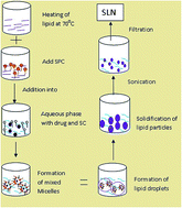
Cytarabine is an anticancer drug used in hematological malignancies and lymphoma but its short biological half life demands continuous intravenous infusion or time spaced injection. This would lead to patient incompliances and discomfort. Therefore it is imperative to look for novel therapeutic systems with lesser side effects urgently to address the underlying causes of poor treatment outcomes associated with conventional therapy. Hence entrapping of cytarabine in a solid lipid nanoparticle (SLN) based formulation could be a better alternative to sustain release and improve therapeutic activity by maintaining the plasma level. We chose tristearin as solid lipid and formulated it in nanoparticulate form by an ultrasonic emulsification method. SLN formulations were optimized and characterized for various physiochemical parameters like size (below 200 nm), charge, morphology and entrapment efficiency. An in vitro drug release study through a dialysis bag revealed a prolonged release of cytarabine. A cell line study on HL60 cell showed a significantly higher efficacy of cytarabine SLN as compared to cytarabine solution. Well characterized carriers were further subjected to hemolytic toxicity. Results showed no hemolytic toxicity and good stability at ambient temperature.


1. Introduction

Cytarabine (cytosine arabinoside, 1-b-D-arabinofuranosyl cytosine) is an anticancer drug belonging to the class of pyrimidine nucleoside analogues. It is one of the mainstays used in the treatment of acute myeloid leukemia,1 non-Hodgkin's lymphoma (blast phase) and erythroleukemia.2 It is also used alone or in combination with other anticancer drugs such as daunorubicin, doxorubicin, thioguanine, or vincristine for the treatment of leukemia and solid tumors.3 Cytarabine is used in induction therapy in combination with anthracyclines and in consolidation therapy at higher dose for acute myeloblastic leukemia (AML) patients.4 The combination of cytarabine with purine nucleoside analogue such as fludarabine and cladribine, has been extensively explored in the treatment of patients with relapsed or refractory lymphoma or combination with other drugs using different regimens, such as DHAP (cytarabine-cisplatin-dexamethasone) and ESHAP (etoposide-methylprednisolone-cytarabine-cisplatin).1,5 Cytarabine is hydrophilic molecule and has a short plasma half life about 10 min (terminal half life 1 to 3 h). The low permeability of cytarabine is due to its low membrane permeability and rapid conversion into inactive metabolite 1-b-D-arabinofuranosyl uracil.6 Hence continuous intravenous infusion is to be needed to achieve constant plasma level of the drug in 24 h. The higher doses of cytarabine lead to toxicity of normal organ, tissue and side effects.7 These toxicity and non specific distribution often results in chemotherapeutic failure.8 Understanding the metabolism and side effects has allowed many investigations to improve low bioavailability and stability. These efforts depend upon formulation and modification, and can be divided into two major categories prodrug and drug delivery system.9–14 The prodrug strategy for cytarabine involves the chemical modification or introduction of a potentiating group on parent drug. Prodrug approach requires all clinical phase as new molecules and such long time journey limits its utility. While drug delivery system involves physical encapsulation or absorption on carrier without any chemical modification, provide site specific and controlled release. Liposomal cytarabine is an example of successful intrathecal delivery of drug for meningitis (meningeal leukemia). But liposomes have low stability due to lipid rancidification and high formulation cost needs other carrier which can overcome such limitations.15

Application of particulate carrier in nanoparticles form is a very attractive possibility to achieve controlled drug release.16 Polymeric nanoparticles are well suited for this purpose but acute and chronic toxicity of polymer17,18 leads shifting to lipid based carriers. The use of solid lipid particles is originated from Speiser and coworker in the beginning of 1980s to retard drug release for peroral administration.8,19 Nanoparticles made from solid lipid are known as solid lipid nanoparticles (SLN). They came in light in the middle of 1990s, as alternative carriers for liposomes, polymeric nanoparticles, nanosuspension and nanoemulsion.20 SLN are controlled release carrier made up of lipids that are solid at room temperature as well as body temperature. They combine the advantage of other novel carrier system (e.g. physical stability, protection of labile compound from degradation, controlled release, excellent tolerability) while at the same time reduce associated problems,21,22 such as membrane stability and drug leaching, large scale production feasibility, liquid state and instability at room temperature.23,24 At the beginning SLN formulations had been proposed for parenteral route and gradually shifted to various routes i.e., oral, dermal, pulmonary, rectal and thoroughly characterized in vitro and in vivo. Recently SLN began to act as topical carrier not only for pharmaceutical molecules, but also for cosmetic products.25 They are made up of bioreducible and biocompatible material, with the capability to incorporate both lipophilic and hydrophilic molecules.26,27 SLN along with controlled release and drug targeting potential, have no biotoxicity and sterilization problem, and do not need use of organic solvents. Furthermore they can decrease the uptake of the drug by mononuclear phagocyte system, hence extended drug circulation in body and increase the targeting potential of the drug.

In this study we have developed tristearin SLN bearing cytarabine in soya phosphatidylcholine and sodium cholate mixed micelles form, and characterized for particle size, zeta potential, X-ray diffraction, differential scanning calorimetry and in vitro drug release. Stability and cell viability studies were also considered for the development of cytarabine loaded SLN.

2. Results and discussion

Organoleptic and morphological features of SLN do not change with time and are stable almost one year from production.8 They are based on physiologically tolerated lipids, so reduces risk of toxicity. Tristearin is well established solid lipid used for the production of SLN and its nanoparticles for oral, parenteral and topical delivery is well documented.32–35 Cytarabine was chosen because it is a schedule dependent antineoplastic drug used clinically in the treatment of leukemia. Cytarabine is water soluble drug and selection of lipid according to drug solubility profile has no value in SLN production. Scheme 1 represents SLN production by ultrasonic emulsification method. Narrow size distribution and high entrapment efficiency was considered for production of optimized SLN formulation. When drug concentration is increased from 0.10 to 0.20% of total lipid, entrapment efficiency was increased but no change in particle size significantly (Table 1). Further increase in drug concentration abrupt increase in particle size and PDI, possibly due to saturation solubility of drug in solvent and crystal of drug interfere with size distribution. Entrapment efficiency also decreased from 95 to 62% on increasing drug concentration from 0.20 to 0.40% wt of total lipid indicates the optimum drug lipid ratio is 0.20/1 that would provide particle size 186 ± 4 nm and high entrapment efficiency (95%). Verifying the influence of emulsifier on SLN size, dispersions containing various amount of emulsifier mixture (1[thin space (1/6-em)]:[thin space (1/6-em)]1 ratio) of SC[thin space (1/6-em)]:[thin space (1/6-em)]Pluronic F68 were prepared. The emulsifier was used in a concentration range of 0.5 to 2.0% with respect to final dispersion. As reported the combined use of one on more emulsifying agents appeared to produce mixed surfactant film at interface having high surfactant coverage as well as sufficient viscosity to promote stability.36
image file: c6ra10111a-s1.tif
Scheme 1 Schematic representation of SLN formation via micelle composition.
Table 1 Effect of drug concentration on size and entrapment
S.No. Drug[thin space (1/6-em)]:[thin space (1/6-em)]lipid ratio Particle size Polydispersity Entrapment efficiency (%)
1 0.1/1 178 ± 3 0.132 ± 0.03 66.3 ± 2
2 0.2/1 186 ± 4 0.141 ± 0.01 95 ± 2
3 0.3/1 207 ± 6 0.148 ± 0.04 90.5 ± 1
4 0.4/1 215 ± 11 0.208 ± 0.03 62 ± 3


Table 2 reports the particle size, poly dispersity index and entrapment efficiency which results after using different concentration of surfactant. Two percent w/v (soya PC: Pluronic F68) emulsifier was found to be optimum with respect to smaller size and high entrapment efficiency. This may be due to high concentrations of surfactant which reduce the surface tension and assist the particle partition during preparation. This decrease in particle size is also connected with a remarkable increase in surface area and entrapment. Table 3 showed that 0.05% SC is sufficient to increase entrapment efficiency with narrow size. Further increased concentration of SC showed no significant effect on size and entrapment efficiency. The decrease in particle size of SLN at high concentration of SC is due to effective reduction of interfacial tension between lipid and aqueous phase leading to formation of emulsion droplets of narrow size. By increasing the concentration of SC, the solubilization of mixed micelles was increased, which resulted in the high entrapment of SLN. Further increase of SC amount did not reveal increase in entrapment efficiency because the highest affinity was achieved at 0.05%.

Table 2 Effect of Pluronic F68 concentration on size and entrapment
S.No. Surfactant concentration Particle size (nm) Polydispersity Entrapment efficiency (%)
1 0.5 252 ± 8 0.211 ± 0.02 68 ± 2
2 1.0 212 ± 6 0.202 ± 0.01 85 ± 2.5
3 1.5 174 ± 5 0.163 ± 0.01 84 ± 2
4 2.0 152 ± 6 0.105 ± 0.03 93 ± 4


Table 3 Effect of sodium cholate concentration on size and entrapment
S.No. Sodium cholate (%) Particle size (nm) Polydispersity Entrapment efficiency (%)
1 0.01 250 ± 5.6 0.132 51 ± 3.2
2 0.025 242 ± 8 0.131 54 ± 1.8
3 0.05 184 ± 4 0.107 93.5 ± 2
4 0.075 181.5 ± 5 0.096 94 ± 2.5
5 0.10 178 ± 2.8 0.094 94 ± 2


Zeta potential is a key factor to evaluate the stability of colloidal dispersion. In general, SLN could be dispersed easily when zeta potential was above 30 mV due to electric repulsion between particles.37 Zeta potential of SLN formulations were found between −29 and −44 mV revealing that the formulations were stable because more repulsive force prevents formation of aggregates and zeta potential was not affected by blending of drug and emulsifier with solid lipid. Morphology study by TEM showed spherical shape nearly 170–190 nm correlated with particle size found by Zetasizer [Fig. 1]. SEM image of developed SLN revealed uniform, smooth surface and spherical shape [Fig. 2].


image file: c6ra10111a-f1.tif
Fig. 1 Characterization of tristearin SLN using TEM. A drop of SLN was placed on copper grid and negatively stained with uranyl acetate. Image was recorded on Philips CM12 TEM at 200 kV.

image file: c6ra10111a-f2.tif
Fig. 2 Surface morphology evaluation of the developed tristearin SLN by SEM. Image was recorded after gold coating at high vacuum on JSM-6490LV SEM.

2.1. X-ray and DSC

An important parameter affecting drug incorporation is the polymorphic modification of the lipid particle matrix. Generally, the production process of the nanoparticles can change the type of modification of their respective fraction. The X-ray diffraction pattern of tristearin, SLN and cytarabine loaded SLN are depicted in Fig. 3. X-ray pattern revealed significant difference between the diffraction of bulk lipid, blank SLN and drug loaded SLN. Tristearin showed a sharp narrow peak at 17.51 degree while SLN gave broader and low intensity peak at the same degree. This shows that in SLN tristearin is partially recrystallized (around 80%). Sharp major peak of pure cytarabine at 27.02 degree did not appear in SLN suggested that cytarabine in SLN is present in amorphous form. Polymorphic transformation of lipid during preparation of SLN occurs which can be easily estimated by DSC.38 It is a popular tool to investigate the crystallinity of colloidal SLN matrices. DSC graph of cytarabine showed a peak at 234.62 °C, in contrast to bulk lipid, melting behaviour of SLN was decreased by about 2 °C. The thermogram of drug loaded SLN did not show melting peak of crystalline cytarabine at around 234.62 degree, which indicated the amorphous state of cytarabine. The tristearin is melted at around 73.31 °C, however cytarabine loaded SLN decrease melting peak by about 2.11 °C suggesting the formation of less ordered lipid crystal due to embedding of drug within (Fig. 4). The decline in the melting point of SLN is compared to that of pure lipid, can be assigned to the colloidal dimension of the particles, particularly to their large surface to volume ratio. The broadening of the heating peak and the reduction of the melting temperature indicate an increased number of lattice defects.35 The small particle size and large surface area being an energetically suboptimal state leads to a decrease of the crystallization point. The DSC results were also correlated with X-ray results in respect of amorphous form of cytarabine.
image file: c6ra10111a-f3.tif
Fig. 3 Determination of crystallinity by XRD of drug, tristearin SLN and drug loaded SLN. Data were recorded at 40 kV and 45 mA on Rigaku Miniflex Diffractometer.

image file: c6ra10111a-f4.tif
Fig. 4 DSC graph of drug, SLN and drug loaded SLN recorded by DSC-60 at 10 °C per min.

2.2. Entrapment efficiency

Low entrapment efficiency of water soluble drugs in SLN is major challenge in pharmaceutical field. As the solidification of lipid in water change their polymorphic state and accommodate surrounding molecules within. The drug loading pattern was supposed to be a core enriched lipid model, as represent in Scheme 2. The entrapment efficiency of cytarabine SLN varied from 66 ± 0.6 to 94 ± 1.2. The highest entrapment efficiency was observed with 0.2/1 drug to lipid ratio and 2% surfactant concentration (Table 4). Entrapment efficiency and drug loading of nanoparticles were determined using ultracentrifugation with some modification according to nature of SLN. Due to higher zeta potential of nanoparticles under aqueous dispersion with pH 6.0, the nanosized particles were difficult to separate from ultracentrifugation. In order to separate SLN easily for further analysis, the pH value of nanoparticles dispersion was adjusted to pH 1.2 by adding 0.1 M hydrochloric acid to reduce the zeta potential and thus the nanoparticles can be easily separated by centrifugation.39
image file: c6ra10111a-s2.tif
Scheme 2 Representation of drug entrapment within SLN.
Table 4 Characterization and results of optimized cytarabine loaded SLN
Parameters Results
Particle size (nm) 174 ± 2.5
Polydispersity 0.082
Zeta potential −40.2
Surface morphology by TEM and SEM Spherical with smooth surface
Entrapment efficiency (%) 95.5 ± 1.68
Loading efficiency (%) 18.28 ± 1.02
In vitro release via dialysis bag Upto 7 days continuous release
Release rate Biphasic initially rapid and then sustained
Hemocompatibility Negligible effect on RBC
Stability at 4–8 °C and 25 °C Stable at both temperature


2.3. In vitro drug release study

In vitro drug release from drug solution was complete within 2 h, but sustained upto 7 days from tristearin SLN. The release profile of cytarabine from SLN is shown in Fig. 5. The nanoparticles released the drug slowly without showing any burst release phenomenon, which is generally reported in case of SLN. The sustained release of drug was attributed to the tristearin property, generally accounted to diffusion of the drug through lipoidal membrane of the SLN. Fast drug release occurred initially from SLN up to 15%. A possible mechanism that may be accounted is the fast release of drug adsorbed on the surface or entrapped in the outer stratum. This may be attributed to the aqueous solubility of cytarabine leading to rapid dissolution of drug molecules present in the surface layer of the particles. Then the release rate became slow after 4 h and remained for 144 h. The present biphasic drug release pattern is also supported by various research groups.40,41
image file: c6ra10111a-f5.tif
Fig. 5 In vitro drug release profile of cytarabine from loaded in tristearin SLN in PBS pH 7.4.

2.4. Cell viability assay

Biocompatibility and safety of nanocarriers is major concern regarding for therapeutic purpose. So, there is necessitate to seem for biocompatible and safe SLN. Blank SLN and cytarabine loaded SLN was tested against normal human lymphocyte by MTT assay. MTT assay results showed that exposure of these SLN did not induce toxicity to the normal human lymphocytes. As shown in Fig. 6 no significant change was observed in the morphology of normal and cytarabine SLN treated lymphocyte. The cell viability was more than 80% at highest dose tested (80 μg ml−1). At higher dose 160 ± 1.8%, 106 ± 0.6% and 92 ± 2.3% cell viability was observed with blank SLN, cytarabine and cytarabine SLN, respectively (Fig. 7). Hence these formulations were found to be safe up to 80 μg ml−1 concentration. Cytarabine rapidly converted to its triphosphate form, which damage DNA when the cell cycle holds in the S phase (synthesis of DNA). Rapidly dividing cells (require DNA replication for mitosis) are more affected. In our experiments exposure of cytarabine for long duration also interfere with cell doubling time as indicated by lower cell proliferation by cytarabine and cytarabine SLN as compared to blank SLN.
image file: c6ra10111a-f6.tif
Fig. 6 Cell morphology of normal human lymphocyte cell 72 h after treatment with different formulations, (A) no treatment positive control, (B) with cytarabine, (C) with blank SLN and (D) with cytarabine loaded SLN.

image file: c6ra10111a-f7.tif
Fig. 7 In vitro cell cytotoxicity study of cytarabine, blank tristearin SLN, and cytarabine loaded tristearin SLN.

2.5. Anticancer potential

To access the cytotoxicity of cytarabine loaded SLN, their lymphocyte killing activity was determined against HL 60 cells. The Fig. 8 shows concentration dependent % growth control of cytarabine and cytarabine SLN on HL 60 cell line and hence inference that cell viability in both formulations is concentration dependent. Blank SLN did not show any cytotoxicity to HL60 cells, cytarabine loaded SLN showed higher cytotoxicity than that of cytarabine solution. This phenomenon may be ascribed to cytarabine in SLN which enhanced cellular uptake, by internalized by cells through an endocytosis process, and then escaped from the endosomes and/or the lysosome to enter the cytoplasm.42 As shown in Fig. 9 there was significant change in number of cell and in the morphology of normal and cytarabine SLN treated lymphocyte.
image file: c6ra10111a-f8.tif
Fig. 8 Anticancer activity of blank cytarabine, tristearin SLN and cytarabine loaded SLN on HL 60 cell at different concentrations.

image file: c6ra10111a-f9.tif
Fig. 9 Cell morphology of HL60 cell 72 h after treatment with different formulations, (A) no treatment positive control, (B) with blank SLN, (C) with cytarabine and (D) with cytarabine loaded SLN.

2.6. Hemolytic toxicity

Cytarabine, blank SLN and drug loaded SLN revealed hemolytic toxicity of 9.46 ± 0.3%, 5.41 ± 0.3% and 5.40 ± 0.4% respectively. Moreover, the blank and drug loaded SLN showed hemocompatibility with increasing concentration of lipid (from 0.1 to 2.0%), suggesting that SLN had excellent blood compatibility and could be suitable for in vivo drug delivery.

2.7. Stability

After 6 months of storage at 4 °C and 25 ± 2 °C, size of SLN increased from 174 ± 1.26 nm to 179 ± 3.42 nm and 181 ± 1.82 nm respectively as presented in Fig. 10. Entrapment efficiency was lowered by 1.82% and 3.23% at 4 °C and 25 °C respectively. Lowering of the entrapment efficiency could be due to transitions of lipid from metastable form to stable form which might occur slowly on storage due to small particle size and the presence of emulsifier that may cause drug expulsion from SLN. However the drug expulsion during storage was slowed due to the existence of reverse mixed micelles.
image file: c6ra10111a-f10.tif
Fig. 10 Stability study of cytarabine loaded tristearin SLN at refrigerator condition (4–8 °C) (A) and room temperature (B) (25 ± 2 °C).

3. Methods

3.1. Materials

Cytarabine was obtained from TCI Chemical, Chennai, India. Tristearin and Pluronic F68 was procured from Himedia, Mumbai, India. Soya phosphatidylcholine (SPC) and sodium cholate (SC) was purchased from Sigma Aldrich (USA). All other chemicals used were of analytical grade.

3.2. Preparation of SLN

SLN were prepared by ultrasonic emulsification method, with slight modification.28,29 Briefly, tristearin (3 g) was melted in a beaker at 70 ± 1 °C and SPC (1 g) added in it, and this melt was dispersed in distilled water (70 ± 1 °C) containing a mixture of emulsifier (sodium cholate and Pluronic F68) at different ratio under magnetic stirring at maximum speed for 30 min. The resulting solution was subjected to ultrasonication in bath sonicator for 15 min. Further the dispersion was immediately dispersed in cold distilled water (4 ± 1 °C) followed by continued mechanical stirring for 10 min. The suspension was then filtered through a 0.45 μm filter (Millipore) to remove impurities from materials. The influence of lipid: drug ratio, surfactants concentration on entrapment efficiency and particle size was also investigated.

3.3. SLN characterization

3.3.1. Particle size, polydispersity and zeta potential. The average size of SLN and polydispersity was measured by photon correlation spectroscopy using Malvern Zetasizer (Malvern, UK) at 25 ± 1 °C. Refractive index was set 1.33 and samples appropriately diluted with phosphate buffer pH 7.4 before measurement. Experiments were carried out in triplicate. Surface charge of SLN was also investigated at the same time by evaluating zeta potential.
3.3.2. Shape and surface morphology. Transmission electron microscopy (TEM; Philips CM12 Electron Microscope, Netherlands) was used to visualize the shape of developed SLN. Samples were dried on copper grid and negatively stained with 1% uranyl acetate. After 1 h grids were viewed under microscope at accelerating voltage of 200 kV. To examine the surface of SLN, scanning electron microscopy (SEM) by JSM-6490LV (JEOL, USA) was performed after lyophilization. The lyophilized SLN were stuck on to a brass stub through double adhesive tape. The stub was put in ion sputter coater and gold coating was applied on the same. Then stub was fixed in sample holder and viewed under high vacuum.
3.3.3. X-ray diffraction study. The internal crystallization structure was investigated by small angle XRD. Copper Kα X-ray were produced by X-ray generation (Rigaku Miniflex Diffractometer, Japan) and operated at 40 kV and 45 mA. Experiments were performed in triplicate.
3.3.4. Differential scanning calorimetry (DSC). DSC was performed with DSC-60 (Shimadzu, Japan). Cytarabine, bulk tristearin, SLN with or without cytarabine were placed in conventional aluminium pan (about 10 mg) and scanned at speed of 10 °C per min.
3.3.5. Entrapment efficiency (EE) and drug loading (LD). The amount of cytarabine present in SLN was determined as follows: 0.1 M hydrochloric acid was added to 10 ml freshly prepared cytarabine SLN and the aggregates was ultracentrifuged at 15[thin space (1/6-em)]000 rpm at 4 °C (Cooling Centrifuge Remi, India) in order to remove unentrapped drug from entrapped drug. The filtrate which contains only free drug was discarded. Suspended (containing aggregates of SLN) was collected and washed with distilled water in duplicate and then heated in methanol at 80 °C and further diluted with water. The amount of cytarabine in water was analysed by HPLC method. The entrapment efficiency (EE) and drug loading (DL) was calculated by following equations:
 
image file: c6ra10111a-t1.tif(1)
 
image file: c6ra10111a-t2.tif(2)

3.4. HPLC analysis

HPLC analysis of cytarabine was done using reported method30 with slight modification. Briefly, mobile phase acetonitrile and purified water previously adjusted to pH 2.8 with orthophosphoric acid (2[thin space (1/6-em)]:[thin space (1/6-em)]98 v/v) was delivered at a flow rate 1 ml per min in C18 column of Younglin (YL 9100, South Korea). The peak was observed at 280 nm after 6.8 min.

3.5. In vitro drug release

As the sustained release formulation of cytarabine is intended to deliver a medication over a long period of time, so it is essential to evaluate release behaviour. The drug release from cytarabine SLN were performed in PBS 7.4 using dialysis bag method. The dialysis bag retains nanoparticles and allows the free drug into the dissolution media with a mol wt cut off 12 KD. The bags were soaked in double distilled water for 12 h, before use. Two ml of cytarabine loaded SLN was poured into bag with one end tied with thread. The bag was placed in a conical flask and 100 ml of receiving fluid was added. The conical flask was then placed into a magnetic stirrer at 37 °C with 100 rpm. After 1, 2, 3, 4, 5, 6, 12, 24 h and then up to 144 h at 24 h interval, the medium in the conical flask was taken and replaced with fresh dialysis medium to maintain sink condition. The filtrate was then analyzed by HPLC method.

3.6. Cell viability assay

To evaluate the safety 10, 20, 40 and 80 μg ml−1 of SLN formulations were incubated with normal human lymphocytes. Isolation of lymphocytes was done by density gradient centrifugation. Lymphocytes were isolated from freshly collected EDTA peripheral human blood. Firstly blood was diluted with phosphate buffer saline at 1[thin space (1/6-em)]:[thin space (1/6-em)]1 ratio. Briefly, 3 ml of Ficoll-HiSep was taken in 15 ml centrifuge and 10 ml of diluted blood was loaded without disturbing the interface and centrifuge at 1800 rpm for 30 min. Lymphocyte ring was separated and washed thrice with PBS by centrifugation at 1500, 1200, and 1000 rpm for 15, 12 and 10 min respectively. Cells were cultured in RPMI medium supplemented with 10% heat inactivated fetal bovine serum (FBS), 100 U ml−1 penicillin and 100 μg ml−1 streptomycin at 37 °C in a humidified atmosphere of 5% CO2. The effect of SLN on mononuclear cells at 10, 20, 40 and 80 μg ml−1 (1 × 10⁵ cells per ml) was evaluated. The culture without any SLN was used as negative control for cell proliferation, while adriamycin (1 μM) was used as positive control. After 72 h, 20 μl of 10 mg ml−1 3-(4,5-dimethylthiazol-2yl)-2,5-diphenyl tetrazolium bromide (MTT) solution was added to the wells. After 4 h incubation at 37 °C in CO2 incubator, plates were centrifuged at 3000 rpm for 25 min. The formazan crystals were dissolved in DMSO (100 μl) and the absorbance of individual wells was noted at 570 nm via an ELISA reader at 25 ± 1 °C.

3.7. Cytotoxicity potential

HL60 acute promyelocytic leukemia (APL) cell line were procured from National Center for Cell Sciences (NCCS), Pune, India and separately cultures in RPMI medium supplemented with 10% heat inactivated fetal bovine serum (FBS), 100 U ml−1 penicillin and 100 μg ml−1 streptomycin at 37 °C in a humidified atmosphere of 5% CO2. After 48 h incubation cell viability was tested through trypan blue exclusion assay. When the cell count reached 5 × 10⁴ cell per ml, in vitro cell line study was initiated. The cytotoxic activity of cytarabine, blank SLN (as increasing total lipid concentration) and cytarabine loaded SLN were assessed by measuring the inhibition of cell growth by MTT based calorimetric assay. Exponentially growing HL60 was seeded at 3 × 10⁴ cells per ml in 24 well plates. The cells were separately treated with various concentrations (10–80 μg ml−1) of cytarabine and cytarabine loaded SLN simultaneously and incubated under controlled environment for 72 h. Subsequently, MTT solution 10 mg ml−1 (20 μl) was added to each well and incubated at 37 °C for 4 h, facilitating MTT to be reduced by viable cells with the formation of purple formazan crystals. The formazan crystals were dissolved in DMSO (100 μl) and the absorbance of individual wells was noted at 570 nm via an ELISA reader at 25 ± 1 °C. The cell morphology was also observed with inverted phase contrast microscope (Fig. 7).

3.8. Hemolytic toxicity

Hemolytic toxicity was performed following the previously reported method with slight modifications. Whole human blood samples were collected in EDTA blood collection vials. The red blood cells (RBC) were separated by centrifugation and resuspended in normal saline. Two ml of RBC suspension separately dispersed in normal saline served as control (producing no hemolysis) and that which was dispersed in distilled water considered being 100% hemolytic. One ml of adequately diluted plain cytarabine solution, blank SLN and cytarabine SLN was incubated with 2 ml RBCs suspension after making up the volume to 10 ml with normal saline. The formulation were taken in separate tubes in such amount that resultant final concentration of cytarabine was equivalent in all the cases so as to facilitate the comparison of extent of hemolysis followed by entrapment of cytarabine in SLN. Further hemolysis produced by unloaded SLN was also observed on RBC suspension. The formulations were incubated at 37 ± 1 °C for 30 min, followed by centrifugation at 3500 rpm for 10 min. Subsequently, the absorbance of supernatants was measured spectrophotometrically (Shimadzu 1600, Japan) at 540 nm using saline as blank.31 The degree of hemolysis was thus determined for each sample against absorbance of distilled water which was assumed to cause 100% hemolysis. The hemolytic ratio (HR) was calculated using equation
 
image file: c6ra10111a-t3.tif(3)
where A means absorbance.

3.9. Stability

The storage stability of SLN was assessed by determining the drug retention potential at two different temperature conditions, i.e. 4–8 °C (refrigerator) and 25 ± 2 °C (room temperature) for six months. The SLN formulations were kept in 10 ml transparent storage vial (Merk) after flushing with nitrogen. Samples were withdrawn weakly up to sixth months and analyzed for particle size and the drug content by HPLC method.

4. Statistical analysis

All the results were articulated as mean values ± standard deviation (SD). The statistical analysis was calculated by using Student's t-test and one-way analysis of variance (ANOVA). Probability level of P < 0.05 was considered to be significant.

5. Conclusions

Ultrasonic emulsification method was successfully used to prepare hydrophilic anticancer drug loaded SLN with high entrapment efficiency (95%) and small particle size (below 200 nm). XRD pattern and DSC results were confirmed that cytarabine entrapped within carrier (core shell model), indicated by disappearance of drug peak in SLN. In vitro drug release study of cytarabine SLN revealed sustained release as compared to drug solution over a weak. Hemolytic toxicity study showed favourable indication for hemocompatibility. The data of in vitro cell viability on human lymphocyte revealed no significant change with developed cytarabine SLN. However, anticancer activity on HL60 cell line indicated higher potency of cytarabine loaded SLN as compared to cytarabine solution. The present work illustrated the higher sensitivity of cell to the drug entrapped in SLN than drug solution. This novel strategy here might be attractive to the development of sustained release formulation of anticancer drug for a weak.

Conflict of interest

Authors declare that they have no conflict of interest.

Acknowledgements

The authors are highly thankful to UGC New Delhi for awarding the fellowship as RGN-SRF during this work. The authors are grateful to AIIMS, New Delhi for providing necessary facilities to carryout Transmission Electron Microscopy (TEM). USIC of Babasaheb Bhimrao Ambedkar University, Lucknow is also acknowledged for Scanning electron microscopy (SEM).

References

  1. T. Robak and A. Wierzbowska, Clin. Ther., 2009, 31(2), 2349–2370 CrossRef CAS PubMed .
  2. P. D. Knoester, W. J. M. Underberg and J. H. Beijnen, Anticancer Res., 1993, 13, 1795–1808 CAS .
  3. E. Jabbour, J. E. Cortes, F. J. Giles, S. O'Brien and H. M. Kantarjian, Cancer, 2007, 109, 2171–2181 CrossRef CAS PubMed .
  4. H. I. El-Subbagh and A. A. Al-Badr, Cytarabine, in Profiles of Drug Substances, Excipients, and Related Methodology, Burlington, ed. Harry G. Brittain, Academic Press, 2009, vol. 34, pp. 37–113 Search PubMed .
  5. H. Abali, Y. Urun, B. Oksuzoglu, B. Budakoglu, N. Yıldırım, T. Guler, G. Ozet and N. Zengin, Cancer Invest., 2008, 26, 401–406 CrossRef CAS PubMed .
  6. M. G. Pallavicini, Pharmacol. Ther., 1984, 25, 207–238 CrossRef CAS PubMed .
  7. R. L. Capizzi, J. C. White, B. L. Powell and F. Perrino, Semin. Hematol., 1991, 28(4), 54–69 CAS .
  8. W. Mehnert and K. Mader, Solid lipid nanoparticles production, characterization and applications, Adv. Drug Delivery Rev., 2001, 47, 165–196 CrossRef CAS PubMed .
  9. Y. Kong, Y. Sun, M. Zhu, Y. Zhang, J. Sun, Z. He and T. Asian, J. Pharm. Sci., 2012, 7, 359–363 Search PubMed .
  10. B. S. Chhikara, D. Mandal and P. Keykavous, Eur. J. Med. Chem., 2010, 45, 4601–4608 CrossRef CAS PubMed .
  11. M. D. Blanco, R. M. Trigo, C. Teijón, C. Gómez and J. M. Teijón, Biomaterials, 1998, 19, 861–869 CrossRef CAS PubMed .
  12. K. Ruckmani, B. Jayakar and S. K. Ghosal, Drug Dev. Ind. Pharm., 2000, 26, 217–222 CrossRef CAS PubMed .
  13. N. Subramanian, A. Yajnik and R. S. R. Murthy, AAPS PharmSciTech, 2004, 5, 1–9 Search PubMed .
  14. E. F. Craparo, G. Cavallaro, M. L. Bondì and G. Giammona, Macromol. Chem. Phys., 2004, 205, 1955–1964 CrossRef CAS .
  15. R. Raj, P. Mongia, S. K. Sahu and A. Ram, Curr. Drug Targets, 2016, 17, 206–228 CrossRef CAS PubMed .
  16. P. Mongia, R. Khatik, R. Raj, N. Jain and A. K. Pathak, J. Biomater. Tissue Eng., 2014, 4, 738–743 CrossRef .
  17. M. S. Taylor, A. U. Daniels, K. P. Andriano and J. Heller, J. Appl. Biomater., 1994, 5, 151–157 CrossRef CAS PubMed .
  18. R. R. M. Bos, F. R. Rozema, G. Boering, A. J. Nijenhuis, A. J. Pennings, A. B. Verwey, P. Nieuwenhuis, H. W. B. Jansen and W. C. Debruijn, Biomaterials, 1992, 10, 405–411 CAS .
  19. R. H. Müller, K. Karsten Mäder and S. Gohla, Eur. J. Pharm. Biopharm., 2000, 50, 161–177 CrossRef .
  20. C. Schwarz, W. Mehnert, J. S. Lucks and R. H. Muller, J. Controlled Release, 1994, 30, 83–96 CrossRef CAS .
  21. V. Jenning and S. Gohla, J. Microencapsulation, 2001, 18, 149–158 CrossRef CAS PubMed .
  22. A. zur Mühlen, C. Schwarz and W. Mehnert, Eur. J. Pharm. Biopharm., 1998, 45, 149–155 CrossRef .
  23. R. H. Muller, S. Maassen, H. Weyhers and W. Mehnert, J. Drug Targeting, 1996, 4, 161–170 CrossRef CAS PubMed .
  24. C. Freitas and R. H. Müller, Int. J. Pharm., 1998, 168, 221–229 CrossRef CAS .
  25. R. H. Muller, M. Radtke and S. A. Wissing, Adv. Drug Delivery Rev., 2002, 54, S131–S155 CrossRef CAS PubMed .
  26. V. Jenning, A. Gysler, M. Schafer-Korting and S. H. Gohla, Eur. J. Pharm. Biopharm., 2000, 49, 211–218 CrossRef CAS PubMed .
  27. C. S. Mai, W. Mehnert and M. Schäfer-Korting, Int. J. Pharm., 2000, 196, 165–167 CrossRef .
  28. Y. Luo, D. Chen, L. Ren, X. Zhao and J. Qin, J. Controlled Release, 2006, 114, 53–59 CrossRef CAS PubMed .
  29. R. Raj, P. Mongia, A. Ram and N. K. Jain, Artif. Cells, Nanomed., Biotechnol., 2015, 1–6 CrossRef PubMed .
  30. A. Bhatnagar, S. Loura and M. Chaudhari, Eurasian J. Anal. Chem., 2012, 7(3), 160–167 CAS .
  31. X. Guo, C. Shi, J. Wang, S. Di and S. Zhou, Biomaterials, 2013, 34, 4544–4554 CrossRef CAS PubMed .
  32. A. J. Domb, Int. J. Pharm., 1995, 124, 271–278 CrossRef CAS .
  33. P. Ahlin, J. Kristl and J. Smid-Kobar, Acta Pharm., 1998, 48, 257–267 Search PubMed .
  34. K. Westesen and H. Bunjes, Int. J. Pharm., 1995, 115, 129–131 CrossRef CAS .
  35. H. Bunjes, K. Westesen and M. H. J. Koch, Int. J. Pharm., 1996, 129, 159–173 CrossRef CAS .
  36. M. Trotta, F. Debernand and O. Caputo, Int. J. Pharm., 2003, 257, 153–160 CrossRef CAS PubMed .
  37. R. H. Muller, R. K. Mader and S. Gohla, Eur. J. Pharm. Biopharm., 2000, 50, 161–167 CrossRef CAS PubMed .
  38. H. Bunjes, F. Steiniger and W. Richter, Langmuir, 2007, 23(7), 4005–4011 CrossRef CAS PubMed .
  39. F. Q. Hu, Y. Hong and H. Yuan, Int. J. Pharm., 2004, 273, 29–35 CrossRef CAS PubMed .
  40. S. J. Lima, M. K. Leeb and C. K. Kim, J. Controlled Release, 2004, 100, 53–61 CrossRef PubMed .
  41. A. Jain, P. Kesharwani, N. K. Garg, A. Jain, S. A. Jain, A. K. Jain, P. Nirbhavane, R. Ghanghoria, R. K. Tyagi and O. P. Katare, Colloids Surf., B, 2015, 134, 47–58 CrossRef CAS PubMed .
  42. S. Q. Liu, N. Wiradharma, S. J. Gao, Y. W. Tong and Y. Y. Yang, Biomaterials, 2007, 28, 1423–1433 CrossRef CAS PubMed .

This journal is © The Royal Society of Chemistry 2016
Click here to see how this site uses Cookies. View our privacy policy here.