Gold nanoparticle-mediated electron transfer of cytochrome c on a self-assembled surface

Roberto A. S. Luz and Frank N. Crespilho*
Instituto de Química de São Carlos, Universidade de São Paulo, 13560-970, São Carlos, SP, Brazil. E-mail: frankcrespilho@iqsc.usp.br

Received 15th April 2016 , Accepted 22nd June 2016

First published on 24th June 2016


Abstract

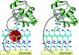
The presence of gold nanoparticles (AuNPs) at the protein/electrode interface has a significant impact on the electrodic microenvironment, and allows the optimization of the activity catalysis as well as electrochemical properties. Here, we report a novel and accurate methodology to observe AuNP mediated electron transfer mechanism from Cytochrome c (Cyt c) to a polycrystalline gold surface. Poly(allylamine hydrochloride) molecules (PAH) were used as spacers between Cyt c and the electrode surface, and the electron rate constant within the PAH layer was measured in the presence and absence of AuNPs. Based on cyclic voltammetric experiments and Marcus theory, a four-fold increase in the electron rate constant was observed in the presence of AuNPs, and the reorganization energy was estimated to be 0.49 eV. Furthermore, AuNPs decreased the effective distance between the redox center of Cyt c and the electrode surface by 20%. These results suggest that the electron transfer properties of Cyt c based protein electrodes are significantly enhanced in the presence of the AuNPs.


Introduction

Electrons do not flow smoothly along a cell-sized wire in living systems that are based on electron transfer dynamics of redox enzymes. They are known to be individually transported, and they jump from protein to protein.1–3 This method of electron transfer implies that the microenvironment of a protein can control electron release as well as the method in which electrons are picked up from a particular point and delivered to the exact point where they are needed. Cytochrome c (Cyt c), a small heme-protein (34 Å) associated with the inner mitochondrial membrane, is an essential component of the electron transport chain in living systems.4–6 It contains a covalently bound heme group that can switch reversibly between the Fe2+ and Fe3+ oxidation states during electron transfer.

In contrast to several redox proteins, the active center of Cyt c is not buried so deeply in amino acid sequence, making it a model protein for studying direct electron transfer (DET). DET reactions between proteins and electrode surfaces have been extensively explored over the past few years, especially for revealing important steps in the kinetics of enzymatic reactions and the mechanistic details of electron transfer.7–10 DET reactions also constitute an important foundation for the development of biodevices such as biosensors11,12 and biofuel cells,13,14 where performance is directly related to a fast and well-defined electron transfer. As described by Marcus,15 the electron tunnelling DET mechanism depends mainly on three factors namely, the protein structure and specifically the redox center location within the protein, the orientation of the protein, and the electron transfer distance. For occurrence of DET in an optimized electrode configuration, the protein must be oriented with its active site facing the electrode in order to ensure the shortest possible electron transfer distance (usually no greater than 14 Å).16 However, the bulky tertiary or quaternary structure of some proteins increases this distance and prevents DET. Further, the denaturation of protein chains and subsequent passivation of the electrode surface are also observed in some cases. In this context, different strategies have been developed for protein immobilization on solid surfaces without damaging their native structures and redox properties, and for maximizing the electronic communication between the active site and the electrode.17,18 Most of these approaches are based on modification of electrodes using various materials that interact with proteins. Electrochemical techniques such as fast scan voltammetry may then be applied to obtain thermodynamic and kinetic variables related to DET of adsorbed proteins.19

Eddowes and Hill,20 and Yeh and Kuwana21 carried out pioneering research on DET of Cyt c. Following this, several important studies reported the modification and structural characterization of electrode surfaces that are active towards Cyt c, where a well-defined direct electrochemistry could be obtained. In this context, the incorporation of nanomaterials at the protein/electrode interface has been one of the main lines of enquiry.22–25 Several authors25–28 reported a favorable microenvironment for protein immobilization upon inclusion of gold nanoparticles (AuNPs) at the protein/electrode interface, where catalytic and electrochemical activity were maintained for considerably longer periods. However, a deeper comprehension of the thermodynamic and kinetic properties, and the nature of DET in the presence of nanoparticles is still required. Here, we propose a novel electrode interface for observation of electron transfer from Cyt c. For this purpose, Cyt c was adsorbed on Au polycrystalline electrodes with and without AuNPs between molecules and the electrode surface. Using these self-assembled electrodes, we demonstrated a four-fold enhanced electron rate constant when AuNPs were present in the proximity of Cyt c, which was in good agreement with the Marcus theory of electron transfer. In summary, our results contribute to the idea that not only the protein structure but also the electron transfer distance between redox centers creates the perfect environment for the electron transfer.

Experimental details

Materials

Bovine heart cytochrome c (MW: 12[thin space (1/6-em)]327 Da), L-cysteine (Cys), chloroauric acid trihydrate (HAuCl4·3H2O), and poly(allylamine hydrochloride) (PAH, MW: 15[thin space (1/6-em)]000 Da) were purchased from Sigma-Aldrich. Sodium borohydride (NaBH4) and hydrogen peroxide (H2O2) were purchased from Vetec Quimica, Brazil. H2O2 solutions were prepared freshly before use. Phosphate buffer (5 mM, pH 7.0) was prepared using NaH2PO4 and Na2HPO4 (obtained from Acros organics), and Cyt c solution was prepared by dissolution in the phosphate buffer. All other solutions were prepared using deionized water (approximate resistivity, 18.2 MΩ cm) in previously cleaned and dried glassware.

Synthesis of gold nanoparticles

AuNPs were synthesized by drop-wise addition of 10 mL aqueous NaBH4 solution (10 mM) in an aqueous solution mix containing 10 mL PAH (10 mM, used as stabilizing agent) and HAuCl4 (1 mM). The stoichiometric ratio between NaBH4 and HAuCl4 was kept at 10[thin space (1/6-em)]:[thin space (1/6-em)]1 to ensure the total reduction of Au3+ to Au0 (zero valence species). The synthetic reaction needed approximately 30 min for completion, upon which an AuNP containing aqueous suspension possessing reddish coloration and featuring a plasmon absorption band at 525 nm in the UV-Vis spectrum was obtained (see Fig. S1 in ESI).29,30 The average particle diameter estimated by transmission electron microscopy (TEM) was 7.9 nm (see Fig. S2 in ESI). Apart from acting as a stabilizing agent in AuNP synthesis, PAH confers a positive charge to the nanoparticle suspension (zeta potential +64 mV) and enables application of the AuNPs in fabrication of self-assembled films.

Preparation of bioelectrodes

A polycrystalline gold electrode (3 mm diameter) was polished with 0.05 μm alumina, cleaned in piranha solution (H2SO4[thin space (1/6-em)]:[thin space (1/6-em)]H2O2: 3[thin space (1/6-em)]:[thin space (1/6-em)]1) and subjected to consecutive voltammetric cycles in 0.1 M H2SO4. The electrode was then modified by incubation in 0.02 M Cys solution (pI = 5.02) for 4 h at 4 °C, which assigned it an anionic character. After extensive washing with deionized water, the electrode modified Au/Cys was immersed in a cationic suspension of AuNPs stabilized in PAH (AuNP-PAH) for 10 h at 4 °C, followed by thorough washing with deionized water to remove non-adsorbed or weakly adsorbed AuNP-PAH. The Au/Cys/AuNP-PAH electrode was immersed again in Cys solution for 4 h at 4 °C, and finally incubated in a cationic solution of 80 μM Cyt c (pI ∼ 10.0–10.5) for 10 h at 4 °C resulting in an Au/Cys/AuNP-PAH/Cys/Cytc electrode. L-Cysteine plays an important role in the electron transfer reaction of Cyt c by providing negatively charged sites that can interact with the hydrophilic surface of the enzyme. Additionally, the carboxylic and amino groups of Cys interact with the lysine residues surrounding the heme edge of Cyt c.4 Schematic fabrication of the Au/Cys/AuNP-PAH/Cys/Cytc electrode is shown in Fig. 1. A similar methodology was employed under identical conditions to construct Au/Cys/PAH/Cys/Cytc modified electrodes (without gold nanoparticles) for comparative studies. All resulting electrodes were washed with deionized water and stored at 4 °C in phosphate buffer when not in use.
image file: c6ra09830d-f1.tif
Fig. 1 Stepwise schematic illustration involved in fabrication of the Au/Cys/AuNP-PAH/Cys/Cytc electrode and photographs of cysteine, AuNP-PAH and Cyt c solutions used to modify the electrodes. Inset: interaction sites between Cys, AuNP-PAH, and Cyt c. Step I represents electrostatic interaction between the carboxylic groups of cysteine and amine groups of AuNP-PAH. In step II, AuNP-PAH interacts with cysteine mainly via a S–Au bond. Step III represents the immobilization of Cyt c, whose interaction with Cys is dominated by electrostatic forces between the carboxylic groups and NH3+ present on Cys and lysine residues around the edge of the heme group of Cyt c. It's important to note that the scheme is merely illustrative and does not take into account realistic length-scales.

Methods

Cyclic voltammetry and chronoamperometry were carried out with a potentiostat/galvanostat Autolab PGSTAT 128N. All measurements were performed using a three-electrode cell using platinum and Ag/AgCl (saturated KCl) electrodes as auxiliary and reference electrodes, respectively. The modified electrode was used as working electrode. Prior to experiments, buffer solutions were deaerated by bubbling argon for 20 min and argon atmosphere was maintained during all measurements. UV-Vis absorption spectra for Cyt c (oxidized species) in phosphate buffer, and immobilized on quartz slides (by using similar methodology employed to prepare the modified gold electrodes) were recorded using a Jasco V-670 spectrophotometer. FTIR spectra were obtained using a Bruker Tensor 27 spectrometer and gold-coated glass substrates were used to prepare films. Zeta potential and TEM images were recorded on a Zetasizer Nano ZS apparatus (Malvern Instruments) and on a FEI Tecnai G2 F20 HRTEM electron microscope, respectively.

Kinetic modelling

Marcus theory applied to heterogeneous systems31–33 was used to interpret the electron transfer kinetics of enzymatic electrodes. The electrochemical rate constants (kred/oxi) were calculated numerically as a function of overpotential (EE0′) on the basis of Chidsey's relation (eqn (1)),34 obtained from the standard Marcus equation35 in combination with the Fermi–Dirac distribution, since electron transfer can occur to or from any Fermi level (Ei) in the electrode:
 
image file: c6ra09830d-t1.tif(1)

E is the applied potential, E0′ the formal potential of the adsorbed couple, λ is the reorganization energy and x = (EEi)F/RT. The terms R, T, and F correspond to the universal gas constant, temperature and Faraday constant, respectively. kmax is the limit of rate constant when the overpotential tends to infinity and is given by:

 
image file: c6ra09830d-t2.tif(2)

V02 represents the maximum electronic coupling, r is the distance between donor and acceptor, and β is the decay coefficient. To calculate the rate constants from Chidsey's relationship we adopted the method described by Armstrong and coworkers.33 In this method, the integral function (eqn (1)) is approximated numerically by summation over a sufficiently wide range of x.

 
image file: c6ra09830d-t3.tif(3)
with Λ = λF/RT, Ψ = (EE0′)F/RT and xi given by:
 
image file: c6ra09830d-t4.tif(4)
where xp is:
 
image file: c6ra09830d-t5.tif(5)
a and s parameters determine the range and the number of steps, respectively, and xp is the “center of gravity” of the peak-shaped integrand. The reduction rate constant (kred) is obtained with Ψ = +(EE0′)F/RT and the oxidation rate constant (koxi) is obtained with Ψ = −(EE0′)F/RT. Using a = 7, s = 100, and Λ < 100, errors ≤ 10−7 are obtained for all Ψ values.33 Butler–Volmer model36 was also used to determine the electron transfer rate constants. All calculations in this study were performed using the Matlab® R2013a software.

Results

Gold nanoparticles and cytochrome c in a self-assembled surface

Polyelectrolyte multilayer (PEM) approach and L-cysteine modified gold surfaces were used to fabricate two types of electrodes, as shown in Fig. 2a. As described in experimental section, the Au electrode was initially modified with L-cysteine molecules (Au/Cys) followed by modification with AuNP-PAH. Another layer of Cys was deposited on Au/Cys/AuNP-PAH resulting in the Au/Cys/AuNP-PAH/Cys electrode. Finally, Cyt c was adsorbed on Au/Cys/AuNP-PAH/Cys resulting in an electrode with the configuration such as Au/Cys/AuNP-PAH/Cys/Cytc. Using the same methodology, an Au/Cys/PAH/Cys/Cytc modified electrode lacking the AuNPs was also obtained.
image file: c6ra09830d-f2.tif
Fig. 2 (a) Two electrode configuration utilized to study the influence of AuNPs on electron transfer between Cyt c and Au electrode surface; (b) visible absorption spectra of Cyt c in phosphate buffer solution (black line) and Cys/AuNP-PAH/Cys/Cytc film on quartz slide (red line); (c) FTIR spectra obtained in the reflection mode for Cys, AuNP-PAH, AuNP-PAH/Cys, Cyt c, and Cys/Cytc films.

The molecular structure of Cyt c was observed to be unaffected by the immobilization method. This was confirmed by appearance of the Q-band at 530 nm and the Soret band in the electronic spectra.27 The Soret band results from the additive effects of the transition dipole moments of the two orbital excitations a1u-eg and a2u-eg of the π–π* transitions of the porphyrin ring in Cyt c. Shifts in this band reflect the conformational changes of the heme microenvironment.37,38 The spectrum of Cyt c in phosphate buffer showed a well-defined Soret band at 410 nm (Fig. 2b, black line). These bands were observed to remain at identical positions also after immobilization on the self-assembled Cys/AuNP-PAH/Cys/Cytc layer. Moreover, FTIR spectra were obtained for Cys, AuNP-PAH, AuNP-PAH/Cys, Cyt c, and Cys/Cytc films constructed on the Au surface (Fig. 2c). When the spectra for Cys, AuNP-PAH, and AuNP-PAH/Cys were compared, the weak intensity band at 2542 cm−1 corresponding to S–H stretching in cysteine39 was observed to disappear in the AuNP-PAH/Cys film. This can be attributed to the cleavage of the S–H bond and the formation of a new S–Au bond40,41 indicating that the interaction between Cys and AuNPs occurs mainly via the formation of this bond. Broadened bands between 2600 and 3400 cm−1, corresponding to stretching modes of NH3+ were also observed for these spectra. Additionally, it was possible to observe CO2 symmetric and asymmetric stretching modes at 1424 and 1582 cm−1 and deformation of NH3+ at 1508 cm−1, respectively in the spectrum of Cys.42 In the Cyt c spectrum, bands resulting from vibrations of peptide backbones could be easily identified at 1535, 1652, and 3300 cm−1, which were respectively attributed to N–H stretching, C[double bond, length as m-dash]O stretching (amide I), and N–H deformation (amide II).43 These stretching bands were displaced to 1510 and 1585 cm−1 in the Cys/Cytc film, indicating that the interaction between Cyt c and Cys was dominated by electrostatic forces between carboxyl and amino groups present in the Cys and lysine residues around the edge of the heme group in Cyt c. This observation provided evidence for consistency in the structural models for electrodes proposed in Fig. 1. Furthermore, the catalytic properties of Cyt c were retained upon immobilization (for details please refer ESI), where H2O2 was easily reduced at low concentrations by the Au/Cys/AuNP-PAH/Cys/Cytc and Au/Cys/PAH/Cys/Cytc bioelectrodes, which implied a continuity in the activity of Cyt c even after immobilization on the modified electrode.

Gold nanoparticles enhance direct electron transfer

Cyclic voltammetry indicated a direct electron transfer between Cyt c and the electrode surface. Herein, a quasi-reversible and well-defined redox couple with a formal potential (E0′) of 65 mV (versus Ag/AgCl) was observed in the phosphate buffer. The electrochemical process is related to the redox couple heme FeIII/FeII. Since no decrease in faradaic currents or peak potential shifts were observed even after subjecting the electrodes to over 100 consecutive voltammetric cycles, Cyt c molecules were concluded to be stably placed on electrode surfaces (Fig. 3a and b).
image file: c6ra09830d-f3.tif
Fig. 3 Cyclic voltammograms (CVs) at 50 mV s−1 for (a) Au/Cys/AuNP-PAH/Cys/Cytc and (b) Au/Cys/PAH/Cys/Cytc (black lines). Green and blue lines are respectively the CVs for bare Au electrode and Au/Cys; CVs for Au/Cys/AuNP-PAH/Cys/Cytc (black line) and Au/Cys/PAH/Cys/Cytc (red line) in phosphate buffer at (c) 5 mV s−1 and (d) 10 V s−1; (e) CVs at 50 mV s−1 for Au/Cys/AuNP-PAH/Cys/Cytc in phosphate buffer varying pH values (3.0–8.0); and (f) effect of pH on the formal potential.

The oxidation and reduction currents for both electrodes, with and without AuNPs at 5 mV s−1, were almost identical (Fig. 3c), which suggested similar amounts of protein adsorption on both electrodes. The surface coverage (Γ) of Cyt c on the electrode surface was estimated based on integration of CVs area obtained according to Γ = Q/nFA at 5 mV s−1 (shown in Fig. 3c), where Q is the charge involved in the electrochemical reaction, n is the number of electrons transferred (n = 1), F is Faraday's constant, and A is the electrode area. As expected, the surface coverage of Cyt c on both electrodes was identical and 3.8 × 10⁻¹¹ mol cm−2. These values are in agreement with previous reports concerning Cyt c on indium tin oxide and Au electrodes.4,44 At this moment, we don't have data regarding varying the particle size because our intention is to show that AuNPs inside of polymeric film can help the charge transfer. In this case, AuNP average diameter is 7.9 nm (see ESI, Fig. S2). However, it is expected the presence of nanoparticles on electrode surface cause increase in electrode surface area promoting as consequence the deposition of greater amounts of enzyme. As a result, it should be observed higher faradaic currents when compared to bare electrode (without AuNPs). Nevertheless, in our study, this behavior was not noted. As described in above, the amount of Cyt c deposited on both electrodes was the same. Even though it is not conclusive this may be related to the fact that AuNPs were covered with a cysteine layer before the immobilization of Cyt c, which may help to minimize the area effect normally promoted by the nanoparticles size.

Further, a swift increase in the faradaic current was observed in case of the electrode with AuNPs at higher scan rates (Fig. 3d). This indicated that the presence of nanoparticles facilitated the electron transfer process of Cyt c. As expected, the electrochemical reaction was found to be pH-dependent. Thus, CVs were observed to change when the pH varied from 3.0 to 8.0, and quasi-reversible CVs with stable and well-defined redox peaks were obtained (Fig. 3e). Increasing E0′ values were observed when the pH was decreased (Fig. 3f). This shift suggested that the electron transfer reaction between Cyt c and the modified electrode was accompanied by proton transport.27,45 Moreover, the slope of E0′ versus pH for the electrode Au/Cys/AuNP-PAH/Cys/Cytc (51 mV per pH), was smaller than the theoretically expected value of 59 mV per pH for the transfer reaction including one electron and one proton.45,46 Although these observations are inconclusive, few authors27,45,47 have attributed this fact to the effect of the protonation states of trans ligands to the heme iron and amino acids around the heme, or the protonation of water molecules coordinated to the iron center. It is noteworthy that changes in the peak potentials and currents of voltammograms at varying pH were reversible in the pH range of 3.0–8.0. For instance, the CVs obtained at pH 7.0 could be reproduced even after immersion of the electrode in a buffer with pH 4.0 followed by transfer into the buffer with pH 7.0. Higher current densities with a lower peak potential separation were observed at pH 7.0 (Fig. 3e), indicating that this pH value (similar to the optimum pH of Cyt c in solution) provided a higher reversibility of the system and an environment that favoured DET. Based on these results, phosphate buffered solution (pH 7.0) was used as an electrolyte for further studies regarding electron transfer kinetics.

An increase in the scan rate promoted an increase in current densities of the oxidation (jpa) and reduction (jpc) peaks (Fig. 4a and b), which showed a linear relationship in the range from 0.005 to 10 V s−1 (Fig. 4c and d). This behavior indicated that the electrochemical processes were governed by electron transfer at the enzyme/electrode interface and that Cyt c was strongly adsorbed onto the modified electrodes.


image file: c6ra09830d-f4.tif
Fig. 4 Cyclic voltammograms with background-subtracted peaks for (a) Au/Cys/AuNP-PAH/Cys/Cytc, and (b) Au/Cys/PAH/Cys/Cytc in 5 mM phosphate buffer recorded at different scan rates (0.01 to 6 V s−1). Typical CVs without background correction for both electrodes are shown in the inset; (c) dependence of current densities of the anodic (jpa) and cathodic (jpc) peaks as a function of scan rate; (d) variation of anodic and cathodic peak overpotential (EE0′) as function of log(v) for (e) Au/Cys/AuNP-PAH/Cys/Cytc; and (f) Au/Cys/PAH/Cys/Cytc. Black and red lines indicate linear fit curves.

kred/oxi was obtained based on Butler–Volmer (BV) theory,36 which describes a semiempirical exponential dependence of the rate constants with the overpotential (EE0′), according to eqn (6) and (7):

 
kred = k0[thin space (1/6-em)]exp(−αnF(EE0′)/RT) (6)
 
koxi = k0[thin space (1/6-em)]exp((1 − α)nF(EE0′)/RT) (7)
where k0 is the standard rate constant (at zero overpotential), n is the number of electrons transferred, and α is the transfer coefficient representing the degree of symmetry of the energy barrier of the redox reaction. In ideal situations, a symmetrical case is represented by α = 0.5. However, α is observed to deviate from 0.5 in several cases due to which the determination of α is crucial for estimating kred/oxi. Generally, α may be experimentally estimated from the slope of the straight lines of Epa and Epc versus log(v) by using the mathematical treatment proposed by Laviron48 (Fig. 4e and f) according to eqn (8) and (9). Once the value of α is known, k0 can be calculated by using eqn (10), where va and vc are the x-intercepts of anodic and cathodic lines, respectively.31
 
image file: c6ra09830d-t6.tif(8)
 
image file: c6ra09830d-t7.tif(9)
 
image file: c6ra09830d-t8.tif(10)

α and k0 were estimated to be 0.62 and 0.87 s−1 for Au/Cys/AuNP-PAH/Cys/Cytc and 0.58 and 0.23 s−1 for Au/Cys/PAH/Cys/Cytc. Despite being widely used to describe electron transfer kinetics, this formalism is limited in application due to a number of constraints.48 For example, the primary assumption of the BV theory states that the activation energies for both anodic and cathodic curves are assumed to be a linear function of overpotential, where the effect of reorganization energy (λ) is neglected. Consequently, results based on BV formalism do not show accurate rate constant values.31,49 As previously demonstrated,33,50 BV equations are equivalent to the limiting case of integrated Marcus equation (eqn (1)), when λ is infinitely high.

Experimentally, this implies that BV equations can only be used to calculate the rate constants with sufficient accuracy in systems where λ is much higher than the maximum applied overpotential. These deviations can be understood from the fact that the BV theory considers reaction surfaces to be linear instead of parabolic (similar to Marcus theory) and ignores contributions to the rate from the electrode states at different applied potential (Fermi level).50 In conclusion, it is valid to put forth that the Marcus theory15,35,51 should be considered for investigating electron transfer reactions of redox proteins.

Marcus theory was originally developed for homogeneous electron transfer reactions. For electron transfers between an electrode and adsorbed redox species, Chidsey34 derived a relation between the rate constants, kred/oxi, and the overpotential (eqn (1)) by integrating the Marcus equation. This relation describes the rate of electron transfer between a donor and an acceptor over all Fermi levels in the electrode, using the Fermi–Dirac distribution to explain the probability of occupancy of each level.32,33,52 The reorganization energy, λ (defined as the energy required to reorient all atoms from equilibrium state to the product state), is very important since it strongly influences the rate constants. The total reorganization energy is composed of two parts where, λ = λi + λo. The inner contribution λi, is related to the energy required to modify bond distances and in some cases the spin states, whereas the outer contribution λo, is related to the energy required to reorganize the solvent.31 One method to estimate λ is to examine the influence of temperature on the rate constants through an Arrhenius plot ln[k0/T1/2] vs. T−1, which is derived from Chidsey's formulation34 based on the Marcus density-of-states model15,35 for electron transfer rate. Assuming the reorganization energy to be independent of temperature, λ can be determined from the slope of the Arrhenius plot, thereby producing an activation energy that is equal to λ/4.15,53,54

 
λ = −4.03dln[k0/T1/2]/d[T−1] (11)
where dln[k0/T1/2]/d[T−1] is the slope of experimental plot of ln[k0/T1/2] vs. T−1. Fig. 5 shows the Arrhenius plot for Au/Cys/AuNP-PAH/Cys/Cytc, where a good linear relationship between ln[k0/T1/2] as a function of T−1 can be observed in the temperature range of 278–308 K. The electrolyte temperature was varied by using a thermostatic bath (GE-MultiTemp IV Thermostatic Circulator, 0.01 °C resolution). The plot was constructed based on the standard rate constants obtained by Laviron's formalism (eqn (10)). For this purpose, CVs were obtained for the Au/Cys/AuNP-PAH/Cys/Cytc electrode at different scan rates (0.005–10 V s−1) and varying temperatures between 278 and 308 K. Based on eqn (11), the reorganization energy of Cyt c immobilized on the modified electrodes was calculated to be 0.49 eV, which was lower than that reported for cytochromes in solution (∼0.6 eV).55 This lower value was attributed to the limited solvent access to the redox center due to enzyme adsorption on the electrode, and is in agreement with previously reported studies.56,57


image file: c6ra09830d-f5.tif
Fig. 5 Arrhenius plot ln[k0/T1/2] vs. T−1 for Au/Cys/AuNP-PAH/Cys/Cytc.

Effective electron transfer distance

After confirming that the significant increase in faradaic current in presence of AuNPs was not related to the amount of electroactive protein on the electrode surface, we investigated the influence of AuNPs on electron transfer. For this purpose, Marcus theory31–33 was applied by using the Chidsey model for heterogeneous systems.34 The rates constants (kred/oxi) were obtained numerically as a function of overpotential (EE0′). The rate constant kmax for the AuNP containing electrode was 5.61 × 10² s−1, which was about 4 times higher than that observed for the electrode without AuNPs (1.48 × 10² s−1) (Fig. 6). Although the explanation for this result is under in discussion, some authors have proposed that the interfacial electron transfer of Cyt c in the presence of AuNPs occurs by two consecutive electrochemical steps equivalent to an electron hopping mechanism, as described in the following equation:58,59
 
image file: c6ra09830d-t9.tif(12)
where k1 and k−1 are the rate constants for electron transfer between AuNPs and Cyt c, i.e., the rate constants of oxidation and reduction of Cyt c. Similarly, k2 and k−2 are the rate constants for electron transfer between the electrode and AuNPs. Interfacial electron transfer between nanoparticles and a metal electrode is intrinsically easier than transfer between nanoparticles and redox system by many orders of magnitude (k2k1).58,60,61 Thus, the two-step process is mainly determined by the electron transfer between Cyt c and the AuNPs, which is also the limiting step. Moreover, it is known that the rate of a heterogeneous ET reaction decreases exponentially with distance (d).62 Thereby, considering only the limiting step (since k2k1 in eqn (12)) and knowing that the ET effective distance between Cyt c and the AuNPs (d2) is lower than that for direct ET to the bare electrode (d1) (Fig. 6), the remarkable increase in rate constants induced by the presence of AuNPs can be attributed to the decrease of ET effective distance.

image file: c6ra09830d-f6.tif
Fig. 6 Electron transfer rate constants, kred/oxi vs. (EE0′) between Cyt c immobilized on the modified electrodes surface and Au/Cys/AuNP-PAH/Cys/Cytc (red line), and Au/Cys/PAH/Cys/Cytc (black line). Curves are calculated based on eqn (1), using λ = 0.49 eV.

For reduction/oxidation of immobilized molecules, the dependence of rate constants, kred/oxi, on the distance (d) between the redox center and electrode surface may be described as following:62

 
kred/oxi(at d) = kred/oxi(at d0)exp[−β(dd0)] (13)

For Cyt c immobilized on Au/Cys/AuNP-PAH/Cys/Cytc and Au/Cys/PAH/Cys/Cytc, eqn (13) can be rewritten according to eqn (14) and (15), where d1 is the distance between the redox center of Cyt c and the modified electrode surface (in absence of AuNPs) and d2 is the distance between Cyt c and the AuNPs surface (Fig. 6). d0 is related to a redox center on the electrode surface (zero distance).

 
kd1 = kd0[thin space (1/6-em)]exp[−β(d1)] (14)
 
kd2 = kd0[thin space (1/6-em)]exp[−β(d2)] (15)

Electron tunnelling constant (β) represents slope of the plot ln(k) vs. d, where d may be controlled through spacer groups (e.g., with the number of CH2 groups in alkyl chain length). β has been determined for different redox systems and has been found to be somewhat insensitive to the identity of spacer group.34,63–65 Thus, assuming β to be equal for both bioelectrodes, we propose the following equation24 for estimating the ratio of d1 and d2.

 
image file: c6ra09830d-t10.tif(16)

Thus, based on the relation d2 = 0.8d1, derived from the above equation and by using the previously calculated electron transfer rate constants for Au/Cys/AuNP-PAH/Cys/Cytc and Au/Cys/PAH/Cys/Cytc, the presence of AuNPs can be clearly demonstrated to decrease the effective distance of electron transfer by 20%.

Conclusions

Here we report the AuNP mediated electron transfer of Cyt c where kred/oxi was obtained as a function of potential. By using Laviron's formalism and based on the plot of ln[k0/T1/2] vs. T−1, the reorganization energy (λ) of Cyt c was estimated to be 0.49 eV. The maximum rate constant (kmax) for Cyt c on the AuNP modified electrode was approximately 4 times higher as compared to that on the AuNP lacking electrode. Thus, electron transfer was facilitated by the incorporation of AuNPs at protein/electrode interface. Further, the presence of AuNPs was observed to decrease the effective electron transfer distance between the redox center of Cyt c and electrode surface by 20% (dAuNP = 0.8dPAH). The high stability and excellent reproducibility displayed by these bioelectrodes enabled us to obtain highly accuracy results presented in this study.

Acknowledgements

The authors gratefully acknowledge Brazilian agencies including FAPESP (F. N. Crespilho, projects numbers: 2015/16672-3, 2013/14262-7 and 2013/04663-4); CNPq (F. N. Crespilho, project numbers: 306106/2013-2 and 478525/2013-3), INEO, and Nanomedicine Networks (NanoBio-Net and NanoBioMed-Brazil, CAPES) for financial support. R. A. S. Luz acknowledges FAPESP (Doctoral Fellowship number 2009/17898-4) for support.

Notes and references

  1. A. B. Ricks, G. C. Solomon, M. T. Colvin, A. M. Scott, K. Chen, M. A. Ratner and M. R. Wasielewski, J. Am. Chem. Soc., 2010, 132, 15427–15434 CrossRef CAS PubMed.
  2. I. Daizadeh, E. S. Medvedev and A. A. Stuchebrukhov, Proc. Natl. Acad. Sci. U. S. A., 1997, 94, 3703–3708 CrossRef CAS.
  3. C. C. Moser, J. M. Keske, K. Warncke, R. S. Farid and P. L. Dutton, Nature, 1992, 355, 796–802 CrossRef CAS PubMed.
  4. Y.-C. Liu, S.-Q. Cui, J. Zhao and Z.-S. Yang, Bioelectrochemistry, 2007, 70, 416–420 CrossRef CAS PubMed.
  5. D. Voet and J. G. Voet, Biochemistry, John Wiley, New Jersey, 3rd edn, 2003 Search PubMed.
  6. F. Salemme, Annu. Rev. Biochem., 1977, 46, 299–330 CrossRef CAS PubMed.
  7. F. A. Armstrong, J. Chem. Soc., Dalton Trans., 2002, 661–671 RSC.
  8. F. A. Armstrong, Curr. Opin. Chem. Biol., 2005, 9, 110–117 CrossRef CAS PubMed.
  9. C. Léger, S. J. Elliott, K. R. Hoke, L. J. Jeuken, A. K. Jones and F. A. Armstrong, Biochemistry, 2003, 42, 8653–8662 CrossRef PubMed.
  10. C. Léger and P. Bertrand, Chem. Rev., 2008, 108, 2379–2438 CrossRef PubMed.
  11. R. M. Iost, J. M. Madurro, A. G. Brito-Madurro, I. L. Nantes, L. Caseli and F. N. Crespilho, Int. J. Electrochem. Sci., 2011, 6, 2965–2997 CAS.
  12. R. A. Luz, R. M. Iost and F. N. Crespilho, in Nanobioelectrochemistry, Springer, 2013, pp. 27–48 Search PubMed.
  13. G. M. Olyveira, R. M. Iost, R. A. Luz and F. N. Crespilho, in Nanoenergy, Springer, 2013, pp. 101–123 Search PubMed.
  14. R. A. S. Luz, A. R. Pereira, J. C. P. Souza, F. C. P. F. Sales and F. N. Crespilho, ChemElectroChem, 2014, 1, 1751–1777 CrossRef CAS.
  15. R. A. Marcus, Rev. Mod. Phys., 1993, 65, 599–610 CrossRef CAS.
  16. C. C. Page, C. C. Moser, X. Chen and P. L. Dutton, Nature, 1999, 402, 47–52 CrossRef CAS PubMed.
  17. C. G. Galan, Á. B. Murcia, R. F. Lafuente and R. C. Rodrigues, Adv. Synth. Catal., 2011, 353, 2885–2904 CrossRef.
  18. A. Sassolas, L. J. Blum and B. D. Leca-Bouvier, Biotechnol. Adv., 2012, 30, 489–511 CrossRef CAS PubMed.
  19. D. H. Murgida and P. Hildebrandt, Phys. Chem. Chem. Phys., 2005, 7, 3773–3784 RSC.
  20. M. J. Eddowes and H. A. O. Hill, J. Chem. Soc., Chem. Commun., 1977, 771b–772 RSC.
  21. P. Yeh and T. Kuwana, Chem. Lett., 1977, 1145–1148 CrossRef CAS.
  22. Y. Liu, Y. Du and C. M. Li, Electroanalysis, 2013, 25, 815–831 CrossRef CAS.
  23. C. Xiang, Y. Zou, L.-X. Sun and F. Xu, Talanta, 2007, 74, 206–211 CrossRef CAS PubMed.
  24. M. V. A. Martins, A. Pereira, R. A. S. Luz, R. M. Iost and F. N. Crespilho, Phys. Chem. Chem. Phys., 2014, 16, 17426–17436 RSC.
  25. L. Wang and E. Wang, Electrochem. Commun., 2004, 6, 49–54 CrossRef CAS.
  26. S. Guo and E. Wang, Anal. Chim. Acta, 2007, 598, 181–192 CrossRef CAS PubMed.
  27. J. J. Feng, G. Zhao, J.-J. Xu and H.-Y. Chen, Anal. Biochem., 2005, 342, 280–286 CrossRef CAS PubMed.
  28. X. Luo, A. Morrin, A. J. Killard and M. R. Smyth, Electroanalysis, 2006, 18, 319–326 CrossRef CAS.
  29. F. N. Crespilho, M. Emilia Ghica, M. Florescu, F. C. Nart, O. N. Oliveira Jr and C. Brett, Electrochem. Commun., 2006, 8, 1665–1670 CrossRef CAS.
  30. M. T. Castañeda, S. Alegret and A. Merkoci, Electroanalysis, 2007, 19, 743–753 CrossRef.
  31. A. L. Eckermann, D. J. Feld, J. A. Shaw and T. J. Meade, Coord. Chem. Rev., 2010, 254, 1769–1802 CrossRef CAS PubMed.
  32. C. Léger and P. Bertrand, Chem. Rev., 2008, 108, 2379–2438 CrossRef PubMed.
  33. H. A. Heering, J. Hirst and F. A. Armstrong, J. Phys. Chem. B, 1998, 102, 6889–6902 CrossRef CAS.
  34. C. E. D. Chidsey, Science, 1991, 251, 919–922 CrossRef CAS PubMed.
  35. R. A. Marcus and N. Sutin, Biochim. Biophys. Acta, 1985, 811, 265–322 CrossRef CAS.
  36. A. J. Bard and R. L. Faulkner, Electrochemical Methods: Fundamentals and Applications, John Wiley & Sons, New York, 1980 Search PubMed.
  37. H.-Z. Zhao, Q. Du, Z.-S. Li and Q.-Z. Yang, Sensors, 2012, 12, 10450–10462 CrossRef CAS PubMed.
  38. H. Liu, Y. Tian and Z. Deng, Langmuir, 2007, 23, 9487–9494 CrossRef CAS PubMed.
  39. R. Silverstein and F. Webster, Spectrometric identification of organic compounds, John Wiley & Sons, 7th edn, 2005 Search PubMed.
  40. S. Wang and D. Du, Sens. Actuators, B, 2004, 97, 373–378 CrossRef CAS.
  41. S. Wang and D. Du, Sensors, 2002, 2, 41–49 CrossRef CAS.
  42. A. Ihs and B. Liedberg, J. Colloid Interface Sci., 1991, 144, 282–292 CrossRef CAS.
  43. L.-C. L. Huang and H.-C. Chang, Langmuir, 2004, 20, 5879–5884 CrossRef CAS PubMed.
  44. A. K. Yagati, T. Lee, J. Min and J.-W. Choi, Colloids Surf., B, 2012, 92, 161–167 CrossRef CAS PubMed.
  45. H.-H. Liu, Z.-Q. Tian, Z.-X. Lu, Z.-L. Zhang, M. Zhang and D.-W. Pang, Biosens. Bioelectron., 2004, 20, 294–304 CrossRef CAS PubMed.
  46. A. M. Bond, Modern Polarographic Methods in Analytical Chemistry, Taylor & Francis, 1980 Search PubMed.
  47. I. Yamazaki, T. Araiso, Y. Hayashi, H. Yamada and R. Makino, Adv. Biophys., 1978, 11, 249–281 CAS.
  48. E. Laviron, J. Electroanal. Chem. Interfacial Electrochem., 1979, 101, 19–28 CrossRef CAS.
  49. L. Tender, M. T. Carter and R. W. Murray, Anal. Chem., 1994, 66, 3173–3181 CrossRef.
  50. K. Weber and S. E. Creager, Anal. Chem., 1994, 66, 3164–3172 CrossRef CAS.
  51. R. A. Marcus, Annu. Rev. Phys. Chem., 1964, 15, 155–196 CrossRef CAS.
  52. J. Hirst and F. A. Armstrong, Anal. Chem., 1998, 70, 5062–5071 CrossRef CAS PubMed.
  53. W. Park and H. Hong, Bull. Korean Chem. Soc., 2006, 27, 381 CrossRef CAS.
  54. J. F. Smalley, S. W. Feldberg, C. E. Chidsey, M. R. Linford, M. D. Newton and Y.-P. Liu, J. Phys. Chem., 1995, 99, 13141–13149 CrossRef CAS.
  55. S. Terrettaz, J. Cheng and C. J. Miller, J. Am. Chem. Soc., 1996, 118, 7857–7858 CrossRef CAS.
  56. S. Song, R. A. Clark, E. F. Bowden and M. J. Tarlov, J. Phys. Chem., 1993, 97, 6564–6572 CrossRef CAS.
  57. D. Alvarez-Paggi, M. A. Castro, V. Tórtora, L. Castro, R. Radi and D. H. Murgida, J. Am. Chem. Soc., 2013, 135, 4389–4397 CrossRef CAS PubMed.
  58. P. S. Jensen, Q. Chi, F. B. Grumsen, J. M. Abad, A. Horsewell, D. J. Schiffrin and J. Ulstrup, J. Phys. Chem. C, 2007, 111, 6124–6132 CrossRef CAS.
  59. M. Bixon, B. Giese, S. Wessely, T. Langenbacher, M. E. Michel-Beyerle and J. Jortner, Proc. Natl. Acad. Sci. U. S. A., 1999, 96, 11713–11716 CrossRef CAS.
  60. J.-N. Chazalviel and P. Allongue, J. Am. Chem. Soc., 2010, 133, 762–764 CrossRef PubMed.
  61. J. Zhao, C. R. Bradbury, S. Huclova, I. Potapova, M. Carrara and D. J. Fermín, J. Phys. Chem. B, 2005, 109, 22985–22994 CrossRef CAS PubMed.
  62. J. F. Rusling, B. Wang and S. E. Yun, Bioelectrochemistry: Fundamentals, Experimental Techniques and Applications, 2008, pp. 39–85 Search PubMed.
  63. C. Miller, P. Cuendet and M. Graetzel, J. Phys. Chem., 1991, 95, 877–886 CrossRef CAS.
  64. C. E. Chidsey, C. R. Bertozzi, T. Putvinski and A. Mujsce, J. Am. Chem. Soc., 1990, 112, 4301–4306 CrossRef CAS.
  65. H. O. Finklea and D. D. Hanshew, J. Am. Chem. Soc., 1992, 114, 3173–3181 CrossRef CAS.

Footnote

Electronic supplementary information (ESI) available: Details of the synthesis of AuNPs and biocatalytic properties of Cytc immobilized on modified electrodes. See DOI: 10.1039/c6ra09830d

This journal is © The Royal Society of Chemistry 2016
Click here to see how this site uses Cookies. View our privacy policy here.