Uniform polymer beads by membrane emulsification-assisted suspension polymerisation

Mohammad Alroaithi and Shahriar Sajjadi*
Faculty of Natural and Mathematical Sciences, Kings College London, Strand, London, WC2R 2LS, UK. E-mail: Mohammad.alroaithi@kcl.ac.uk; Shahriar-sajjadi-emami@kcl.ac.uk; Tel: +44 (0)20 7848 2322

Received 15th April 2016 , Accepted 10th August 2016

First published on 11th August 2016


Abstract

This work focuses on a two-stage polymerisation process for the production of uniform polymer beads. Highly uniform droplets were firstly produced by a stirred-vessel membrane emulsification device. Methyl methacrylate (MMA) and a specific grade of polyvinyl alcohol (PVA) were used as monomer and stabiliser, respectively. The effects of various process parameters affecting the droplet size and uniformity including feeding policy, agitation speed, stabiliser concentration, and flowrate were investigated. The evolution of droplet size and its coefficient of variation (CV) were monitored over the course of emulsification. A new start-up policy, validated by monitoring droplet formation at the membrane surface, was introduced that eliminated the non-uniformity in the size of droplets formed early during emulsification. The mechanisms contributing to droplet size distribution broadening at the membrane surface during formation were decoupled from those acting in the emulsification vessel during circulation. The high CV obtained at low PVA concentration and high agitation speed was attributed to drop breakup and coalescence occurring in the emulsification vessel, respectively, after droplets formed. The emulsification was followed by a shear-controlled suspension polymerisation to convert the discrete droplets of monomer to polymer beads. A wide range of reactor impeller speeds and PVA concentrations was studied to find the conditions under which the droplets formed via membrane emulsification would not undergo further break-up or coalesce during polymerisations and a one-to-one copy of the initial droplets with the same CV can be achieved.

1. Introduction

Polymeric beads with a narrow size distribution have been shown to be potentially important for a wide range of applications, including ion exchange resins,1 drug delivery,2,3 support materials for enzymes,4 chromatographic packing materials,5 as well as other medical and pharmaceutical applications.6 The traditional method to produce polymer beads is based on dispersing monomers in a stirred tank containing an aqueous solution of stabilising agents; suspension polymerisation.7 The resulting droplets are transformed to polymer beads with the help of mixing, initiator and reaction temperature.8 The effect of important parameters on particle size including stabiliser concentration, impeller speed, reaction temperature and initiator concentration have been studied in great detail in the literature.7–12 These studies have shown that suspension polymerisation often results in particles with wide size distributions, mainly due to varying rate of droplet breakage and coalescence in the stirred tank reactors. Because of continuous droplets coalescence, there is a significant difference between the size of initial monomer droplets and resulting polymer beads in conventional suspension polymerisation reactors.13 The coefficient of variation (CV) as high as 35% has been reported for polymer beads resulting from conventional suspension polymerisation reactors.14

Membrane emulsification, which is capable of producing droplets with a narrow size distribution, has received increasing interest over the past decades.15 It benefits from the ability to control droplets size and lends itself easily to high throughput production.16 Cross-flow membrane emulsification is the most common type of membrane emulsification techniques, in which the dispersed phase is directly passed into another immiscible liquid (the continuous phase) through uniform pores. The droplets are then detached at the surface of the membrane under certain conditions.17 Cross-flow systems benefit from a constant shear stress across the membrane surface and a reliable scalability. However, the major disadvantages associated with cross-flow systems are the cleaning requirement for membranes after each use and the associated costs of maintenance.17 Recently a new type of membrane emulsification, which is based on stirred cell-flat membrane (SCFM), has received an increasing attention as a viable alternative to other membrane emulsification methods.17 While in principle SCFM may be classified under cross-flow membrane emulsification, it differs in the sense that it uses the shear stress generated by a stirring impeller to rupture droplets. The device is much easier to operate, and provides good control over the droplets size and size distribution.16

The factors controlling the size and uniformity of emulsion droplets in SCFM are the type of membranes and their characteristic properties including pore size and distance between the pores, emulsion formulation, and hydrodynamic conditions (flow rate, shear stress, viscosity of the liquid phases and interfacial tension).18 In addition, the surface property of the membrane is another important factor; for example, a hydrophilic membrane is required for making oil-in-water (o/w) emulsions whereas a hydrophobic membrane is required for water-in-oil (w/o) emulsions. The shear stress is the most influential parameter affecting droplets size and size distribution. Despite it is possible to obtain relatively uniform droplets in the absence of shear stress,17 several researchers concluded that a shear stress must be applied at the surface of the membrane if highly uniform droplets are desired. A simple paddle stirrer has been found to induce a uniform shear stress at the membrane surface and provide more uniform droplets.16–18 Increasing the shear stress will stop the growth of the emerging droplets and result in a faster detachment of droplets, leading to smaller droplets with a higher degree of uniformity.19,20 Uniform emulsions can be created via SCFM using two types of membranes; standard and ringed membrane, with smaller number of pores for the latter one. A ringed membrane appears to have a remarkable advantage over a standard membrane. The degree of uniformity of droplets created by a ring membrane, expressed in terms of coefficient of variation (CV), has been reported to be approximately one-half of that of the standard membrane at the same operating condition.16

The application of conventional membrane emulsification methods to suspension polymerisation has been reported in the literature.10,21–24 However, we are not aware of any report on the use of SCFM in suspension polymerisation. The application of SCFM to suspension polymerisation is particularly important because both processes, SCFM and suspension polymerisation reactions, occur in a similar environment (i.e. stirred vessel) via which the extent of shear can be correlated. The main aim of this research is to evaluate the possibility of producing highly uniform polymer beads by SCFM-assisted suspension polymerisation. Once uniform droplets are produced via SCFM, they are polymerised in a shear-controlled suspension polymerisation reactor. We used a wide range of flowrate, impeller speed and stabiliser concentration in order to arrive at the optimum conditions. We also developed a different start-up approach, from what has been used by other researchers, which led to an enhanced droplet uniformity.

2. Experimental

2.1 Materials

Methyl methacrylate (MMA), lauryl peroxide (LPO) and poly(vinyl alcohol) (PVA) (Mw = 85[thin space (1/6-em)]000–146[thin space (1/6-em)]000; degree of hydrolysis 87–89%) were purchased from Sigma Aldrich. The monomer was purified with ion-exchange resins to remove inhibitors (Sigma Aldrich) prior to use.

2.2 Set up

Emulsions were produced using a stirred vessel membrane device provided by Micropore Technology Ltd. The Micropore Dispersion Cell (MDC) consisted of a polytetrafluoroethylene (PTFE) base with an injection tube (PEEK/stainless steel), an emulsification vessel (3.5 cm in diameter), a sealing gasket to prevent the leaking, a stirrer motor (24 volts) and a shaft unit (stainless steel) with power supply and vortex breaker made of PTFE/stainless steel. A hydrophilic-ringed membrane with an array of pores with 10 μm diameter and 200 μm pitches (distance between pore centres) was used. The paddle impeller length and width were 3.1 cm and 1.2 cm, respectively. The membrane was made of nickel with glass based coating with a total area and porosity of 2.8 cm2 and 0.20%, respectively. Prior to use, the membrane disc was cleaned by the following procedure. The disc was initially cleaned with a commercial detergent and subsequently flushed thoroughly with large quantities of distilled water. The washed disc was dipped in an ultrasonic bath containing distilled water for 1 min. The disc was then subjected to a sequence of chemical treatment in the ultrasonic bath, starting with the addition of 4 M NaOH solution, followed by the addition of 2.0 wt% citric acid solution (to remove the oxide layer) and methanol, and finally rinsed again with the continuous aqueous phase.

2.3 Procedure

A total of 20 ml of methyl methacrylate (MMA) containing 1.0 wt% of lauryl peroxide (LPO) as initiator was injected through the pores of the membrane at a constant rate, using a syringe pump (Harvard), into 80 ml of the continuous aqueous phase (distillate water) containing a given concentration of poly(vinyl alcohol) (PVA) as stabiliser. This gives a dispersed phase ratio (ϕ) of 0.20. All emulsification experiments were carried out at room temperature. Two start-up techniques were used to feed the monomer into the emulsification vessel. These will be discussed in detail in the results section. The resulting emulsions were collected in a hydrophilic borosilicate glass beaker and poured gently into the reactor vessel while being stirred at a given rpm, from a close distance to avoid any unwanted impact on the emulsion. The reactor content, kept under a blanket of nitrogen, was then heated up to the reaction temperature for polymerisation to start. We monitored the emulsion uniformity during this transfer and ensured that there was no detectable change in the CV of droplets.

The reactor used for polymerisation was a 0.5 l jacketed glass reactor with a diameter of 10 cm equipped with four 90° baffles. A four-bladed flat turbine impeller with diameter and width of 3.8 cm and 1.1 cm, respectively, was installed inside the reactor for agitation. The temperature of the reactor content was controlled at the desired temperature (75.0 ± 0.5 °C) by water at appropriate temperature being pumped through the reactor jacket. Fig. 1 shows a schematic illustration of the stirred vessel membrane emulsification and reaction units used.


image file: c6ra09807j-f1.tif
Fig. 1 Schematic illustration of (a) stirred vessel membrane emulsification, with a micrograph image showing the regular array of pores on the membrane used, and (b) polymerisation reactor units used.

2.4 Characterisation of emulsion droplets and polymer particles

Conversions were measured gravimetrically. Approximately 3.0 g of the dispersion was withdrawn from the reactor and placed in an aluminium foil dish. The samples were dried in an oven at 80 °C for 24 hours. The monomer conversions were calculated as the weight ratio of the polymer produced to the total monomer in the recipe.

Surface tensions were measured using Du-Nouy ring method. Droplets/particles sizes were measured by using a calibrated optical microscope (Kyowa Tokyo, Japan) with a camera (Moticam 2300) connected to a computer. The Sauter-mean diameter (D32) of droplets/particles, the coefficient of variation (CV), standard deviation (σ) and the number-average diameter (Dn) were calculated, as defined in eqn (1)–(4). N is the number of particles/droplets counted for size measurements. Particles size and size distributions of some samples were also analysed by laser diffraction particle sizer (Malvern, Coulter LS130) to confirm the particle sizes determined by the optical microscope.

 
image file: c6ra09807j-t1.tif(1)
 
image file: c6ra09807j-t2.tif(2)
where;
 
image file: c6ra09807j-t3.tif(3)
 
image file: c6ra09807j-t4.tif(4)

3. Results and discussion

This section is presented in two parts; emulsification and polymerisation. The first stage was carried out to investigate various process parameters that can affect the size and uniformity of the droplets including feeding policy, flowrates, shear stress, and stabiliser concentration. The second stage of the process is the polymerisation of the dispersions made by membrane emulsification in a stirred tank reactor. A wide range of reactor's impeller speeds was studied to find the optimum conditions for the polymerisation process.

3.1 Stage one: droplet formation by membrane emulsification

3.1.1 Feeding policy. Two start-up techniques were performed to feed the monomer into the emulsification vessel. In method C, the continuous aqueous phase was poured into the emulsification vessel, which was withdrawn through the pores of the membrane and pumped back repeatedly to remove the air bubbles. Afterward, the dispersed phase was injected through the membrane pores into the emulsification vessel. This start-up technique has been recommended by the manufacture of the device (Micropore Technology), and has been used by other researchers.8,16,20,25,26

We introduce an alternative start-up method, method D, in which only to-be-dispersed phase is fed through the membrane. This policy did not allow any intermixing between the phases in the membrane reservoir. Prior to emulsification, the reservoir was void. The water continuous phase, placed in the emulsification vessel, is prevented from drawing into the reservoir, through the membrane pores, during pumping if the injection tube is connected to the pump. This guaranteed smooth filling of the reservoir by the dispersed phase as air was pushed out through the membrane, and allowed monomer to come into contact with the water continuous phase only at the membrane surface and during droplet formation.

We investigated the effects of both start-up methods on droplet size in order to maximise the uniformity of droplets. Fig. 2a and b shows the droplet size distribution (DSD) of the emulsions made at two typical feeding rates. While the average size of droplets was the same for both methods at low flow rates, represented by Q = 0.5 ml min−1, the size distribution of drops was significantly broader for method C. At high feed rates, represented by Q = 5.0 ml min−1, the average droplet size was again similar, but the size distribution of droplets formed by method C was broader though to a lesser extent.


image file: c6ra09807j-f2.tif
Fig. 2 Droplet size distribution of emulsions made via methods C and D at flowrate of (a) Q = 0.50 ml min−1 and (b) Q = 5.0 ml min−1. Insets are corresponding micrographs. Scale bar is 100 μm (rpmE = 1000; ϕ = 0.20; [PVA] = 1.0 g l−1). The top view of the membrane showing the formation of droplets in the absence of stirring for (c) method D at the flow rate of 2 ml min−1, and (d–f) for method C at the flow rates of 0.5, 2.0, 5.0 ml min−1, respectively. The corresponding schematic illustrations of the filling pattern in the reservoir are also shown in (c–f) for methods D and C. (g) Variations in t/τ with flowrate for methods D and C.

In order to explain these differences in droplet size distributions, we studied the filling pattern of the reservoir at different flowrates for both methods. Fig. 2c–f shows the schematic illustration of the filling pattern in the reservoir as well as the top views of the membrane surface over a period of time during feeding. The total volume of the reservoir under the membrane, is Vb = 4.40 ml. The residence time of the oil in the reservoir is simply given by τ = Vb/Q, where Q is the flowrate (ml min−1). The variations in t/τ versus Q, with t being the time at which the first droplet appears on the membrane, are given in Fig. 2g. For method D (Fig. 2c) all pores became active within a second at t/τ = 1.0 regardless of Q values, as expected, indicating that the monomer had to fill the reservoir before it could pass through the pores (residence times of 528, 132 and 52 s were obtained for the flow rate of 0.5, 2.0 and 5.0 ml min−1, respectively). The pore activation pattern for method C differs appreciably from that for method D, depending on the flowrate. The dimensionless time t/τ continuously decreased with increasing Q. Fig. 2d–f shows the schematic illustration of the oil–water mixing pattern in the reservoir and the top view photographs of the membrane for three flowrates using method C. At a low feed rate (Q = 0.5 ml min−1), the filling pattern for method C is similar to that of method D, with air being replaced by water. This appears to be due to the surface affinity of the monomer towards the hydrophobic polymeric base which exceeds the gravity effect and keeps the monomer under water. As a result, the first monomer droplet appeared almost at the same time as that in method D (t/τ = 1.0). This long exposure of the monomer phase to the water phase during filling time at low Q, which lasted around 9.0 min for Q = 0.5 ml min−1, could lead to the adsorption of PVA by the monomer phase. The adsorption of surfactants on expanding interfaces usually starts within seconds.27 It has been experimentally shown that even polymeric PVA molecules can be adsorbed on the surface of hydrophobic oils within a fraction of minute.28,29 Therefore, it is likely that the earlier droplets were produced at lower interfacial tension, resulting in the formation of a large number of small droplets (satellite), as shown in the micrograph images in Fig. 2a. The formation of satellite droplet at low Q has been reported before,30 but not elucidated. It was also noticed that some of the pores were not active (dead zone), which is likely to be due to the presence of small pockets of the water continuous phase trapped underneath the membrane and across the holes at low Q. A smaller number of active pores (see Fig. 2d) also implies a higher volumetric flux, and as a result larger droplets are formed later during emulsification when the effect of PVA adsorption has subsided. It has been reported in the literature31 and also confirmed in the following section that the size of droplets increases with increasing flux (i.e. flow rate). The formation of small and large droplets at low Q, at early and late emulsification time, respectively, causes a wide droplet size distribution to evolve, as shown in Fig. 2a, with the average size remaining almost constant. At intermediate Q, 2.0 ml min−1, the first droplet appeared at t = 76 s and more pores became active in comparison to low Q. At high Q, 5.0 ml min−1, however, the monomer jet produced in the reservoir channeled through the water phase towards the membrane so that the first droplet appeared at t = 18 s at the membrane surface. The PVA adsorption by the monomer during transition period in method C is of little significance at high Q because of short contact time. More pores became active with time until most pores became active at around 52 s (see Fig. 2f). During this transition period, the flux underwent a continuous decrease due to the increase in the number of active pores with time, which can subsequently affect the uniformity of droplets by forming increasingly smaller droplets with decreasing flux. This resulted in slight positive skewness of droplets distribution (Fig. 2b), but a similar average drop size with that of method D was obtained.

We can conclude that adopting a right start-up protocol can remarkably improve the uniformity of resulting droplets. The CV of drops produced using method D was smaller than the CV values of droplets obtained by using conventional start-sup method C in this research and in those reported by other investigators.30 However, the difference between the two becomes less noticeable at high Q, and where a large volume fraction of monomer is to be produced thus alleviating the effect of small droplets formed early during emulsification on the overall CV. Therefore, in view of the improved results, only the second method (D) was selected as the start-up policy for the rest of experiments.

3.1.2 Effect of flowrate. Fig. 3a indicates the effects of flowrate on the droplet size and coefficient of variation. The droplet size increases with the flowrate, a trend which has also been reported before.16,20,32 For the conditions used in Fig. 3, the velocity of the continuous phase (vc) is around 160.0 cm s−1 at 1000 rpm (vc = πNID where NI is the impeller speed; NI = 1000 rpm, and D is the impeller diameter; D = 3.1 cm) while the dispersed phase velocity (vd) ranged from 0.60 cm s−1 to 13.0 cm s−1 for the disperse phase flowrate (Q) ranging from 0.20 ml min−1 to 5.0 ml min−1, respectively (vd = Q/nπr2) where n is the number of pores, n = 8800, and r is the pore radius; (r = 5.0 μm). This means that by increasing the flow rate, the velocity gradient (Δv = vcvd) between phases always decreased. Therefore, the increase in the droplet size with flowrate can be attributed to the decrease in the velocity gradient between the dispersed and continuous phases. A smaller velocity gradient between the phases reduces the shear stress on the forming droplets and results in an increase in the droplet size.32
image file: c6ra09807j-f3.tif
Fig. 3 (a) Effect of flowrate on the Sauter-mean droplet diameter (D32) and the CV. (b) The droplet formation time at different flowrates (method D; rpmE = 1000; ϕ = 0.20; [PVA] = 1.0 g l−1).

Another factor that can contribute to the increase in the droplet size with increasing flowrate is the dynamic interfacial tension. Fresh interfaces are created as a droplet expands during formation. The rate of surface expansion increases with the flowrate. The transport of surfactant molecules from the aqueous phase to the surface of droplets is controlled by diffusion. The higher is the flow rate, the shorter is the droplet formation time, calculated by tf = nV/Q, where n is the number of pores, V is the volume of the droplet and Q is the flowrate. The droplet formation time versus flowrate is shown in Fig. 3b, which indicates that at low flow rates the emerging droplets remained in contact with water during their formation for relatively long time. However, at higher rates droplets formed rather quickly, leaving little time for PVA adsorption on droplets. The lower the amount of surfactant adsorbed at the expanding droplet interface, the larger the dynamic interfacial tension will be at the moment of droplet detachment. The increased dynamic interfacial tension augments the cohesive force on the forming droplet, resulting in a larger droplet size.18

The CV initially decreased with increasing flowrate (Fig. 3a). The high CV at low Q is possibly due to the longer contact time between the monomer droplets and the surfactant and the resulting reduced interfacial tension, as stated above, which favours the formation of droplets with non-uniform sizes.30 Increasing the flowrate to 1.0 ml min−1 improved the uniformity of the droplets, and kept CV almost constant until the flowrate of 3.0 ml min−1. A further increase in the flowrate slightly increased the CV. The optimum conditions therefore were found to be within the flow rate of 1.0–3.0 ml min−1. We therefore selected the flowrate of 2.0 ml min−1 for the rest of this study.

3.1.3 Effect of impeller speed. The effect of impeller speed within NI = 500–2500 rpm on the resulting droplets diameter, CV and DSD are shown in Fig. 4. Note that the NI specific to emulsification and polymerisation stages has also been shown in the following text by rpmE and rpmR, respectively. The droplets form at the pore spacing of the membrane and are detached by the dragging force. Increasing the impeller speed therefore shortens the droplet formation time on the membrane, due to the higher shear stress acting on the droplets at the surface, and as a result reduces their average size (see Fig. 4a).19 The DSD remained almost constant within rpm = 500–1500, but broadened with further increase in the impeller speed (see Fig. 4b).
image file: c6ra09807j-f4.tif
Fig. 4 Variations in (a) the Sauter-mean droplet diameter (D32) and (b) the droplet size distribution with the emulsification impeller speed (NI or rpmE). (c) The variations in the CV with impeller speed for emulsification times of 2.0 min and 10.0 min. The inset illustrates the increase in CV due to drop break up in the emulsification vessel. (d) Micrographs of the emulsions obtained with emulsification time at (i) 2000 rpm and (ii) 2500 rpm. Scale bar is 100 μm (method D; Q = 2 ml min−1; ϕ = 0.20; [PVA] = 1.0 g l−1).

In order to find the reason for the DSD broadening with increasing rpm, we monitored droplets size and CV with emulsification time; at t = 2.0 min into emulsification and at final emulsification time of t = 10.0 min. The average droplet sizes obtained at t = 2.0 and 10.0 min for rpmE = 500–1500 were similar and within the experimental error. The results for CV are shown in Fig. 4c. It is obvious from this figure that the CVs at t = 2.0 and 10 min were similar for the runs within the range of rpmE = 500–1500, indicating that droplets formed early during emulsification were stable and remained so. However, one could see the effect of high shear stress, represented by rpmE = 2000–2500, on CV with time (Fig. 4c and d). The initial CV at t = 2.0 min mainly represents the droplet formation mechanism and the interaction of the droplets on the membrane surface, whereas the final CV embeds information regrading drops interaction in the course of emulsification. The comparison implies that the uniformity of the droplets degraded with emulsification time due to a continuous break-up and coalescence in the emulsification vessel. The shear stress at the membrane surface, where droplets are formed, is only a fraction of the maximum shear stress generated at the vicinity of the impeller. With increasing rpm, the likelihood of droplet rupture at the impeller tip increases, leading to the formation of droplets with a wide size distribution.

3.1.4 Effect of stabiliser concentration. Stabilisers play a major role in balancing the rates of droplets break-up and coalescence in the process.33–37 Studies have suggested polymeric water-soluble stabilisers, such as Poly Vinyl Alcohol (PVA), for use in suspension polymerisation processes. The stability of the droplets by PVA depends largely on its degree of hydrolysis. The optimum droplets stabilisation in suspension polymerization process is usually achieved when the degree of hydrolysis of PVA is between 80 and 90% and molecular weight is of above 70[thin space (1/6-em)]000. Using PVA with a different degree of hydrolysis may adversely affect the polymer morphology.7,8,38,39 We used a PVA with a molecular weight and degree of hydrolysis between = 85[thin space (1/6-em)]000 and 146[thin space (1/6-em)]000 and 87–89%, respectively in this research. The effects of PVA concentration on the droplets size and CV were studied within a wide range at 0.25, 0.5, 1.0, 2.0 and 4.0 g l−1. As shown in Fig. 5a and b the droplets size and CV initially decreased with increasing PVA concentration, but both reached a plateau at the PVA concentration of around 1.0 g l−1. The decrease in droplet size with increasing stabiliser concentration could be easily explained by the associated decrease in the interfacial tension (see Fig. 5c), which assists droplet rupture from the membrane. The concentration of PVA at the onset of the plateau for the Sauter-mean droplet diameter (D32) is around 1.0 g l−1, which is close to the critical micellar concentration (CMC) of the PVA.39 We should note that increasing the concentration of the stabiliser will improve the stability of the droplets against coalescence only if they are not yet fully covered by the stabiliser. A full surface coverage is usually attained at the condition of CMC in the aqueous phase. Any further increase above the CMC will only form micelles (aggregation of stabiliser molecules in the liquid phase), with no significant effect on droplet coverage by the surfactant. The presence of micelles should be avoided in suspension polymerisation as they can act as the locus of micellar nucleation.7,8,39
image file: c6ra09807j-f5.tif
Fig. 5 (a) Effects of PVA concentration on the Sauter-mean diameter of droplets (D32). (b) CV of the droplets obtained at emulsification time of 2.0 min and 10.0 min. The inset illustrates the increase in CV due to drop coalescence in the emulsification vessel. (c) Interfacial tension versus PVA concentration. (d) Micrographs of the droplets obtained at emulsification time of 2.0 min and 10.0 min using (i) 0.25 g l−1 and (ii) 1.0 g l−1 PVA. Scale bar is 100 μm (method D, rpmE = 1000; ϕ = 0.20; Q = 2 ml min−1).

In order to find the reason for DSD broadening with decreasing stabiliser concentration, we monitored the size and CV of drops with emulsification time. The results for t = 2.0 and 10 min are also shown in Fig. 5b. It is evident from Fig. 5b and the micrographs shown in Fig. 5d for the two typical stabilizer concentrations that the CVs of drops were similar for all stabiliser concentrations at early time (i.e. when they just formed) but a difference gradually developed which widened with time and decreasing PVA concentration, particularly when PVA concentration was below 0.50 g l−1. The increase in CV for [PVA] = 0.25 g l−1 was associated with an increase in the Sauter mean diameter of drops from approximately 60.0 μm at t = 2.0 min to 70.0 μm at t = 10.0 min, as seen in Fig. 5d. An increasing CV with time for the lower PVA concentrations, associated with an increase in D32, implies that coalescence occurred during stirring in the emulsification vessel and probably at the membrane surface with emerging drops. Droplets were stable at stabiliser concentrations equal to or greater than 1.0 g l−1 and as a result their CV did not change significantly with time.

3.2 Stage 2: suspension polymerisation of resulting droplets

3.2.1 Pre-polymerisation stage. Before starting with the production of polymeric particles via suspension polymerisation, it should be ensured that droplets break-up and coalescence are minimised under the mixing conditions employed in the reactor. The emulsification vessel and polymerisation reactor featured different vessel and impeller diameters, and baffling systems and as a result they had different flow regimes. Mixing in the polymerisation reactor occurred under turbulent conditions at NI >100 rpm. Two common policies for scale-up in turbulent mixing of liquid–liquid dispersions are constant power input per unit mass of fluid (which is proportional to NI3D2 with NI as the impeller speed and D the impeller diameter) and constant impeller tip speed (∝NID). It has been also reported that the maximum energy dissipation rate occurring at the vicinity of the impeller, which can be 100 times greater than the average value, should be used for scale up.14 However, the membrane emulsification occurred under laminar or transitional conditions as indicated by the formation of vortices, making it difficult to perform a theory-guided comparative analysis. Therefore, we experimentally studied a wide range of polymerisation reactor impeller speeds (rpmR) to find the optimum mixing condition, which would not degrade the degree of uniformity of droplets resulting from the membrane emulsification during polymerisation. An intermediate concentration of PVA, 1.0 g l−1, from the range of PVA concentrations used in this study, was chosen. A dispersion was formed in the emulsification vessel at 1000 rpm and 2.0 ml min−1 flowrate and gently placed inside the reactor vessel. The impeller speed was raised stepwise at the rate of 50 rpm per 10 min (dNI/dt = 5 sI−1). Droplets size data were recorded at the end of each stage.

Fig. 6 indicates the effect of the reactor impeller speed on the CV of the droplets with time. As one can see there is a little change in the CV when the impeller speed is lower than 300 rpm, however, the degree of uniformity of droplets started to degrade with further increase in the impeller speed beyond the critical speed of 300 rpm. In a typical suspension polymerisation, an optimum rpm is usually required not only to preserve droplet sizes during the reaction but to provide rapid heat transfer from the reactor too. An impeller speed of lower than 100 rpm was found to be impractical because of creaming and phase separation of the droplets, and lack of control on the reaction temperature.


image file: c6ra09807j-f6.tif
Fig. 6 The effects of the reactor impeller speed (NI or rpmR) on the CV of droplets formed by membrane emulsification (method D, rpmE = 1000, Q = 2 ml min−1; ϕ = 0.20; [PVA] = 1.0 g l−1).

We concluded from Fig. 6 that the safe band for conducting polymerisation reactions is within 100–300 rpm, from which we selected rpmR = 250. This prudent NI provided a minimum drop break up and coalescence in the polymerisation reactor. It was also sufficient to enhance mixing and heat transfer during reactions so that the reaction temperature could be easily controlled.

3.2.2 Polymerisation stage. After the optimum impeller speed was identified, the reaction was carried out at the same rpm in order to study the effect of PVA concentration on the size of monomer droplets and final polymer particles. Our aim was to be able to control the growth of droplets during polymerisation and obtain similar droplet/particle size distributions. A suspension polymerisation reaction typically passes through four stages which are; transition, quasi steady-state, growth or sticky and identification stages.39 In the current work, the transition stage did not exist, as droplets had already been formed. The conversions were measured versus time for polymerisation runs using a low and high concentration of PVA, and are plotted in Fig. 7a. From this figure it is clear that the rate of polymerisation was not affected by the PVA concentration and droplet size, which agrees with the literature.39 It also shows that there is a dramatic increase in the rate of polymerisation around the conversion of 0.30, which is known to be due to the gel effect. The onset of the gel effect occurred at about 40 min and as a result conversion reached a value as high as 90% in few minutes.39 So we can conclude that the kinetics of polymerisation, and as a result droplets properties such as viscosity, were not influenced by the PVA concentration used.
image file: c6ra09807j-f7.tif
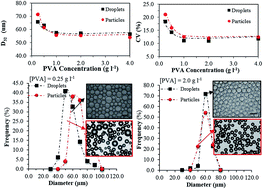
Fig. 7 (a) Variations in conversion versus time for two different PVA concentrations. (b and c) The Sauter-mean diameter (D32) and CV of monomer droplets/final particles versus PVA concentration, respectively. (d and e) Size distribution of monomer droplets and final particles at two PVA concentrations of 0.25 g l−1 and 2.0 g l−1, respectively. Insets are corresponding micrographs. Scale bar is 100 μm (method D, rpmE = 1000, rpmR = 250, Q = 2 ml min−1; ϕ = 0.20).

Fig. 7b illustrates the comparisons of the D32 of monomer droplets and final polymer particles versus PVA concentration. As can be clearly seen, the average size of droplets was slightly above that of particles when [PVA] ≥ 0.5 g l−1. We take a note that droplets shrink around 10% in diameter when they transform from MMA monomer droplets to PMMA particles (ρMMA = 0.940 g cm−3, ρPMMA = 1.18 g cm−3). This suggests that there was no significant droplet coalescence or break up during polymerisation within this range of PVA concentration and as a result a similar CV was obtained for polymer particles and monomer droplets. However, for [PVA] < 0.5 g l−1, particles size became larger than their initial droplet size, despite their shrinkage, and their CV increased, indicating a significant coalescence occurring in the course of polymerisation, as shown in Fig. 7c. The droplet/particle size distributions shown in Fig. 7d and e confirm that wide and narrow PSDs were obtained at typical low and high concentrations of PVA, respectively.

This clearly suggests that the degree of uniformity of droplets was effectively maintained during polymerisation if a sufficient amount of stabiliser, via which the growth stage could be hindered, was used. This minimum or critical concentration was found to be 0.5 g l−1 for the current polymerisation system. We showed in a previous section that in the absence of reaction in the membrane emulsification vessel, the critical PVA concentration is 1.0 g l−1 and droplets formed using PVA concentrations lower than this value underwent coalescence.

Fig. 7c shows that the CV of polymer beads from assisted-suspension polymerisation is smaller than 20%, which shows a significant improvement over that from conventional polymerisation, which is usually greater than 30%.40,41 To confirm this, we also made polymer beads using a similar formulation to that used in this research via a conventional polymerisation system in which both emulsification and polymerisation occurred simultaneously in the reactor ([PVA] = 1.0 g l−1, ϕ = 0.20, T = 75.0 ± 0.5 °C, rpmR = 700). The CV of the final beads was 42.0%, which is much greater than the CV of those obtained from the membrane-assisted polymerisation.

4. Conclusion

Highly uniform monomer droplets obtained via stirred cell-flat membrane (SCFM) were successfully converted to uniform polymer beads via suspension polymerisation. The similarity between the environments where drops form in the SCFM vessel and those of the polymerisation reactors where droplets undergo polymerisation facilitates the controllability of drops uniformity. We introduced a novel start-up method that did not allow intermixing of phases prior to emulsification and any associated mass transfer involved, thereby enhancing the uniformity of resulting drops.

Highly uniform droplets were obtained via membrane emulsification at an impeller speed range 500–1500 rpm and flowrates within 1.0–3.0 ml min−1. The size distribution of monomer droplets underwent broadening at high impeller speed due to droplet break up in the emulsification vessel. The monomer droplet size and CV increased with further increase in the flowrate above the optimal range. PVA helped to stabilise the droplets; however, there was not any apparent advantage in increasing the amount of stabiliser above its CMC. The CV significantly increased at low PVA concentration due to droplet coalescence in the emulsification vessel during circulation. Safe ranges of the reactor impeller speed and PVA concentration, within which the degree of uniformity of monomer droplets formed by membrane emulsification could be preserved during polymerisation, were found. One important highlight of this research is that we were able to decouple factors responsible for degradation of drops uniformity during circulation (in the emulsification vessel) from those affecting drops during formation at the membrane surface from the outset. The main conclusion drawn is that to achieve maximum drop uniformity the phase ratio of dispersed drops should be kept to minimum and drops leave the emulsification vessel as soon as they are formed. This suggests that an optimum range of operations for the SCFM device will be achieved in a continuous mode.

Acknowledgements

One of the authors gratefully acknowledges the generous financial sponsorship provided by the government of Saudi Arabia.

References

  1. S. Maass, F. Metz, T. Rehm and M. Kraume, Prediction of Drop Sizes For Liquid–Liquid Systems In Stirred Slim Reactors-Part I: Single Stage Impellers, Chem. Eng. J., 2010, 162, 792–801 CrossRef CAS.
  2. T. Uchida, K. Shiosaki, Y. Nakada, K. Fukada, Y. Eda, S. Tokiyoshi, N. Nagareya and K. Matsuyama, Pharm. Res., 1998, 15, 1708–1713 CrossRef CAS.
  3. J. L. Cleland, A. Lim, A. Daugherty, L. Barron, N. Desjardin, E. T. Duenas, D. J. Eastman, J. C. Vennari, T. Wrin and P. Berman, J. Pharm. Sci., 1998, 87, 1489–1495 CrossRef CAS PubMed.
  4. S. Brahim, D. Narinesingh and A. Guiseppi-Elie, J. Mol. Catal. B: Enzym., 2002, 18, 69–80 CrossRef CAS.
  5. M. Tang, X. J. Cao, Z. Z. Liu, X. Y. Wu and D. Gance, Process Biochem., 1999, 34, 857–862 CrossRef CAS.
  6. S. Mitragotri and J. Lahann, Nat. Mater., 2009, 8, 15–23 CrossRef CAS PubMed.
  7. B. W. Brooks, Angew. Makromol. Chem., 1990, 35–6, 121–140 Search PubMed.
  8. E. Vivaldolima, P. E. Wood, A. E. Hamielec and A. Penlidis, Ind. Eng. Chem. Res., 1997, 36, 939–965 CrossRef CAS.
  9. J. Alvarez and R. E. Martinez, J. Appl. Polym. Sci., 1999, 209–221 Search PubMed.
  10. P. J. Dowding, J. W. Goodwin and B. Vincent, Colloids Surf., A, 2001, 180, 301–309 CrossRef CAS.
  11. M. Konno, K. Arai and S. Saito, J. Chem. Eng. Jpn., 1982, 15, 131–135 CrossRef CAS.
  12. D. Maggioris, A. Goulas, A. H. Alexopoulos, E. G. Chatzi and C. Kiparissides, Chem. Eng. Sci., 2000, 55, 4611–4627 CrossRef CAS.
  13. F. Jahanzad, S. Sajjadi and B. W. Brooks, Ind. Eng. Chem. Res., 2005, 44, 4112–4119 CrossRef CAS.
  14. Z. Genwen and K. M. Suzanne, Chem. Eng. Sci., 1998, 53, 2063–2079 CrossRef.
  15. S. M. Joscelyne and G. Tragardh, J. Membr. Sci., 2000, 169, 107–117 CrossRef CAS.
  16. S. R. Kosvintsev, G. Gasparini, R. G. Holdich, I. W. Cumming and M. T. Stillwell, Ind. Eng. Chem. Res., 2005, 44, 9323–9330 CrossRef CAS.
  17. G. T. Vladisavljevic, I. Kobayashi and M. Nakajima, Microfluid. Nanofluid., 2012, 13, 151–178 CrossRef CAS.
  18. S. R. Kosvintsev, G. Gasparini and R. G. Holdich, J. Membr. Sci., 2008, 313, 182–189 CrossRef CAS.
  19. S. J. Peng and R. A. Williams, Chem. Eng. Res. Des., 1998, 76, 894–901 CrossRef CAS.
  20. G. T. Vladisavljevic and H. Schubert, J. Membr. Sci., 2003, 225, 15–23 CrossRef CAS.
  21. R. Wang, Y. Zhang, G. Ma and Z. Su, J. Appl. Polym. Sci., 2006, 102, 5018–5027 CrossRef CAS.
  22. G. H. Ma, C. J. An, H. Yuyama, Z. G. Su and S. Omi, J. Appl. Polym. Sci., 2003, 89, 163–178 CrossRef CAS.
  23. R. Nuisin, S. Omi and S. Kiatkamjornwong, J. Appl. Polym. Sci., 2003, 90, 3037–3050 CrossRef CAS.
  24. S. Omi, Colloids Surf., A, 1996, 109, 97–107 CrossRef CAS.
  25. R. G. Holdich, M. M. Dragosavac, G. T. Vladisavljevic and S. R. Kosvintsev, Ind. Eng. Chem. Res., 2010, 49, 3810–3817 CrossRef CAS.
  26. J. Santos, G. T. Vladisavljevic, R. G. Holdich, M. M. Dragosavac and J. Munoz, Chem. Eng. Res. Des., 2015, 98, 59–69 CrossRef CAS.
  27. V. Schröder, O. Behrend and H. Schubert, Colloids Surf., A, 1998, 202, 334–340 Search PubMed.
  28. M. Zerfa and B. W. Brooks, Colloids Surf., A, 1998, 132, 267–273 CrossRef CAS.
  29. J. D. Litster, T. Howes and Y. He, J. Chem. Eng. Jpn., 2004, 2, 181–186 Search PubMed.
  30. E. Egidi, G. Gasparini, R. G. Holdich, G. T. Vladisavljevic and S. R. Kosvintsev, J. Membr. Sci., 2008, 323, 414–420 CrossRef CAS.
  31. Y. Hong and F. Wang, Microfluid. Nanofluid., 2007, 3, 341–346 CrossRef CAS.
  32. P. Garstecki, M. J. Fuerstman, H. A. Stone and G. M. Whitesides, Lab Chip, 2006, 6, 437–446 RSC.
  33. I. Kobayashi, M. Yasuno, S. Iwamoto, A. Shono, K. Satoh and M. Nakajima, Colloids Surf., A, 2002, 207, 185–196 CrossRef CAS.
  34. V. Schroder, O. Behrend and H. Schubert, J. Colloid Interface Sci., 1998, 202, 334–340 CrossRef CAS.
  35. K. Kandori, K. Kishi and T. Ishikawa, Colloids Surf., 1991, 269–279 CrossRef CAS.
  36. K. Kandori, K. Kishi and T. Ishikawa, Colloids Surf., A, 1991, 55, 73–78 CAS.
  37. H. Yuyama, T. Watanabe, G. H. Ma, M. Nagai and S. Omi, Colloids Surf., A, 2000, 168, 159–174 CrossRef CAS.
  38. M. Zerfa and B. W. Brooks, Chem. Eng. Sci., 1996, 51, 3223–3233 CrossRef CAS.
  39. F. Jahanzad, S. Sajjadi and B.W. Brooks, Chem. Eng. Sci., 2005, 60, 5574–5589 CrossRef CAS.
  40. P. Cheremisinoff, Handb. Eng. Polym. Mater., 1997, 15, 185–186 Search PubMed.
  41. P. J. Dowding and B. Vincent, Colloids Surf., A, 2000, 161, 259–269 CrossRef CAS.

This journal is © The Royal Society of Chemistry 2016
Click here to see how this site uses Cookies. View our privacy policy here.