Ammonium ion detection in solution using vertically grown ZnO nanorod based field-effect transistor

Rafiq Ahmad*a, Nirmalya Tripathyb, Muhammad Yasir Khana, Kiesar Sideeq Bhata, Min-sang Ahna and Yoon-Bong Hahn*a
aSchool of Semiconductor and Chemical Engineering, Nanomaterials Processing Research Center, Chonbuk National University, 567 Baekjedaero, Deokjin-gu, Jeonju-si, Jeollabuk-do 54896, Republic of Korea. E-mail: ahmadrafiq38@gmail.com; ybhahn@chonbuk.ac.kr
bDepartment of BIN Fusion Technology, Chonbuk National University, 567 Baekjedaero, Deokjin-gu, Jeonju-si, Jeollabuk-do 54896, Republic of Korea

Received 15th April 2016 , Accepted 29th May 2016

First published on 2nd June 2016


Abstract

Vertically aligned ZnO nanorods were directly grown on a seeded glass substrate between a pre-deposited source–drain to fabricate a field-effect transistor (FET) based ammonium ion sensor. Controlled growth of aligned nanorods provided a well-defined large surface area for the detection of ammonium ions in solution.

Accurate detection of liquid ammonia (i.e. ammonium ion) level in environmental pollution control, food, clinical analyses and other industry applications has immense scientific importance.1 In particular, ammonia is one of the most important substances encountered in our daily lives. Specifically, the major industrial application is in agriculture, in fertilizer production. Its utilization for developing synthetic fibres, plastics and explosives is also very important. However, ammonia usage in chemical and fertilizer factories comprise the main sources of ammonium ion discharge in running water. In clinical samples, the dosage of ammonia concentration is very important because an abnormal status of ammonia is directly connected to organism dysfunction, like kidney and hepatic deficiency and diabetes.2 On the other hand, ammonia levels in the blood are measured with interest in sports medicine. During activity, the human body produces ammonia that can diffuse out of the blood into the lungs when the ammonia levels are higher than that in the air.3 Thus, a good sensor that can be used for continuous ammonium ion level monitoring is needed.

In this regard, various sensors such as electrochemical sensor, optical sensor, chemiresistive sensor, polymer and metal oxide based sensors, biomaterials based sensors and IV based sensors have been used for the detection of ammonia in gas and liquid forms.4 Among them, the IV technique is one of the simplest and easy techniques for an efficient detection of various hazardous chemicals and environmental pollutions. Several studies have been reported for the fabrication of IV based chemical sensor using various nanostructures.5 To further enhance the performance of sensing devices, a gate electrode over a channel region between the source and drain were used i.e. metal-oxide semiconductor FET (MOSFET). The MOSFET is one of the most widely used devices, which utilizes an electric field modulated by the size and shape of the source–drain channel. During analyte detection, a gate electrode controls the carrier (electrons and holes) flow through the channel formed between the source and drain that leads to a change in the drain current. As a better conduction channel, both conducting polymers such as polypyrrole (PPy) and semiconducting metal oxide nanostructures have been widely utilized as promising sensing materials due to their unique sensing abilities. Especially, in metal oxides family, zinc oxide (ZnO) possesses a special place due to its easy synthesis, distinctive optical, electrical and chemical sensing properties. The various important properties of ZnO includes its direct and wide band-gap (∼3.37 eV), high exciton binding energy (60 meV) much larger than other semiconducting materials, piezoelectric, easy and cost effective synthesis, high electron features, optical transparency and biocompatible nature and so on.6 But, there are only few reports of using ZnO nanostructures for ammonia chemical sensor applications.4f,7 However, in most of them, binders/polymers were used to fix the electrocatalysts on the electrode surface, which inevitably reduce electrocatalytic activity, poor reproducibility, and low stability.

Herein, we propose to grow vertically aligned ZnO nanorods directly on the seeded glass substrate between pre-deposited source–drain to fabricate a field-effect transistor (FET) based ammonium ion sensor. The directly grown nanorods not only provided much higher surface area but also improved reproducibility and stability for ammonium ion detection in solution.

Fig. 1 shows the schematic of fabrication strategy of the FET based ammonium ion sensor. In the first step, a cleaned glass substrate was taken and thin layers of silver source–drain electrodes (∼100 nm thick) were sputtered on the unmasked region having 3 mm length and 2 mm width (a). Then, in the same way channel region was defied using mask for the deposition of ∼60 nm ZnO seed layer between the source–drain electrodes with a channel length and width of 5 mm and 2 mm, respectively (b). In the next step, vertically aligned ZnO nanorods were grown on the seeded region (c). In a typical synthesis process, an equimolar (0.04 M) of Zn(NO3)2·6H2O and HMTA were dispersed in distilled water (50 mL); transferred into Pyrex glass bottle with suspended seeded substrate upside down; and heated at 85 °C for 4 h. After the completion of reaction, the electrodes were rinsed with distilled water to remove impurities and dried in air. Finally, the electrodes were passivated with PDMS leaving active area to reduce the leak current and eliminate the effect of metal-nanorods contact region (d), to makes sure that the entire conductance changes originate only from the nanorods. The fabricated FET device was connected to a data acquisition system for real-time data measurement (HP 4155A, semiconductor parameter analyzer) using a typical IV technique. To measure ammonium ion in a phosphate buffered solution (PBS, pH = 7.0), 50 μL of various concentrations of liquid ammonia (ammonium hydroxide solution in PBS buffer) was deposited on a pre-defined area by PDMS to make sure the same coverage for different solutions during each measurement. Then, the drain current (ID) dependence on drain–source voltage (VDS) was measured in the absence/presence of ammonium ion solution in a fixed gate–source voltage (VGS) range (1.0–5.0 V) through an Ag/AgCl reference electrode (diameter = 2 mm).


image file: c6ra09731f-f1.tif
Fig. 1 Schematic illustration fabrication process of the FET based ammonium ion sensor. (a) Silver source–drain deposition on glass substrate; (b) ZnO seed layer deposition by sputter; (c) ZnO nanorods growth and (d) PDMS covering.

Fig. 2 shows the representative field emission scanning electron microscopy (FESEM, Hitachi S4700) and transmission electron microscopy (TEM, JEOL-JEM-2010) images. From the surface and cross-sectional images (Fig. 2a and b), ZnO nanorods can be clearly seen in high density, uniformly grown and vertically aligned to the substrate. From the detailed structural characterization of as-synthesized ZnO nanorods (Fig. 2c–f), the low-magnification (c) and high-resolution (d) TEM images with corresponding diffraction pattern (e) exhibits full consistency with the obtained FESEM results in terms of morphology, shape and size of the ZnO nanorods. From the SAED image (f), the distance between two lattice fringes is about 0.52 nm, which corresponds to the d-spacing of [0001] crystal plans, consistent with that of the bulk wurtzite crystal of ZnO and confirms that the products are single crystalline and grown along the c-axis direction. The energy dispersive X-ray spectroscopy (EDX) confirms that the as-grown ZnO nanorods were composed of zinc and oxygen elements only without any impurity (inset of Fig. 2a).


image file: c6ra09731f-f2.tif
Fig. 2 (a) Top-view and (b) cross-sectional FESEM images, (c) TEM image of ZnO nanorods, (d) HRTEM image, (e) SAED pattern and (f) the corresponding FFT pattern of ZnO nanorods. Inset of a shows the EDX spectra of ZnO nanorods.

Further, we examined chemical composition and state of elements with X-ray photoelectron spectroscopy analysis (XPS) (Fig. 3). The spectrum showed C, Zn and O elements with no impurity (Fig. 3a). The C 1s peak at broad binding energy (284.6 eV) is due to use of carbon as reference for calibration analysis of other binding energies to avoid the specimen charging (Fig. 3b).8 The high-resolution Zn 2p spectrum shows a doublet whose binding energy peaks are at ∼1022.2 eV and ∼1045.28 eV, which corresponds to Zn 2p3/2 and Zn 2p1/2 core levels, respectively (Fig. 3c). The binding energy difference (∼23 eV) calculated from the XPS study confirms that Zn atoms were in Zn2+ oxidation state.8 The high-resolution XPS spectra of O 1s (Fig. 3d) showed the higher binding energy at ∼531.3 eV, corresponds to O2− ions on the wurtzite structure of hexagonal Zn2+ ions, surrounded by Zn atoms with the full supplement of nearest-neighbour O2− ions.9 The strong binding energies of Zn 2p and O 1s confirms the formation of pure ZnO materials by the association of Zn2+ and O2− ions to form Zn–O bonds in ZnO crystals.


image file: c6ra09731f-f3.tif
Fig. 3 (a) Wide scan XPS survey spectra of ZnO nanorods with high-resolution XPS spectra of (b) C 1s, (c) Zn 2p, and (d) O 1s.

The crystallinity of as-synthesized ZnO nanorods were characterized by X-ray diffractometer (XRD) measured with Cu-Kα radiations (λ = 1.54178 Å) in the range of 30–60° with 8° min−1 scanning speed and Raman-scattering measurements carried out at room temperature with 514.5 nm line of an Ar+ laser as the excitation source (Fig. 4). All the diffraction peaks were indexed to the wurtzite-structured hexagonal phase of single crystalline bulk ZnO with lattice constant of a = 3.249 and c = 5.206 Å (JCPDS card no. 35-1451). The clear and strong peak (002) indicates the growth of ZnO nanorods along the c-axis direction without any impurities (a). Additionally, the Raman spectra (b) shows a dominant and sharp peak at 437 cm−1 and a weak peak at 380 cm−1, which are assigned to the Raman active optical phonon E2 mode for the wurtzite ZnO and E2H–E2L (multi phonon process) and A1T modes, respectively. No peaks for E1L mode are found in the spectra.


image file: c6ra09731f-f4.tif
Fig. 4 (a) XRD pattern and (b) Raman spectra of the as-synthesized ZnO nanorods.

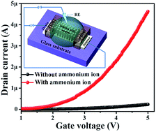
The electrical characterization of the fabricated FET based ammonium ion sensor was characterized by a typical IV technique (overall experimental setup is schematically shown in inset of Fig. 5). The electrical response of our sensing device measured in the absence and presence of 50 μM ammonium ion in PBS solution are shown in Fig. 5, confirms the high selectivity nature of ZnO nanorods to the ammonium ion in PBS solution. Also, as ZnO possesses good electro-catalytic and fast electron exchange properties, hence it is believed that due to these properties a rapid increase in the current was observed in the presence of ammonium ion.4f


image file: c6ra09731f-f5.tif
Fig. 5 IV response of FET based ammonium ion sensor without (a) and with (b) 50 μM ammonium ion in 0.1 M PBS solution (pH = 7.0).

To investigate the sensing properties of the fabricated FET ammonium ion sensor, the devices were exposed to the ammonium ion solutions of varying concentrations ranging from 0 to 2.5 mM (Fig. 6a). The sensor response gradually increases with ammonium ion concentration corresponding to the rapid electrons transfer relay into conduction band and results into an enhancement in the sensor response.4,10 A calibration curve was plotted by taking average of noticeable current changes over a potential range (i.e., 2.0 to 5.0 V; Fig. 6b), which exhibited a wide response range from 0.01 μM to 2.5 mM. A clear linear relationship of ammonium ion concentrations to average current response was presented with a high regression coefficient value of 0.9898, indicating a high degree of accuracy throughout the assay range. From the plot (Fig. 6b), a high sensitivity of 93.16 μA cm−2 mM−1 was obtained, which was the highest value as reported for ammonia sensors.4e,4f,11 In addition, the detection limit was also calculated to be 0.07 μM, using standard deviation of the response (SD) and the slope of the calibration curve (S), i.e. LOD = 3.3(SD/S). The better sensing performance of our sensors can be ascribed to the high surface area and direct electron mobility feature of the vertically grown ZnO nanorods on electrode active area.


image file: c6ra09731f-f6.tif
Fig. 6 (a) IV response of FET based ammonium ion sensor with increasing concentration of ammonium ion in 0.1 M PBS solution (pH = 7.0) at a fixed gate–source voltage range of 1.0–5.0 V through an Ag/AgCl reference electrode and (b) their calibration curve showing average current (i.e., 2.0 to 5.0 V) vs. ammonium ion in log scale (b).

As it is reported, the surface of ZnO nanorods are sensitive to both oxidizing and reducing analytes due to the presence of adsorbed oxygen species on the outer layer, which gets ionized into different oxygen species. The predominant most active surface oxygen species (O2−), leads to chemisorb oxygen onto the surface and decrease in the conductance of sensor or increase in resistance.12

 
O2 + 2e → 2O2−(ads) (1)

In addition to this, the increase in current could be attributed to the discharge of the trapped electron into the conduction band. Also, the decomposition of liquid ammonia molecule is an exothermic reaction, releasing the sufficient amount of energy to the electrons to overcome the barrier.

 
NH4OHliquid → NH4+ + OH (2)

As indicated in eqn (2), catalytic reaction of liquid ammonia results in the production of NH4+, which reacts with the surface adsorbed oxygen (Oads) and releases the trapped electrons to the conduction band of ZnO nanorods. The energy released during decomposition of adsorbed ammonia molecules would be sufficient for electrons to jump up into the conduction causing on increase in the conductivity of the sensor.12b

Anti-interference and stability, the other essential parameters of sensor were studied. At first, the sensor response was measured in only 50 μM of ammonium ion without any potential interfering reagents. Then, 100 μM of each major interference ions (Na+, K+, Cl) were added to 50 μM ammonium ion followed by sensors response measurements. In the presence of interfering ions with 50 μM ammonium ion, the sensor showed almost similar response to that of without any interference. Furthermore, stability of the fabricated devices was tested intermittently in a period of 10 weeks, and a good operational stability was obtained. The good anti-interference activity and stability suggested that the fabricated sensing devices were quite reliable for ammonium ion detection in solution.

Conclusions

In summary, we have successfully fabricated the FET based ammonium ion sensor by growing vertically aligned ZnO nanorods directly on the seeded glass substrate between pre-deposited source–drain. The direct synthesis of ZnO nanorods on active area provides high surface area and easy substrate penetration structures, which were considered as the key factors for improved sensing performances. As a result, fabricated FET sensor showed excellent sensing performance including high sensitivity, wide linear range, low concentration detection ability, good selectivity and storage stability. The vertically aligned ZnO nanorods provided direct and efficient pathways for analyte activity within the conductive channel, and contributed to both high sensitivity and stability. Hence, this study provides an efficient strategy for the fabrication of a low-cost, fast, and portable device for environmental monitoring and disease diagnosis.

Acknowledgements

This work was supported by National Leading Research Laboratory program (NRF-2011-0028899) through the National Research Foundation of Korea funded by the Ministry of Science, ICT and Future Planning.

Notes and references

  1. (a) P. W. Alexander, T. Dikitrakopoloulos and D. B. Hibbert, Electroanalysis, 1997, 9, 1331 CrossRef CAS; (b) L. Y. Heng, S. Alva and M. Ahmad, Sens. Actuators, B, 2004, 98, 160 CrossRef CAS; (c) R. Ghosh, A. Midya, S. Santra, S. K. Ray and P. K. Guha, ACS Appl. Mater. Interfaces, 2013, 5, 7599 CrossRef CAS PubMed; (d) R. Subramanian, K. Crowley, A. Morrin and A. J. Killard, Anal. Methods, 2013, 5, 134 RSC.
  2. (a) M.-Z. Dai, Y.-L. Lin, H.-C. Lin, H.-W. Zan, K.-T. Chang, H.-F. Meng, J.-W. Liao, M.-J. Tsai and H. Cheng, Anal. Chem., 2013, 85, 3110 CrossRef CAS PubMed; (b) F. Nicolao, C. Efrati, A. Masini, M. Merli, A. F. Attili and O. Riggio, J. Hepatol., 2003, 38, 441 CrossRef CAS PubMed; (c) B. Maranda, J. Cousineau, P. Allard and M. Lambert, Clin. Biochem., 2007, 40, 531 CrossRef CAS PubMed.
  3. C.-Y. Shen, H.-C. Huang and R.-C. Hwang, J. Chemom., 2008, 22, 548 CrossRef CAS.
  4. (a) S. Koul, R. Chandra and S. K. Dhawan, Sens. Actuators, B, 2001, 75, 151 CrossRef CAS; (b) A. Oprea, E. Simon, M. Fleischer, H.-P. Frerichs, C. Wilbertz, M. Lehmann and U. Weimar, Sens. Actuators, B, 2005, 111–112, 582 CrossRef CAS; (c) L. Li, P. Gao, M. Baumgarten, K. Müllen, N. Lu, H. Fuchs and L. Chi, Adv. Mater., 2013, 25, 3419 CrossRef CAS PubMed; (d) S. Chen and G. Sun, ACS Appl. Mater. Interfaces, 2013, 5, 6473 CrossRef CAS PubMed; (e) K. Crowley, E. O'Malley, A. Morrin, M. R. Smyth and A. J. Killard, Analyst, 2008, 133, 391 RSC; (f) G. N. Dar, A. Umar, S. A. Zaidi, S. Baskoutas, S. W. Hwang, M. Abaker, A. Al-Hajry and S. A. Al-Sayari, Talanta, 2012, 89, 155–161 CrossRef CAS PubMed.
  5. (a) A. Umar, M. S. Akhtar, G. N. Dar and S. Baskoutas, Talanta, 2003, 116, 1060 CrossRef PubMed; (b) G. N. Dar, A. Umar, S. A. Zaidi, A. A. Ibrahim, M. Abaker, S. Baskoutas and M. S. Al-Assiri, Sens. Actuators, B, 2012, 173, 72 CrossRef CAS; (c) R. Ahmad, N. Tripathy, D.-U.-J. Jung and Y.-B. Hahn, Chem. Commun., 2014, 50, 1890 RSC; (d) A. Umar, F. Al-Hazmi, G. N. Dar, S. A. Zaidi, R. M. Al-Tuwirqi, F. Alnowaiser, A. A. Al-Ghamdi and S. W. Hwang, Sens. Actuators, B, 2012, 166–167, 97 CrossRef CAS.
  6. (a) A. Rafiq, N. Tripathy, J.-H. Park and Y.-B. Hahn, Chem. Commun., 2015, 81, 11968 Search PubMed; (b) N. Tripathy, A. Rafiq, H. A. Ko, G. Khang and Y.-B. Hahn, Chem. Commun., 2015, 7, 4088 CAS; (c) N. Tripathy, A. Rafiq, H. A. Ko, G. Khang and Y.-B. Hahn, Chem. Commun., 2015, 51, 2585 RSC; (d) Y.-B. Hahn, R. Ahmad and N. Tripathy, Chem. Commun., 2012, 48, 10369 RSC; (e) N. Tripathy, A. Rafiq, E. Y. Kim and G. Khang, RSC Adv., 2014, 4, 46049 RSC; (f) N. Tripathy, A. Rafiq, S. H. Bang, J. Min and Y.-B. Hahn, Chem. Commun., 2014, 80, 9298 RSC; (g) R. Ahmad, N. Tripathy and Y.-B. Hahn, Biosens. Bioelectron., 2013, 45, 281 CrossRef CAS PubMed; (h) S. Kumar and R. Thangavel, J. Sol-Gel Sci. Technol., 2014, 70, 506 CrossRef CAS; (i) S. Kumar, P. Kaur, C. L. Chen, R. Thangavel, C. L. Dong, Y. K. Ho, J. F. Lee, T. S. Chan, T. K. Chen, B. H. Mok, S. M. Rao and M. K. Wu, J. Alloys Compd., 2014, 588, 705 CrossRef CAS.
  7. (a) X. Wang, J. Zhang and Z. Zhu, Appl. Surf. Sci., 2006, 252, 2404 CrossRef CAS; (b) V. B. Raj, A. T. Nimal, Y. Parmar, M. U. Sharma, K. Sreenivas and V. Gupta, Sens. Actuators, B, 2001, 147, 517 CrossRef.
  8. C. D. Wagner, W. M. Riggs, L. E. Davis, J. F. Moulder and G. E. Muilenberg, Handbook of X-ray Photoelectron Spectroscopy, Perkin Elmer, Eden Prairie, 1979 Search PubMed.
  9. (a) J. C. C. Fan and J. B. Goodenough, J. Appl. Phys., 1977, 48, 3524 CrossRef CAS; (b) R. Escudero and R. Escamilla, Solid State Commun., 2011, 151, 97 CrossRef CAS; (c) H. Yin, K. Yu, C. Song, Z. Wang and Z. Zhu, Nanoscale, 2014, 6, 11820 RSC.
  10. (a) A. Umar, M. M. Rahman, S. H. Kim and Y. B. Hahn, Chem. Commun., 2008, 166 RSC; (b) A. O. Dikovska, G. B. Atanasova, N. N. Nedyalkov, P. K. Stefanov, P. A. Atanasov, E. I. Karakoleva and A. T. Andreev, Sens. Actuators, B, 2010, 146, 331 CrossRef CAS.
  11. (a) M. M. Rahman, A. Jamal and S. M. Faisal, J. Nanopart. Res., 2011, 13, 3789 CrossRef CAS; (b) S. B. Khan, M. M. Rahman, E. S. Jang, K. Akhtar and H. Han, Talanta, 2011, 84, 1005 CrossRef CAS PubMed; (c) M. M. Rahman, A. Jamal and M. Faisal, ACS Appl. Mater. Interfaces, 2011, 3, 1346 CrossRef CAS PubMed; (d) R. K. Bedi and I. Singh, ACS Appl. Mater. Interfaces, 2010, 2, 1361 CrossRef CAS PubMed; (e) J.-M. Tulliani, A. Cavalieri, S. Musso, E. Sardella and F. Geobaldo, Sens. Actuators, B, 2011, 152, 144 CrossRef CAS; (f) T. L. Ling, M. Ahmad and L. Y. Heng, Sens. Actuators, B, 2012, 171–172, 994 Search PubMed.
  12. (a) G. H. Jain, L. A. Patil, P. P. Patil, U. P. Mulik and K. R. Patil, Bull. Mater. Sci., 2007, 30, 9 CrossRef CAS; (b) S. G. Ansari, Z. A. Ansari, H.-K. Seo, G.-S. Kim, Y.-S. Kim, G. Khang and H.-S. Shin, Sens. Actuators, B, 2008, 132, 265 CrossRef CAS.

This journal is © The Royal Society of Chemistry 2016
Click here to see how this site uses Cookies. View our privacy policy here.