Improving detection sensitivity by oriented bioconjugation of antibodies to quantum dots with a flexible spacer arm for immunoassay

Bingbo Zhanga, Jiani Yua, Chang Liub, Jun Wanga, Huanxing Han*b, Pengfei Zhang*b and Donglu Shicd
aInstitute of Photomedicine, Shanghai Skin Disease Hospital, The Institute for Biomedical Engineering & Nano Science, Tongji University School of Medicine, Shanghai 200443, China
bCenter for Translational Medicine, Changzheng Hospital, Second Military Medical University, Shanghai 200433, China. E-mail: ashuizhang@gmail.com; hanhuanxing@sohu.com; Tel: +86-021-81871908 Tel: +86-021-81871907
cThe Institute for Translational Nanomedicine, Shanghai East Hospital, The Institute for Biomedical Engineering & Nano Science, Tongji University School of Medicine, Shanghai 200092, China
dThe Materials Science and Engineering Program, College of Engineering and Applied Science, University of Cincinnati, Cincinnati, OH 45221, USA

Received 11th April 2016 , Accepted 15th May 2016

First published on 17th May 2016


Abstract

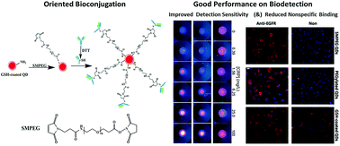
Antibodies with high targeting capability after being labelled by quantum dots (QDs) have been of great importance for in vitro diagnostic, cellular imaging and in vivo theranostic applications. However, such work concentrating on the conjugation of QDs and antibodies is still insufficient. It is encouraged to develop reliable bioconjugation strategies to improve detection sensitivity of QD–antibody bioconjugates. In this study, two bioconjugation approaches were used for antibody and QDs covalent coupling. Their detection sensitivity was parallelly determined and compared. It was experimentally found that succinimidyl valerate-PEG-maleimide (SMPEG) as the cross linker for oriented bioconjugation of antibodies to QDs with a flexible spacer arm can significantly improve the detection sensitivity of the resulting QD–antibody bioconjugates, compared with those prepared with traditional carbodiimide chemistry using EDC·HCl as the cross linker. Gel electrophoresis suggests antibodies can be successfully conjugated with QDs by both the EDC method and the SMPEG method, but immunofiltration assay shows QD–antibody bioconjugates obtained by the SMPEG strategy have lower detection limits and better linear scope patterns for in vitro molecular diagnosis. In vitro cancer cell imaging further shows that QD–antibody bioconjugates prepared by the SMPEG strategy have superior cell targeting capability without observable nonspecific binding, while the EDC-mediated QD–antibody bioconjugates have low signal to noise ratios with obvious nonspecific cellular binding. Moreover, the number of antibodies conjugated on the surface of QDs was determined by the chemiluminescent dot blot assay, showing 2.13 ± 0.5 and 0.76 ± 0.2 of antibodies per QD by EDC-mediated and SMPEG-mediated coupling, respectively. It further verifies that the SMPEG-mediated coupling strategy can significantly maintain the biological activity of the linked antibodies, even though it has low labelling efficiency. It could be therefore claimed that oriented bioconjugation via the SMPEG coupling strategy is favored by immunoassays, with improved detection sensitivity.


1. Introduction

In the last two decades, a variety of inorganic nanocrystals have been designed, synthesized, and functionized, with the aim of utilizing their unique nanoscale properties to either improve existing biosensing methods or to develop new sensing strategies.1–4 Quantum dots (QDs) are among the most widely used nanocrystals owing to their unique optical properties (e.g. high quantum yields, narrow and size tunable emission wavelengths, broad excitation spectra and resistant to photobleaching) for developing various biosensors.4–7 Although QDs-based immunoprobes are frequently reported for biomedical detection applications, their low binding efficiency, poor colloidal stability and nonspecific binding can lead to low sensitivity and false positive results, which undoubtedly spoils their extensive applications.8–11 There is therefore a great need to improve the detection sensitivity of nanoprobes, particularly for some trace substances/quantitative determinations.

QDs-based immunoprobes can be generally prepared by coupling antibodies to QDs via crosslinkers.7,12,13 Antibody coupling or bioconjugation is considered an essential process for the development of immunosensors. The bioconjugation strategy significantly affects their detection sensitivities, due to the concentration and orientation of antibodies on the surface of QDs.14–16 Also, the physical absorption of the resulting bioconjugates can further disturb immunoassays, namely, nonspecific binding can easily arise a false positive.8,9,11

The conjugation approach of QDs and antibodies can be varied by means of different crosslinkers and antibody modifications used.16–19 And the resulting targeting capability of corresponding QD–antibody bioconjugates can be diverse, since the linked antibodies could lose their targeting capabilities with antigen recognition epitope blocked upon conjugation with nanoparticles.16,19,20 This targeting loss can directly demoralize detection sensitivity. 1-Ethyl-3-(3-dimethyllaminopropyl)carbodiimide hydrochloride (EDC·HCl), for instance, a commonly used crosslinker for activating carboxyl groups of nanoparticles and then coupling amine groups of antibodies, is considered mediocre in their resulting targeting performance.21,22 This mature conjugation method is quite straightforward and cost effective, yet it is non-site-directed attachment, using the chemical groups both of the Fab region and the Fc region, and could damage antigen-binding site of antibody when Fab region touched. Active groups, such as amine groups and carboxyl group are generally distributed fully in antibodies, including Fab regions and Fc regions. As such, advanced strategies to oriented bioconjugation should be encouraged and developed to avoid antigen-binding sites.23–26

Efforts on oriented coupling of antibodies are increasingly reported in recent years.24,25,27–29 These site-directed methods aim to keep the antigen-binding sites of the antibodies away from the adherent surfaces and expose outward, thereby increasing the interaction probability with antigens. One approach is to oxidize the carbohydrate moieties on the antibody's Fc region with periodate to form aldehyde groups, which are then chemically bound to hydrazide-activated surfaces.30 As antibodies are only glycosylated at a single site in the CH2 domain of each heavy chain, this approach leaves the two Fab regions of the antibodies completely intact to interact with antigens. This method does not disconnect the framework of antibody. While another approach involves reduction of disulfide bonds between the heavy chain and the light chain. N-Succinimidyl-3-(2-pyridyldithiol)propionate (SPDP) and succinimidyl 4-(N-maleimidomethyl)cyclohexane-1-1-carboxylate (SMCC) are two frequently used crosslinker in this procedure.31,32 Although such work have been reported, other preferable crosslinkers with flexible spacer arms should be encouraged to eliminate nonspecific binding in biomedical detection, in addition to improving detection sensitivity.

In this study, two conjugation strategies and three different spacers were employed for QDs and antibodies bioconjugation. The resulting detection sensitivity, cell targeting and anti nonspecific binding capability were experimentally investigated and compared with each other.

2. Materials and methods

2.1 Main reagents

Cadmium oxide (99.99%), selenium powder (99.99%), zinc oxide (ZnO, 99.99%), sulfur (99.98%), octadecylamine (ODA, 90%), oleic acid (OA, 90%), 1-octadecene (ODE, 90%), tri-n-octylphosphine oxide (TOPO, 90%) and 1-ethyl-3-(3-dimethyllaminopropyl) carbodiimide hydrochloride (EDC·HCl) and N-hydroxysulfosuccinimide sodium salt (sulfo-NHS) were purchased from Sigma-Aldrich. Succinimidyl valerate-PEG-maleimide (SMPEG, Mw = 3400) and bifunctional carboxyl PEG (Mw = 2000) were bought from Laysan Bio Inc., USA. Dithiothreitol (DTT, 98%) and glutathione (GSH, reduced, 98+ %) were purchased from Sigma-Aldrich. Anti-epidermal growth factor receptor (Anti-EGFR) and anti-C-reactive protein (anti-CRP) antibodies were purchased from Millipore Corporation. Bovine serum albumin (BSA) was purchased from Beijing Dingguo Biotechnology. Trichloromethane, acetone, sodium hydrogen carbonate, dimethylsulfoxide (DMSO), and argon were purchased from local suppliers. Deionized water (18.2 MΩ cm resistivity at 25 °C) was used in this study. All the chemicals were used without further purification.

2.2 QDs phase transfer and surface engineering

Hydrophobic CdSe/ZnSe/ZnS core/shell QDs were prepared according to our previously reported methods.33 The synthesized hydrophobic core/shell QDs were transferred into water phase by replacing hydrophobic ligands with glutathione (GSH) molecules.34 Briefly, 25 mg of GSH and 20 mg of sodium hydroxide were dissolved in 1 mL of methanol, and mixed with 0.5 mL of 5 μM of oil soluble QDs solution in chloroform. After stirring overnight, pure water was added to disperse the precipitates. Then, the QDs solution was filtered through a 0.22 μm syringe filter to remove the aggregates, and dialyzed against pure water for 2 days to remove the excess GSH molecules. Finally, GSH modified QDs (GSH-coated QDs) solution was concentrated to about 5 μM by rotary evaporation under reduced pressure.

To improve the stability of GSH-coated QDs, bifunctional carboxyl PEG molecules were used as spacer arms.35 Typically, 5 μmol of NHS and 5 μmol of EDC·HCl were added into 10 μmol of carboxyl-PEG-carboxyl in 5 mL dimethylsulfoxide (DMSO) solution, and allowed to react for 30 min at room temperature to activate the carboxyl groups. Afterwards, DTT was added into the solution to quench the excess EDC·HCl, and GSH-coated QDs were then added to the solution, and the reaction continued for another 4 h. The resultant mixture was dialyzed against pure water for 48 h to thoroughly remove the unbound PEG and other small molecules using a 14 kDa MWCO dialysis tube, and then the PEGylated QDs solution was finally concentrated by rotary evaporation under reduced pressure.

2.3 Conjugation of antibodies to QDs via carbodiimide chemistry

QD–antibody bioconjugates prepared by the classical carbodiimide chemistry were used as the control group. Carboxyl groups on surface of QDs were firstly activated by incubating with EDC·HCl and sulfo-NHS, and then formed amide bonds with the amino groups of antibodies. Sulfo-NHS was used in the reaction since NHS has been known to stabilize the intermediates. Breifly, 500 μL of GSH-coated QDs (1 μM) or PEGylated QDs (1 μM) were mixed EDC·HCl and sulfo-NHS with the mole ratio of QDs[thin space (1/6-em)]:[thin space (1/6-em)]EDC·HCL[thin space (1/6-em)]:[thin space (1/6-em)]sulfo-NHS is 1[thin space (1/6-em)]:[thin space (1/6-em)]1000[thin space (1/6-em)]:[thin space (1/6-em)]1000. After 30 min incubation, excess crosslinkers were removed by ultrafiltation (MWCO, 100 kDa). Afterward, purified antibody were added and incubated for another 1 h, and blocked with BSA for another 1 h. Finally, QD–antibody bioconjugates were purified by centrifugation at 32[thin space (1/6-em)]000 rpm for 1 h and washing with PBST (0.01 M, pH 7.4, 0.02% Tween-20) for 3 times.

2.4 Oriented conjugation of antibodies to QDs via SMPEG with a flexible spacer arm

Succinimidyl valerate-PEG-Maleimide (SMPEG) was used for preparation of QD–antibody bioconjugates. Typically, amine groups on surface of GSH-coated QDs reacted with 1000 equivalent of SMPEG for 1 h, and the formed maleimide functionalized QDs were purified via ultrafiltration (MWCO, 100 kDa) to remove the excess crosslinkers. Antibody fragments with sulfhydryl groups were obtained by reduction of antibodies with 20 mM DTT for 1 h. The reduced antibodies were purified from DTT with ultrafiltration (MWCO, 30 kDa). Afterwards, maleimide functionalized QDs were immediately mixed with the above purified reduced antibody fragments. After 1 h incubation, BSA was added to block the unreacted sites and incubated for another 1 h. The resulting bioconjugates were centrifuged at 32[thin space (1/6-em)]000 rpm for 1 h and washed with PBST (0.01 M, pH 7.4, 0.02% Tween-20) for three times.

2.5 Immunofiltration assay with QD–antibody bioconjugates

Immunofiltration assay as a kind of sandwich immunoassays was used to test the detection sensitivity of the prepared QD–antibody bioconjugates. The system was mainly composed of a thin, porous nitrocellulose (NC) membrane on which anti-CRP monoclonal antibody was immobilized. The membrane was positioned over an absorbent paper and sealed in a plastic cassette containing a hole to expose the membrane. The setup details can be found in our published paper.36 Sample was firstly mixed with QD–antibody bioconjugates, and antigens in sample will be captured by QD–antibody bioconjugates. Subsequently, the assay was typically performed as following: 5 μL of serum sample was added into QD–antibody conjugates solution and gently pipetted for at least 3 times. Next, 120 μL of conjugates solution was added into the hole of immunofiltration device. After the conjugates solution totally soaked in, 200 μL of washing buffer added to rinse the unbound conjugates. The whole assay can be finished within 5 min. The test results can be read with naked eyes under a UV light (365 nm) illumination, and the corresponding fluorescent images were captured by a Canon G12 digital camera. The fluorescent intensities of QDs on test spots were measured by an optical fiber spectroscopy couple with a UV laser (405 nm).

2.6 Cell imaging

A549 tumor cells were cultured in RPMI supplemented with 10% fetal bovine serum, 100 U mL−1 penicillin, 0.1 mg mL−1 streptomycin at 37 °C in 5% CO2. After 24 h seeding cells on coverslips, the cells were washed with 20 mM PBS (pH 7.2) and fixed with 4% paraformaldehyde and 0.1% Triton X-100 in PBS for 15 min each. The fixative was then aspirated, and cells washed with PBS three times. Cells in coverslips were blocked with 1% BSA in PBS, and incubated for 1 h with rabbit anti-EGFR antibody (Epitomics, #1902-1, CA, USA) as the primary antibody followed by QDs conjugated with goat-anti-rabbit antibody as the second antibody (Boster Biotech Inc, Wuhan, China) for 1 h. Cell nuclei were stained with 4′,6-diamidino-2-phenylindole dihydrochloride (DAPI).

2.7 Chemiluminescence dot blot assay

To determine the amount of antibody conjugated with QDs, chemiluminescent dot blot assay was used. 10 μL of QD–antibody bioconjugates and anti-CRP antibody dilution were directly spotted on a methanol activated PVDF membrane (Millipore, USA), and dried at 37 °C for 2 h. Dried membrane was further blocked with 5% BSA in PBS buffer for 1 h at room temperature. After washing with PBST buffer for several times, the membrane was stained with anti-mouse horseradish peroxidase (HRP) conjugated secondary antibody (1[thin space (1/6-em)]:[thin space (1/6-em)]5000 dilution, Jackson ImmunoResearch, USA) in 5% BSA buffer and washed with PBST buffer for three times. Immunodetection was performed using enhance chemiluminescence (ECL) substrate (Pierce technology) and detected with a ChemiDoc XRS imaging system (Biorad, USA).

We measured the density (signal) of the protein spot on the PVDF membrane and compared them to controls of known antibody concentrations. Using image analysis software that measures the spot density of chemiluminescence image obtained by dot blot assay, we fitted curve to known concentrations of unconjugated antibody to obtain standard curve of antibody spot densities. And by using this curve, we then determined the concentration of antibody spot associated with covalently bound antibody. The average number (N) of antibody conjugated with QDs was calculated by comparing the molar concentration of antibody (Cab) and QDs (CQDs) of the purified QD–antibody conjugates, i.e., N = Nab/NQDs. For more details, one can refer to the published paper.37 The concentration of QDs was determined by recording the absorbance at peak wavelength using Beer's law and the molar absorbance coefficient calculated by the published paper to calculate the number of QDs.38

2.8 Characterizations

Absorption spectra of QDs were acquired with a UV-vis spectrophotometer (Shimazu 2450, Japan), and photoluminescence (PL) spectra and intensities of QDs were recorded on a fluorescence spectrometer (PerkinElmer LS-55, USA) or a multimode microplate reader (Tecan Infinite M1000, Switzerland). Surface modification of QDs was characterized by gel electrophoresis. 0.5% agarose gels in Tris–EDTA acetate (TEA) buffer were run at 80 V for 20 min.

3. Results and discussions

3.1 Preparation and characterization of QD–antibody bioconjugates

QDs and antibody conjugation is a complicated chemical process due to the varying physico-chemical properties of nanoparticles, including their water solubility, sizes, quantum yields, and surface microenvironments, and the involved antibodies with different molecular structures. As such, the resulting QD–antibody bioconjugates could be aggregated, or quenched, and in most cases lose their binding affinities toward biomolecules due to the steric hindrance of the nanoparticles and failure of the antigen-binding sites of antibodies.39

Conjugation of biomolecules to QDs is vital for their biomedical applications. It is still a challenge to seek for simple, effective and repeatable conjugation procedures.39 Generally, prerequisite is that QDs should hold good colloidal stability and photostability, active chemical groups and flexible spacer arms for an effective antibody coupling. Furthermore, cross linkers also play crucial roles in QDs and antibody conjugation process. Taking these issues into consideration, in this study, home-made high quality core/shell QDs with high quantum yield (72%) were first synthesized. To endow hydrophilicity, water soluble amino acid GSH was applied to exchange the hydrophobic ligands of QDs, since it has a free thiol for ligand exchange and also a carboxyl group and an amino group for bioconjugation. This surface engineering method was reported by our early study and other group.34,35 The phase transfer was found effective, straightforward and reproducible. It was experimentally found ligand exchange by GSH have little impact on the resulting size and distribution, which is mainly attributed to the high affinity of GSH to QDs and its low molecular weight. And their high quantum yield is nearly maintained, favoring for fluorescent detection and imaging. The colloidal stability of PEGylated QDs based on GSH-coated QDs was found excellent, showing hydrodynamic diameters (HDs) nearly keep constant in pH 3–11 range without photobleaching. More details can be found in our previously published paper.35 It's worth pointing out the prerequisite of the stability of QDs is guaranteed, and this superior stability can significantly benefits the conjugation process in return in this study.

In this study, two kinds of crosslinkers were used for covalent conjugation, namely, EDC·HCl/sulfo-NHS and SMPEG. Both GSH-coated QDs and PEGylated QDs with carboxy groups were activated by EDC·HCl and sulfo-NHS, and then coupled with antibodies, respectively. In parallel, GSH-coated QDs with amino group were functionalized by SMPEG, followed linked with antibody fragments with free thiols. The QDs and antibody conjugation is illustrated in Scheme 1. The main stages of conjugation process were investigated via gel electrophoresis technique, as shown in Fig. 1A. Gel electrophoresis was considered a vivid and visual method to study the size and surface charge distribution of nanoparticles.40,41 This analysis can demonstrate the changes of particle size and surface charges along with the coupling process. In gel running buffer (pH 8.5), GSH-coated QDs and their PEGylated QDs both moved toward the anode trimly with very narrow bands. It is worth noting that PEGylated QDs swim more slowly when compared with GSH-coated QDs, which could be attributed to the increased size after PEGylation of the GSH-coated QDs. SMPEG-functionalized GSH-coated QDs exhibit similar electrophoresis migration pattern with GSH-coated QDs, locating at the same distance from the wells. The fact that SMPEG attaching can enlarge the size of QDs but can also boost the negative charges of QDs by neutralizing their own effects on gel migration. Upon conjugation with antibodies, the moving distances become much shorter, owing to the pronounced increase of sizes. And migrating tails appear in QD–antibody bioconjugates bands, mainly due to the uneven migrations of the large sized bioconjugates. This evidence clearly demonstrates the successful conjugation of QDs with antibodies.


image file: c6ra09279a-s1.tif
Scheme 1 Schematic illustration of the QD–antibody conjugation strategies using two different crosslinkers (EDC·HCl/sulfo-NHS, and SMPEG); and PEG as the spacer arm was used in these two conjugation strategies. The impacts of crosslinker and spacer arm on the affinity of the resulting QD–antibody bioconjugates were investigated in this study.

image file: c6ra09279a-f1.tif
Fig. 1 Gel electrophoresis analysis of QDs, including GSH-coated QDs, PEGylated GSH-coated QDs, SMPEG functionalized GSH-coated QDs and their antibody-coupled counterparts from left to right (A); UV-vis absorption spectrum (B) and fluorescent emission spectrum (C) of GSH-coated QDs, SMPEG functionalized GSH-coated QDs and their antibody-coupled counterparts.

Spectroscopic analysis of GSH-coated QDs, SMPEG-functionalized GSH-coated QDs and antibody-coupled counterparts are presented in Fig. 1B and C. Their UV-vis absorption and fluorescent emission spectrum are basically the same. It declares conjugation process have little impact on fluorescent emission.

3.2 Detection sensitivity determination

To evaluate and compare the immuno function of the QD–antibody bioconjugates prepared with different conjugation strategies, the immunofiltration assays using QDs as label instead of traditional gold nanoparticles were respectively carried out with different QD–antibody bioconjugates.36 As shown in Fig. 2A, non-oriented QD–antibody bioconjugates prepared from GSH-coated QDs via EDC strategy show irregular signals for CRP detection with severe nonspecific binding. PEGylated QDs derived from GSH-coated QDs show much better performance on reduction of nonspecific binding and their signals exhibit CRP concentration dependent increasing. This detection improvement mainly benefits from PEGylation of QDs. PEG in this study acting as the spacer arm with around 2k molecular weight has flexible chain and excellent water solubilization, and is considered good material for reducing nonspecific binding, lowering steric hindrance and enhancing hydrophily.42,43 And its detection limit of QD–antibody bioconjugates via EDC-mediated approach is found around 1.56 mg L−1 for CRP detection. While oriented QD–antibody bioconjugates prepared from GSH-coated QDs via SMPEG strategy presents higher detection limit around 0.39 mg L−1 for CRP detection. This increase in detection sensitivity is mainly attributed to the orientation coupling of antibodies on QDs. The labeled antibodies maintained their activity for antigen specific targeting without destroying their antigen-binding sites.44
image file: c6ra09279a-f2.tif
Fig. 2 (A) Fluorescent images and (B and C) quantitative results of the QD–antibody based immunofiltration assay for rapid detection of CRP. QD–antibody bioconjugates obtained by different conjugation strategies were respectively used for CRP detection under the same concentration (1 μM) and detection procedures (200 μL QD–antibody bioconjugates mixed with 5 μL samples, then loading 120 μL of the conjugates into the immunofiltration pad).

Particularly, linear detection ranges of the above three different QD–antibody bioconjugates for CRP is varying (Fig. 2B and C). Results show that SMPEG-based QD–antibody bioconjugates hold the best linear detection range with the greatest slope, QD–antibody bioconjugates based on PEGylated QDs via EDC coupling method followed, and GSH-coated QDs via EDC coupling method actually failed in biodetection. A wide linear detection range is conducive to establishing equation and calculation for biodetection. Interestingly, nonspecific binding resulted from SMPEG functionalized QDs is also significantly reduced. And with increasing of CRP concentrations, the signals are found rising regularly. In this study, SMPEG, as a novel cross linker, contains a length of PEG flexible chain, which favors for antifouling and also decreasing steric hindrance for antigen targeting.42,43 It is obvious that the oriented QD–antibody bioconjugates with a flexible arm are more promising for the immunoassay detection with a wider dynamic range and higher detection sensitivity.44

3.3 Cell-targeted imaging and anti-nonspecific binding study

In this study, we concentrate on developing an effective QD–antibody coupling method to improve detection sensitivity of the resulting bioconjugates. The impressive performance on protein rapid detection of SMPEG-mediated bioconjugation strategy is convincingly displayed above. To further explore its applications, tumor cell diagnosis and imaging were conducted. EGFR expression on A549 cells were detected by immuno-QDs nanoprobes conjugated by different methods in this study (Fig. 3). EGFR is reported overexpressed in most cancer cells and has been recognized as a biomarker of cancer cells since activation of EGFR is associated with the tumorigenic mechanisms such as autonomous cell growth, invasion, angiogenesis, and metastasis.45 Therefore, accurate and quantitative determination of EGFR on cells has been of great importance. In this cell imaging procedure, A549 cells were first incubated with rabbit anti-EGFR as the primary antibody, and followed by goat-anti-rabbit second antibody coupled QDs, and were washed for imaging.
image file: c6ra09279a-f3.tif
Fig. 3 Cell-targeted imaging (A) and its quantitative evaluation (B) by QDs on EGFR-positive A549 cells. A549 cells were first treated with or without primary antibody (rabbit anti-EGFR antibody) for 1 h, and followed by goat-anti-rabbit second antibody conjugated by QDs for 1 h. GSH-coated QDs, PEGylated GSH-coated QDs, and SMPEG-functionalized GSH-coated QDs were compared in this cell targeted imaging. Cell nucleus were stained by DAPI.

Fig. 3 shows SMPEG-mediated bioconjugation strategy provides the most persuasive evidence for cancer cell diagnosis. The other two QD–antibody bioconjugates via the classical carbodiimide chemistry cause unnegligible nonspecific binding. And this physical absorption in most cases can get diagnosis in trouble. A549 cells were stained with the highest contrast by the anti-rabbit second antibody coupled QDs via SMPEG as the crosslinker. And there is no obvious nonspecific binding in this case. Furthermore, their corresponding fluorescent intensity were recorded on a Thermo Fluoroskan Ascent FL plate reader (excitation at 355 nm, emission at 620 nm). Fig. 3B shows that the lowest signal to noise ratio of GSH-coated QDs labelled antibodies via EDC method with serious nonspecific cellular binding, while highest signal to noise ratio is found on QDs via SMPEG-mediated antibody conjugation with nearly no nonspecific cellular binding in quantitative manner.

Nonspecific cellular binding of QDs is a complex issue. It can be attributed to hydrophobic interactions between the ligands of QDs and lipids on the cell membranes. It is also associated with the electrostatic interactions between the cells and the charged groups on the surfaces of QDs.46 The complete coating of QDs and further reduction of charges is critical to nonspecific cellular binding. In this study, hydrophobic surfactants on the surfaces of QDs are inclined to interact with the lipids of cells, which can result in strong nonspecific binding. The high level nonspecific cellular binding in GSH-coated QDs is mainly attributed to the incomplete exchange and rigidness of QD–antibody conjugates. PEGylation of GSH-coated QDs can hide the hydrophobic area and lower the rigidness via PEG spacer arm to some extent.43 This modification as expected decreases their nonspecific cellular binding. Previous studies also showed that pegylated QDs with nearly neutral surface charges had significantly reduced nonspecific binding.11 And best of all in this study, SMPEG, both as the flexible spacer arm and the crosslinker, obliterates these concerns and exert strong antifouling capability. Moreover, its detection sensitivity is much improved on account of avoiding contact with the antigen-binding sites of antibodies. These cellular targeting imaging data indicate excellent reduction of nonspecific binding and significant improvement of detection sensitivity by SMPEG-mediated antibody conjugation.

3.4 Antibody conjugation effciency determination

Above in vitro protein and cell detection demonstrate SMPEG-mediated antibody bioconjugation is superior to EDC-mediated antibody bioconjugation. From the point of view of conjugation mechanism, the biological activity of antibodies coupled via SMPEG strategy can be well maintained, yet the number of anchored antibodies on QDs is encouraged to determined. As such, to determine the amount of antibody covalently conjugated on QDs, a chemiluminescent dot blot immunoassay with HRP enzyme linked secondary antibody for CRP antibody detection was carried out. As shown in Fig. 4, the amount of the antibody in the bioconjugates was detected with a secondary HRP antibody, and calibrated with known concentrations of CRP antibodies. The calculated results suggest that 2.13 ± 0.5 and 0.76 ± 0.2 of antibodies per a QD by EDC-mediated and SMPEG-mediated coupling, respectively. This difference on labeling efficiency could be mainly attributed to the reactivity of chemical groups and steric hindrance. PEGylated QDs have flexible chains and very active terminal carboxy groups for antibody coupling, while the amino groups on GSH-coated QDs are hided inside and near to the surfaces of QDs with steric hindrance for SMPEG functionalization. Nevertheless, the oriented bioconjugates prepared by SMPEG strategy even with less antibodies per particle have higher affinity for immunoassay, which suggests that the oriented bioconjugation process really has less influence on the immuno properties of the anchored antibodies.
image file: c6ra09279a-f4.tif
Fig. 4 Fluorescence image (A) and chemiluminescence image (B) of dot blot assay for detection of the amount of antibody conjugated on QDs. Oriented and non-oriented QD–antibody bioconjugates were spot on the PVDF membrane with a series of known concentration of CRP antibody for calibration. (C) Standard curve constructed from a range of CRP antibody with known concentration. The average amount of antibody in each conjugate was calculated using the calibration line and the known concentration of QDs.

4. Conclusions

In summary, the impacts of conjugation method and spacer arm for antibody covalent coupling on their biodetection performance are experimentally disclosed. Non-oriented and oriented antibody conjugation were carried out and compared in parallel by using EDC/NHS and SMPEG as the cross linkers respectively for investigating the biological activity of the resulting QD–antibody bioconjugates in this study. PEG with flexible chain as the spacer arm were used to reveal the benefits of flexible spacer arm in antibody coupling. It was found that the linked antibodies by QDs show significantly different biological activities, even though they can be successfully conjugated with QDs via both the EDC method (non-oriented bioconjugation) and SMPEG method (oriented bioconjugation). SMPEG-mediated QD–antibody bioconjugates have higher detection sensitivity without nonspecific binding, while EDC-mediated ones show poor performance on biodetection with severe nonspecific binding, evidence from the in vitro protein immunoassay and cell imaging experiments. PEG participation was also found that it can benefit for maintaining the biological activity of the conjugated antibodies and reducing the nonspecific binding. The antibody labeling efficiency further confirm that SMPEG strategy is a reliable oriented conjugation approach without damaging the antigen-binding sites on the coupled antibodies. These findings not only offer insights into the oriented bioconjugation mechanism but also help the design and development of future immunonanoprobes for biodetecting, biosensing and therapeutic applications.

Acknowledgements

The authors are grateful for the financial support of the National Natural Science Foundation of China (81401754, 81571742, 81371618), Science and Technology Commission of Shanghai Municipality (12nm0500800), Shanghai Innovation Program (14ZZ039), International Science & Technology Cooperation Program of China (2014DFA33010), Natural Science Foundation of Shanghai (14ZR1413600), and Shanghai Municipal Health Bureau Research Grant (2013Y106).

References

  1. R. Li, F. Feng, Z. Z. Chen, Y. F. Bai, F. F. Guo, F. Y. Wu and G. Zhou, Talanta, 2015, 140, 143–149 CrossRef CAS PubMed.
  2. Z. Olcer, E. Esen, A. Ersoy, S. Budak, D. Sever Kaya, M. Yagmur Gok, S. Barut, D. Ustek and Y. Uludag, Biosens. Bioelectron., 2015, 70, 426–432 CrossRef CAS PubMed.
  3. K. Van Nguyen and S. D. Minteer, Chem. Commun., 2015, 51, 4782–4784 RSC.
  4. W. S. Guo, N. Chen, C. H. Dong, Y. Tu, J. Chang and B. B. Zhang, RSC Adv., 2013, 3, 9470–9475 RSC.
  5. R. Buiculescu, M. Hatzimarinaki and N. A. Chaniotakis, Anal. Bioanal. Chem., 2010, 398, 3015–3021 CrossRef CAS PubMed.
  6. T. R. Pisanic 2nd, Y. Zhang and T. H. Wang, Analyst, 2014, 139, 2968–2981 RSC.
  7. B. B. Zhang, X. H. Liu, D. N. Li, H. Tian, G. P. Ma and J. Chang, Chin. Sci. Bull., 2008, 53, 2077–2083 CAS.
  8. B. Zhang, X. Wang, F. Liu, Y. Cheng and D. Shi, Langmuir, 2012, 28, 16605–16613 CrossRef CAS PubMed.
  9. V. V. Breus, C. D. Heyes, K. Tron and G. U. Nienhaus, ACS Nano, 2009, 3, 2573–2580 CrossRef CAS PubMed.
  10. E. L. Bentzen, I. D. Tomlinson, J. Mason, P. Gresch, M. R. Warnement, D. Wright, E. Sanders-Bush, R. Blakely and S. J. Rosenthal, Bioconjugate Chem., 2005, 16, 1488–1494 CrossRef CAS PubMed.
  11. B. A. Kairdolf, M. C. Mancini, A. M. Smith and S. Nie, Anal. Chem., 2008, 80, 3029–3034 CrossRef CAS PubMed.
  12. B. B. Zhang, J. Cheng, D. N. Li, X. H. Liu, G. P. Ma and J. Chang, Mater. Sci. Eng., B, 2008, 149, 87–92 CrossRef CAS.
  13. Q. Yang, X. Gong, T. Song, J. Yang, S. Zhu, Y. Li, Y. Cui, B. Zhang and J. Chang, Biosens. Bioelectron., 2011, 30, 145–150 CrossRef CAS PubMed.
  14. K. Boeneman Gemmill, J. R. Deschamps, J. B. Delehanty, K. Susumu, M. H. Stewart, R. H. Glaven, G. P. Anderson, E. R. Goldman, A. L. Huston and I. L. Medintz, Bioconjugate Chem., 2013, 24, 269–281 CrossRef CAS PubMed.
  15. K. Brazhnik, I. Nabiev and A. Sukhanova, Methods Mol. Biol., 2014, 1199, 129–140 CAS.
  16. J. Lee, Y. Choi, K. Kim, S. Hong, H. Y. Park, T. Lee, G. J. Cheon and R. Song, Bioconjugate Chem., 2010, 21, 940–946 CrossRef CAS PubMed.
  17. M. J. Kim, J. Y. Lee, U. Nehrbass, R. Song and Y. Choi, Analyst, 2012, 137, 1440–1445 RSC.
  18. T. Jin, D. K. Tiwari, S. Tanaka, Y. Inouye, K. Yoshizawa and T. M. Watanabe, Mol. BioSyst., 2010, 6, 2325–2331 RSC.
  19. A. Chopra, in Molecular Imaging and Contrast Agent Database (MICAD), Bethesda MD, 2004 Search PubMed.
  20. M. Tasso, M. K. Singh, E. Giovanelli, A. Fragola, V. Loriette, M. Regairaz, F. Dautry, F. Treussart, Z. Lenkei, N. Lequeux and T. Pons, ACS Appl. Mater. Interfaces, 2015, 7, 26904–26913 CAS.
  21. Y. Shan, L. Wang, Y. Shi, H. Zhang, H. Li, H. Liu, B. Yang, T. Li, X. Fang and W. Li, Talanta, 2008, 75, 1008–1014 CrossRef CAS PubMed.
  22. H. Shen, A. M. Jawaid and P. T. Snee, ACS Nano, 2009, 3, 915–923 CrossRef CAS PubMed.
  23. S. Kumar, J. Aaron and K. Sokolov, Nat. Protoc., 2008, 3, 314–320 CrossRef CAS PubMed.
  24. C. Parolo, A. de la Escosura-Muniz, E. Polo, V. Grazu, J. M. de la Fuente and A. Merkoci, ACS Appl. Mater. Interfaces, 2013, 5, 10753–10759 CAS.
  25. J. Z. Hui, A. Al Zaki, Z. Cheng, V. Popik, H. Zhang, E. T. Luning Prak and A. Tsourkas, Small, 2014, 10, 3354–3363 CrossRef CAS PubMed.
  26. P. L. Domen, J. R. Nevens, A. K. Mallia, G. T. Hermanson and D. C. Klenk, J. Chromatogr., 1990, 510, 293–302 CrossRef CAS PubMed.
  27. S. Puertas, P. Batalla, M. Moros, E. Polo, P. Del Pino, J. M. Guisan, V. Grazu and J. M. de la Fuente, ACS Nano, 2011, 5, 4521–4528 CrossRef CAS PubMed.
  28. N. Iwamoto, M. Takanashi, Y. Umino, C. Aoki, A. Hamada and T. Shimada, Bioanalysis, 2016, 8(10), 1009–1020 CrossRef CAS PubMed.
  29. R. Raghav and S. Srivastava, Biosens. Bioelectron., 2016, 78, 396–403 CrossRef CAS PubMed.
  30. C. R. Keener, C. A. Wolfe and D. S. Hage, BioTechniques, 1994, 16, 894–897 CAS.
  31. W. H. Kuijpers, E. S. Bos, F. M. Kaspersen, G. H. Veeneman and C. A. van Boeckel, Bioconjugate Chem., 1993, 4, 94–102 CrossRef CAS PubMed.
  32. J. Mahmoudian, M. Jeddi-Tehrani, H. Rabbani, A. R. Mahmoudi, M. M. Akhondi, A. H. Zarnani, L. B. Goli, M. Babaei and R. Ghods, Avicenna J. Med. Biotechnol., 2010, 2, 87–91 CAS.
  33. B. Zhang, X. Gong, L. Hao, J. Cheng, Y. Han and J. Chang, Nanotechnology, 2008, 19, 465604 CrossRef PubMed.
  34. W. Liu, H. S. Choi, J. P. Zimmer, E. Tanaka, J. V. Frangioni and M. Bawendi, J. Am. Chem. Soc., 2007, 129, 14530–14531 CrossRef CAS PubMed.
  35. P. F. Zhang and H. X. Han, Colloids Surf., A, 2012, 402, 72–79 CrossRef CAS.
  36. P. Zhang, H. Lu, J. Chen, H. Han and W. Ma, Theranostics, 2014, 4, 307 CrossRef PubMed.
  37. S. Pathak, M. C. Davidson and G. A. Silva, Nano Lett., 2007, 7, 1839–1845 CrossRef CAS PubMed.
  38. W. W. Yu, L. Qu, W. Guo and X. Peng, Chem. Mater., 2003, 15, 2854–2860 CrossRef CAS.
  39. P. Zrazhevskiy and X. Gao, Nat. Commun., 2013, 4, 1619 CrossRef PubMed.
  40. T. Pellegrino, L. Manna, S. Kudera, T. Liedl, D. Koktysh, A. L. Rogach, S. Keller, J. Rädler, G. Natile and W. J. Parak, Nano Lett., 2004, 4, 703–707 CrossRef CAS.
  41. B. Zhang, D. Xing, C. Lin, F. Guo, P. Zhao, X. Wen, Z. Bao and D. Shi, J. Nanopart. Res., 2011, 13, 2407–2415 CrossRef CAS.
  42. Q. He, J. Zhang, J. Shi, Z. Zhu, L. Zhang, W. Bu, L. Guo and Y. Chen, Biomaterials, 2010, 31, 1085–1092 CrossRef CAS PubMed.
  43. M. Zheng, F. Davidson and X. Huang, J. Am. Chem. Soc., 2003, 125, 7790–7791 CrossRef CAS PubMed.
  44. N. Tajima, M. Takai and K. Ishihara, Anal. Chem., 2011, 83, 1969–1976 CrossRef CAS PubMed.
  45. S. Kalyankrishna and J. R. Grandis, J. Clin. Oncol., 2006, 24, 2666–2672 CrossRef CAS PubMed.
  46. B. Honig and A. Nicholls, Science, 1995, 268, 1144–1149 CAS.

This journal is © The Royal Society of Chemistry 2016
Click here to see how this site uses Cookies. View our privacy policy here.