DOI:
10.1039/C6RA08776K
(Communication)
RSC Adv., 2016,
6, 48231-48236
Isolation and biomimetic total synthesis of tomentodiones A–B, terpenoid-conjugated phloroglucinols from the leaves of Rhodomyrtus tomentosa†
Received
5th April 2016
, Accepted 9th May 2016
First published on 11th May 2016
Abstract
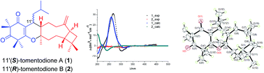
Tomentodiones A (1) and B (2), a pair of C-11′ epimers of caryophyllene-conjugated phloroglucinols with an unprecedented skeleton, were isolated from the leaves of Rhodomyrtus tomentosa. Their structures were elucidated through the application of extensive spectroscopic measurements with the absolute configuration of 1 determined by single-crystal X-ray diffraction analysis and electronic circular dichroism (ECD) calculations. The biogenetic pathways of 1 and 2 were proposed to involve an intermolecular, inverse electron demand Diels–Alder cycloaddition reaction as the key step, and their biomimetic total synthesis was accomplished.
Introduction
Rhodomyrtus tomentosa (Aiton) Hassk, a member of the family Myrtaceae, also better-known as Rose myrtle, is an abundant evergreen shrub native to the tropical areas of southeastern Asia.1 All the stems, leaves, and fruits of this plant can be used as herbal folk medicinal materials. Especially, the leaves of R. tomentosa have been reputed as being a remedy for a variety of symptoms including dysentery, colic diarrhea, gynecopathy, abscesses, and hemorrhage in traditional Chinese medicine.2 Several previous phytochemical studies of R. tomentosa have indicated the presence of triterpenoids,3–5 flavonoids,6 acylphloroglucinols,6–8 tannins,9 and anthracenoids.10,11 We have recently disclosed the isolation and elucidation of two novel meroterpenoids tomentosone C and tementosenol A with significant growth inhibitory activity toward a panel of human cancer cell lines and microbials from the EtOH extract of the leaves.12,13 Based on the speculation and inspection of other novel meroterpenoids in this species, a further extensive phytochemical investigation on the nonpolar portion of this extract resulted in the isolation of two novel meroterpenoid epimers, tomentodiones A (1) and B (2) (Fig. 1), which comprise both β-triketone and caryophyllene moieties. Herein, the isolation and structural elucidation of 1 and 2, including their absolute configuration assignments, as accomplished by single crystal X-ray diffraction analysis coupled with ECD calculations, and their biomimetic synthesis are described.
 |
| Fig. 1 The chemical structures of compounds 1 and 2. | |
Results and discussion
Compound 1 was isolated as colorless crystal and its molecular formula was established as C30H46O3 based on the HRESIMS (positive mode) wherein a protonated molecule [M + H]+ was evident at m/z 455.3459 (calcd 455.3447), indicating the presence of eight indices of hydrogen deficiency. The 13C NMR spectrum exhibited 30 carbon signals, which is in agreement with the molecular formula. The 1H NMR data of 1 (Table 1) showed signals at δH 4.91 and at 4.88, indicating a terminal methylene, nine methyl groups (δH 0.75, 0.84, 0.98, 0.99, 1.06, 1.32, 1.33, 1.35, and 1.39), and a group of overlapping aliphatic signals in the range between 1.40 and 2.40 ppm. The HMBC correlations from methyl singlets at δH 1.33/1.35 to C-1′ (δC 197.5), C-2′ (δC 52.1), and C-3′ (δC 213.9), as well as from methyl singlets at δH 1.32/1.49 to C-3′, C-4′ (δC 47.6), and C-5′ (δC 170.3) (Fig. 2) suggested the presence of an enolized 1,1,3,3-tetramethylcyclohexatrione (syncarpic acid) moiety.14
Table 1 1H (500 MHz) and 13C (125 MHz) NMR data of 1–2 in CDCl3
No. |
1 |
2 |
δH |
δC |
δH |
δC |
1 |
2.05 (m) |
52.1, CH |
1.48 (m) |
58.1, CH |
2α |
1.75 (m) |
33.0, CH2 |
1.59 (m) |
24.1, CH2 |
2β |
1.45 (m) |
|
1.37 (m) |
|
3α |
1.68 (m) |
36.3, CH2 |
1.58 (m) |
36.1, CH2 |
3β |
1.65 (m) |
|
1.73 (m) |
|
4 |
|
83.0, C |
|
86.3, C |
5 |
2.10 (m) |
40.3, CH |
1.75 (m) |
39.8, CH |
6α |
2.09 (m) |
37.4, CH2 |
2.04 (m) |
46.2, CH2 |
6β |
1.90 (m) |
|
1.38 (m) |
|
7α |
2.48 (m) |
35.3, CH2 |
2.13 (m) |
35.3, CH2 |
7β |
2.10 (m) |
|
2.34 (m) |
|
8 |
|
152.2, C |
|
150.8, C |
9 |
2.40 (m) |
42.4, CH |
2.41 (m) |
42.3, CH |
10a |
1.79 (m) |
22.1, CH2 |
1.66 (m) |
25.2, CH2 |
10b |
1.40 (m) |
|
1.66 (m) |
|
11 |
|
33.5, C |
|
34.4, C |
12 |
0.99 (s) |
30.3, CH3 |
0.96 (s) |
29.7, CH3 |
13 |
0.98 (s) |
22.2, CH3 |
0.97 (s) |
21.8, CH3 |
14 |
1.06 (s) |
20.9, CH3 |
1.35 (s) |
25.4, CH3 |
15a |
4.91 (s) |
110.7, CH2 |
4.91 (s) |
111.0, CH2 |
15b |
4.88 (s) |
|
4.90 (s) |
|
1′ |
|
197.5, C |
|
197.2, C |
2′ |
|
55.2, C |
|
55.4, C |
3′ |
|
213.9, C |
|
213.9, C |
4′ |
|
47.6, C |
|
47.6, C |
5′ |
|
170.3, C |
|
170.3, C |
6′ |
|
113.6, C |
|
116.4, C |
7′ |
1.35 (s) |
26.0, CH3 |
1.32 (s) |
25.4, CH3 |
8′ |
1.33 (s) |
23.3, CH3 |
1.33 (s) |
23.3, CH3 |
9′ |
1.39 (s) |
24.0, CH3 |
1.33 (s) |
24.9, CH3 |
10′ |
1.32 (s) |
25.4, CH3 |
1.35 (s) |
25.4, CH3 |
11′ |
2.28 (m) |
34.5, CH |
2.88 (m) |
28.3, CH |
12′a |
1.82 (m) |
38.8, CH2 |
1.39 (m) |
40.9, CH2 |
12′b |
1.33 (m) |
|
1.10 (m) |
|
13′ |
1.44 (m) |
25.4, CH |
1.43 (m) |
26.6, CH |
14′ |
0.84 (d, 6.6) |
24.0, CH3 |
0.82 (d, 6.6) |
24.6, CH3 |
15′ |
0.75 (d, 6.6) |
24.0, CH3 |
0.97 (d, 6.6) |
21.6, CH3 |
 |
| Fig. 2 1H, 1H-COSY and key HMBCs of compound 1. | |
A COSY experiment revealed the presence of two fragments (Fig. 2): a (C-14′/C-15′/C-13′/C-12′/C-11′/C-5/C-6/C-7), and b (C-3/C-2/C-1/C-9/C-10), indicating the presence of an isobutyl group. The HMBC correlations between the methine proton signal at δH 2.28 with C-6′ (113.6), C-5′, C-4 (δC 83.0), and C-5 (δC 40.3) suggested that the isobutyl group was attached to C-11′ of the syncarpic acid moiety. In addition, based on fragments a and b, the HMBC correlations between the two methylene protons at δH 4.91/4.88 and C-7 (δC 35.3), C-8 (δC 152.2), and C-9 indicated that the two fragments are connected through the terminal double bond. Furthermore, the HMBC correlations of the methine at δH 2.40 with C-1 (δC 52.1), C-2 (δC 33.0), C-7 (δC 35.3), and C-11 (δC 33.5), and based on the correlations between the gem-dimethyl group signals at δH 0.99/0.98 with the 13C signals at C-1, C-10, and C-11, suggested that 1 contained a caryophyllene moiety considering an earlier report.15 The only uncertainty for the plane structure of 1 was one remaining indices of hydrogen deficiency. Based on the molecular formula, the remaining oxygen atom was assigned to bridge between C-5′ and C-4, which resulted in the formation of a dihydropyran ring thereby completely accounting for the hydrogen deficiency requirements. In light of the aforementioned evidence, the plane structure of 1 was concluded to be as shown.
The relative configuration of 1 was determined through interpretation of the proton coupling constants and NOESY correlations (Fig. 3). The relative stereochemistry of C-1 and C-9 in the caryophyllene unit was deduced to be identical to that of natural β-caryophyllene. This conclusion was drawn from the informative NOE correlations observed between H-9/H-2α, which suggested that the two protons were cofacial, and were arbitrarily assigned as having an α-orientation. Whereas the observed NOE interactions between H-5/H-1 and H-5/H-2β indicated that H-5 was β-oriented. Finally, the correlation between H3-14/H-11′ revealed that the isobutyl group at C-11′ was also β-oriented.
 |
| Fig. 3 The key NOESY correlations of 1 and 2. | |
Conclusive evidence confirming the stereochemistry was obtained from a single-crystal X-ray diffraction experiment. After many attempts with different solvent combinations, a single-crystal of compound 1 suitable for X-ray crystallography was obtained. The X-ray crystallographic measurements were completed using CuKα with a Flack parameter of 0.2 (3) that clarified the molecular structure of 1 (Fig. 4). In addition, the X-ray diffraction experiment also provided some conformational information regarding the ring conjunctions within 1. With respect to the caryophyllene moiety, the dimethylcyclobutane ring was trans-fused with the seven-membered ring, whereas the isobutyl group had the same orientation as H-1 in the dimethylcyclobutane ring.
 |
| Fig. 4 ORTEP drawing of the X-ray structure of 1. | |
Compound 2 was obtained as a colorless oil. The molecular formula was assigned as C30H46O3 based on the positive ion mode HRESIMS ([M + H]+ m/z 455.3530, calcd 455.3525), indicating the presence of eight indices of hydrogen deficiency. A close comparison of its 1H and 13C NMR spectra (Table 1) with those of 1 suggested that they share the same plane structure (Table 1), which was re-confirmed by the HMBC correlations, such as those from H3-12 to C-1, C-10, C-11; H3-13 to C-1, C-10, C-11; H3-14 to C-3, C-4, C-5; H-15 to C-7, C-8, C-9; and H-11′ to C-4, C-5′. However, differences were detected for the chemical shifts between 1 and 2, particularly those associated with the chiral centers at C-1, C-5, and C-11 [C-1 (δC 52.1, δH 2.05), C-5 (δC 40.3, δH 2.10), and C-11′ (δC 34.5, δH 2.28) for 1], [C-1 (δC 58.1, δH 1.48), C-5 (δC 39.8, δH 1.75), and C-11′ (δC 28.3, δH 2.88) for 2], possibly caused by the strong spatial steric interactions due to a different orientation of the bulky functionality. In addition, all of the proton signals close to C-11′ were shifted to higher field, implying that compounds 1 and 2 were epimers in view of the ‘shielding effect’ of the isobutyl group at C-11′. The relative configuration of 2 was further confirmed by the NOESY spectrum (Fig. 3), wherein the NOE correlations of H-1/H-5, H-5/H-2β, and H-2α/H-9 definitively established the relative configuration of the sesquiterpenoid moiety in 2 as being identical to that in 1. The observed correlations of H-11′/H-5, H-12′/H3-14 indicated that the isobutyl moiety was α-oriented. Therefore, compound 2 was an epimer at C-11′ of 1 and was given the trivial name of tomentodione B.
Considering that tomentodiones A (1) and B (2) are the first representatives of this natural product class with an unprecedented molecular framework that bear a stereogenic center at C-11′, it is not feasible to assign their absolute configuration by empirical comparison of the electronic circular dichroism (ECD) spectrum with that of any structurally similar, configurationally established analogue. In such circumstance, quantum chemical calculations to predict the electronic circular dichroism (ECD) appeared to be the alternative approach. ECD calculations were performed according to standard protocols with slight modifications (see ESI† for details). In summary, conformational analysis was carried out using MacroModel 2010 to locate the conformers with low energies. All of the conformations within 10 kcal mol−1 of the lowest were optimized at the B3LYP/6-31G(d) level of theory.16,17 For those within 3.0 kcal mol−1 relative to the minimum after DFT optimization, the electronic transitions and rotational strengths were calculated at the same theoretical level through time-dependent, density-functional theory (TDDFT) calculations in Gaussian 09. The solvent effect in methanol was considered using the SMD solvent model.18 The Boltzmann-population-weighted calculated ECD curves were generated by SpecDis software.19 Subsequently, the calculated ECD spectra of 1 and 2 were compared with the experimental results, as depicted in Fig. 5, which revealed a good agreement between the experimental and calculated ECD curves. As a consequence, the absolute configurations of C-11′ in 1 and 2 were assigned as S and R, respectively.
 |
| Fig. 5 Experimental and calculated ECD spectra of 1 and 2 in methanol (1_exp and 2_exp refer to the experimental from results of 1 and 2, and 1_calc and 2_calc means the results calculation). | |
Tomentodiones A (1) and B (2) are two novel meroterpenoids biogenetically derived through the conjugation between a β-caryophyllene moiety and a β-triketone, resulting in the formation of a new, unique skeleton. To account for this biotransformation, a plausible biogenetic route was proposed involving an intermolecular, inverse electron demand, hetero-Diels–Alder cycloaddition reaction as the key step (Scheme 1). Briefly, leptospermone, a metabolite frequently isolated from Myrtaceous plants, is a postulated precursor.20 Selective reduction and dehydroxylation would transform it to the key intermediate 5, which can undergo a Diels–Alder [4 + 2] cycloaddition reaction with β-caryophyllene to afford both 1 and 2, respectively, due to different regioselectivity options.
 |
| Scheme 1 Proposed biogenetic pathway of 1 and 2. | |
Based on this speculation and analysis of the associated biogenetic transformation, the biomimetic total synthesis of tomentodiones A (1) and B (2) was conducted (Scheme 2). With the acetylphloroglucinol 3 as the starting material, the syncarpic acid 4 was readily accessed in 67% yield through C-methylation and retro-Friedel–Crafts acylation.21 The proline-catalyzed Knoevenagel condensation further transformed 4 to the key intermediate 5 in almost quantitative yield, and was successfully characterized by NMR. As expected, the biomimetic Diels–Alder cycloaddition occurred smoothly by treatment of 5 and (−)-β-caryophyllene neat or in refluxing toluene to achieve the total synthesis of tomentodiones A (1) and B (2) in excellent efficiency (62% or 57% yield). Gratifyingly, 1 and 2 were identical spectroscopically and chromatographically with the isolated natural products. Moreover, their physical data including melting points and optical rotation data were also in good agreement with those recorded for naturally occurring ones. The aforementioned results further confirmed their absolute configurations and validated the quantum chemical calculations. The effectiveness of this reaction, in spite of the required conditions, may also provide positive evidence that a similar transformation could explain the biosynthesis of these metabolites.
 |
| Scheme 2 Biomimetic total synthesis of 1 and 2. | |
Compounds 1 and 2 were evaluated for their antimicrobial activity against the bacteria Escherichia coli and Staphylococcus aureus. They were devoid of significant activity, but exhibited marginal inhibitory potency at a concentration of 100 μM.
Experiment section
General experimental procedures
Melting point was determined on a Yanagimoto Seisakusho MD-S2 micro-melting point apparatus and was uncorrected. Optical rotations were measured on a Perkin-Elmer 341 polarimeter (Perkin-Elmer, Boston, MA, USA). ECD spectra were recorded on an Applied Photophysics Chirascan spectrometer. UV spectra were recorded in MeOH on a Perkin-Elmer Lambda 35 UV-vis spectrophotometer (Perkin-Elmer, Boston, MA, USA). 1D- and 2D-NMR spectra were recorded on a Bruker Advance-500 spectrometer with TMS as internal standard (Bruker BioSpin AG, Fällanden, Switzerland). ESIMS data were obtained on a MDS SCIEX API 2000 LC/MS/MS mass spectrometer (AB MDS Sciex, Framingham, MA, USA). HRESIMS data were obtained on a Bruker Bio TOF IIIQ mass spectrometer (Bruker Daltonics, Billerica, MA, USA). All solvents were of analytical grade (Shanghai Chemical Plant, Shanghai, China). C18 reversed-phase silica gel (150–200 mesh, Merck), MCI gel (CHP20P, 75–150 μm, Mitsubishi Chemical Industries Ltd., Tokyo, Japan) were used for column chromatography. Precoated silica gel GF254 plates (Qingdao Marine Chemical Inc., Qingdao, China) were used for TLC, and spots were visualized under UV light and by dipping into 5% H2SO4 in alcohol followed by heating.
Plant material
Rhodomyrtus tomentosa leaves were collected from Nankang County, Jiangxi Province, in the South China in June 2012. A voucher specimen (No# SCBG-NPL-120012), which was identified by Prof. Fa-Guo Wang at South China Botanical Garden, has been deposited in South China Botanical Garden (SCBG).
Extraction and isolation
The powdered air-dried and powdered leaves of R. tomentosa (20 kg) were extracted with 95% EtOH (20 L × 3) for 24 h. After removal of the pooled solvents by evaporation under vacuum, a crude extract (2.5 kg) was obtained, which was suspended in H2O and then partitioned successively with n-hexane (3 L × 3), ethyl acetate (3 L × 3) respectively to yield n-hexane-soluble (400 g) and ethyl acetate-soluble (1100 g) residues. The n-hexane-soluble portion was then subjected to silica gel CC eluting with a solvent mixture of n-hexane/ethyl acetate (100
:
0 → 0
:
100, v/v) to afford seven fractions (A–G). Fraction B (30 g) (n-hexane/ethyl acetate, 95
:
5, v/v) was further chromatographed over RP-C18 silica gel chromatography (MeOH/H2O, 95
:
5 → 100
:
0, v/v) to obtain sub-fractions B1 and B2. Sub-fractions B1 (2.5 g) was separated by a Sephadex LH-20 (MeOH) chromatography, followed by repeated silica gel chromatography (n-hexane/ethyl acetate, 50
:
1 → 10
:
1, v/v) to afford compound 1 (250 mg) and 2 (280 mg).
Tomentosone A (1). Mp 80–81 °C; colorless crystals (MeOH); [α]20D +49 (c 0.8, MeOH); UV (MeOH) λmax (log
ε) 268.9 (3.07) nm; IR (KBr) νmax 3675, 2953, 1716, 1651, 1464, 1382, 889 cm−1; 1H- (500 MHz) and 13C- (125 MHz) NMR data were summarized in Table 1; ESIMS: positive ion mode [M + H]+, m/z 455; HRESIMS: positive ion mode [M + H]+, m/z 455.3459 (calcd for C30H47O3, 455.3525).
Tomentosone B (2). Colorless oil; [α]20D +12 (c 0.4, MeOH); UV (MeOH) λmax (log
ε) 267.8 (3.74) nm; IR (KBr) νmax 3675, 2953, 1715, 1617, 1492, 1382, 890 cm−1; 1H- (500 MHz) and 13C- (125 MHz) NMR data were summarized in Table 1; ESIMS: positive ion mode [M + H]+, m/z 455; HRESIMS: positive ion mode [M + H]+, m/z 455.3530 (calcd for C30H47O3, 455.3525).
X-ray crystallographic data for tomentodione A (1). C30H47O3, M = 455.68, orthorhombic, size 0.28 × 0.24 × 0.23 mm3, space group P212121, a = 10.0146 (3) Å, b = 10.5472 (2) Å, c = 26.3161 (5) Å, α = γ = β = 90.00°, V = 2779.66 (10) Å3, T = 292.55(10) K, Z = 4, ρcalcd = 1.089 g cm−3, F(000) = 1004.0, 19
027 reflections in −11 ≤ h ≤ 11, −12 ≤ k ≤ 10, −31 ≤ l ≤ 31, measured in the range 6.72° ≤ θ ≤ 133.84°, completeness θmax = 66.92%, GOOF = 1.024, final R indices [I > 2σ(I)]: R1 = 0.0461, wR2 = 0.1177, final R indices (all data): R1 = 0.0514, wR2 = 0.1232, Flack parameter 0.2(3), largest difference peak and hole = 0.24 and −0.23 e Å−3. Data were collected on an Agilent Xcalibur Nova single-crystal diffractometer using Cu Kα radiation. The full-matrix least-squares calculation was used to refine the crystal structure. The reported crystallographic data (CCDC 1431823) in this paper for the structure of tomentodione A (1) has been deposited in the Cambridge Crystallographic Data Centre.†
Preparation of intermediates 4 and 5. The synthesis of the key intermediates 4 and 5 was conducted according to our recently reported paper,13 and the copies of their NMR spectra could be found in the ESI.†
Preparation of tomentodiones A (1) and B (2).
Method 1. (−)-β-Caryophyllene (1.0 g, 5.0 mmol) and the α,β-unsaturated ketone 5 (238 mg, 0.95 mmol) were dissolved in dry toluene (5.0 mL). After heating at 120 °C for 3 h under N2, the reaction mixture was cooled to ambient temperature and purified directly by flash column chromatography (silica gel; n-hexane/EtOAc, from 100
:
1 to 10
:
1) to afford 1 (112 mg, 26%), as well as a mixture of 2 and by-products (270 mg) as a colorless solid. The mixture was further purified using a 10 cm C18 reversed-phase column with 100% MeOH as eluent to give 2 (133 mg, 31%). Both products were identical in all aspects to the natural isolates. 1: colorless crystals; mp 81–82 °C; [α]20D +38 (c 0.1, MeOH); 2: colorless oil; [α]20D +19 (c 0.2, MeOH). 1H- (500 MHz) and 13C- (125 MHz) NMR spectral data were identical to those of the natural compounds.
Method 2. (−)-β-Caryophyllene (1.0 g, 5.0 mmol) and the α,β-unsaturated ketone 5 (250 mg, 1.0 mmol) were mixed neatly and allowed to stand at ambient temperature for 24 h under nitrogen. After the starting material disappeared (TLC analysis), the reaction products were purified by flash column chromatography (n-hexane/EtOAc, from 100
:
1 to 10
:
1) to yield 1 (127 mg, 28%), as well as a mixture of 2 and by-products (300 mg) as a colorless solid. The mixture was further purified using a 10 cm C18 reversed-phase column with 100% MeOH as eluent to afford 2 (146 mg, 34%). The products were identical in all aspects to the natural isolates.
Antibacterial activity assay. Compounds 1 and 2 were assayed against two bacteria, Staphylococcus aureus (ATCC6538) and Escherichia coli (ATCC8739), which were purchased from Guangdong Microbiology Culture Center (Guangzhou, China). Assays were performed by the published microdilution method for the estimation of minimum inhibitory concentration (MIC) values to evaluate the antimicrobial activity.22,23 Vancomycin was used as positive control. Optical density values at 600 nm were recorded using a UV-6000 spectrophotometer (METASH, Shanghai, China).
Acknowledgements
This research was funded by the National Science and Technology Major Projects of China (2014ZX10005002-005) and the National Natural Science Foundation of China (No. 30973635, 81373293, 81502949). We acknowledge support of computational source from lab of computational chemistry and drug design in Peking University Shenzhen Graduate School and Prof. O. Wiest (University of Notre Dame, IN, USA) for help with MacroModel. We also thank Dr X.-L. Feng (Sun Yat-sen University) for the X-ray diffraction analysis and Dr G.-Q. Li (Jinan University) for helpful discussions. Moreover, we are grateful to Prof. Geoffrey Cordell at the University of Illinois for editing the English.
Notes and references
- J. Chen and L. A. Craven, Flora of China, Science Press/Missouri Botanical Garden Press, Beijing/St. Louis, 2007, vol. 13, pp. 330−331 Search PubMed.
- Editorial Board of ‘Zhonghua Bencao’, State Administration of Traditional Chinese Medicine of the People's Republic of China, Zhonghua Bencao, Shanghai Science and Technology Publishers, Shanghai, China, 2000, vol. 5, pp. 643−646 Search PubMed.
- W. H. Hui, M. M. Li and K. Luk, Phytochemistry, 1975, 14, 833–834 CrossRef CAS.
- W. H. Hui and M. M. Li, Phytochemistry, 1976, 15, 1741–1743 CrossRef CAS.
- J. Xiong, Y. Huang, Y. Tang, M. You and J. F. Hu, Chin. J. Org. Chem., 2013, 33, 1304–1308 CrossRef CAS.
- A. Hiranrat and W. Mahabusarakam, Tetrahedron, 2008, 64, 11193–11197 CrossRef CAS.
- A. Hiranrat, W. Chitbankluoi, W. Mahabusarakam, S. Limsuwan and S. P. Voravuthikunchai, Nat. Prod. Res., 2012, 26, 1904–1909 CrossRef CAS PubMed.
- A. Hiranrat, W. Mahabusarakam, A. R. Carroll, S. Duffy and V. M. Avery, J. Org. Chem., 2012, 77, 680–683 CrossRef CAS PubMed.
- Y. Z. Liu, A. J. Hou, C. R. Ji and Y. J. Wu, Nat. Prod. Res. Dev., 1997, 10, 14–19 Search PubMed.
- N. H. Tung, Y. Ding, E. M. Choi, P. V. Kiem, C. V. Minh and Y. H. Kim, Arch. Pharmacal Res., 2009, 32, 515–520 CrossRef CAS PubMed.
- T. Chen, C. G. Yu and B. L. Yang, Chem. Nat. Compd., 2011, 47, 524–526 CrossRef CAS.
- H. X. Liu, H. B. Tan and S. X. Qiu, J. Asian Nat. Prod. Res., 2016 DOI:10.108/10286020.2015.1121997.
- H. X. Liu, W. M. Zhang, Z. F. Xu, Y. C. Chen, H. B. Tan and S. X. Qiu, RSC Adv., 2016, 6, 25882–25886 RSC.
- B. P. S. Khambay, D. G. Beddie and M. D. J. Simmonds, Phytochemistry, 2002, 59, 69–71 CrossRef CAS PubMed.
- F. Bohlmann and C. Zdero, Phytochemistry, 1978, 17, 1135–1153 CrossRef CAS.
- A. D. Becke, J. Chem. Phys., 1993, 98, 5648–5652 CrossRef CAS.
- C. Lee, W. Yang and R. G. Parr, Phys. Rev. B, 1988, 37, 785–789 CrossRef CAS.
- A. V. Marenich, C. J. Cramer and D. G. Truhlar, J. Phys. Chem. B, 2009, 113, 6378–6396 CrossRef CAS PubMed.
- T. Bruhn, A. Schaumlöffel, Y. Hemberger and G. Bringmann, SpecDis 1.53, University of Wuerzburg, Germany, 2012 Search PubMed.
- J. W. V. Klink, J. J. Brophy, N. B. Perry and R. T. Weavers, J. Nat. Prod., 1999, 62, 487–489 CrossRef PubMed.
-
(a) H. Müller, M. Paul, D. Hartmann, V. Huch, D. Blaesius, A. Koeberle, O. Werz and J. Jauch, Angew. Chem., Int. Ed., 2010, 49, 2045–2049 CrossRef PubMed;
(b) M. Morkunas, L. Dube, F. Götz and E. M. Maier, Tetrahedron, 2013, 69, 8559–8563 CrossRef CAS.
- A. M. Drummond and R. D. Waigh, Recent research developments in phytochemistry, ed. S. G. Pandalai, Research Signpost, India, 2000, pp. 143−152 Search PubMed.
- L. He, C. F. Zhu, Y. Yuan, Z. F. Xu and S. X. Qiu, Phytochem. Lett., 2014, 9, 175–178 CrossRef CAS.
Footnote |
† Electronic supplementary information (ESI) available. CCDC 1431823. For ESI and crystallographic data in CIF or other electronic format see DOI: 10.1039/c6ra08776k |
|
This journal is © The Royal Society of Chemistry 2016 |
Click here to see how this site uses Cookies. View our privacy policy here.