DOI:
10.1039/C6RA08481H
(Paper)
RSC Adv., 2016,
6, 46366-46371
Targeted delivery of vincristine to T-cell acute lymphoblastic leukemia cells using an aptamer-modified albumin conjugate
Received
2nd April 2016
, Accepted 4th May 2016
First published on 6th May 2016
Abstract
Clinical application of vincristine in treatment of cancer is restricted because of its poor solubility and neuropathy. Targeted delivery of cytotoxic drugs could improve their therapeutic efficacy and reduce their severe side effects. Biocompatibility and high accumulation of human serum albumin nanoparticles (HSA) in tumors make this substance an ideal candidate for biomedical usage. Here, a Vincristine–HSA–Sgc8c aptamer (Apt) complex was designed and assessed for the treatment of Molt-4 cells (human acute lymphoblastic leukemia T-cell, target). Vincristine–HSA conjugate and Vincristine–HSA–Apt complex formations were analyzed by particle size analyzer, transmission electron microscopy (TEM) and gel retardation assay. Internalization of the Vincristine–HSA–FAM (3′-fluorescein)-labeled Apt complex into Molt-4 (target) and U266 cells (B lymphocyte human myeloma, nontarget) was monitored by confocal imaging and flow cytometry analysis. For cell viability (MTT assay), both cell lines were treated with vincristine, Vincristine–HSA conjugate, HSA–Apt conjugate and Vincristine–HSA–Apt complex. Vincristine was efficiently loaded (8.5%) into HSA. The results of confocal imaging and flow cytometry analysis indicated that the Vincristine–HSA–FAM-labeled Apt complex was effectively internalized into target cells (Molt-4) but not into nontarget cells (U266). The results of MTT assay also confirmed the internalization data. The Vincristine–HSA–Apt complex had less cytotoxicity in U266 cells compared to vincristine alone and Vincristine–HSA conjugate. In conclusion, the developed drug delivery system acquired properties of high drug loading, high cancer cell accumulation and cancer cell targeting.
1. Introduction
Acute lymphoblastic leukemia (ALL) is a type of blood cancer in which bone marrow and blood cells are involved.1,2 It is the most common type of malignant disease in children.3,4
Vincristine is a chemotherapeutic agent which is frequently used for treatment of leukemia in both children and adults.5 However, clinical administration of vincristine has been limited, because of its neuropathy and poor solubility.6,7
In targeted tumor therapy using nanocarriers, anticancer drugs are preferentially localized in the tumor site and consequently decrease their adverse effects.8,9
Newly, aptamers have been broadly applied as targeting units for targeted delivery of therapeutic agents. Aptamers are synthetic single-stranded nucleic acids, selected by an in vitro process, named SELEX (systematic evolution of ligands by exponential enrichment).10,11 They possess the capability to bind to a broad diversity of targets, ranging from metal ions to proteins and even whole cells.12,13 Aptamers present promising advantages over antibodies, such as excellent stability, low cost, high reproducibility, simple synthesis, no or low toxicity and immunogenicity.14–17 Moreover, relative to antibodies, aptamers have smaller size which leads to more and faster penetration of aptamers into cancer tissues.18 Owing to these characteristics, aptamers have been broadly utilized in diagnostic and therapeutic applications.19,20
Sgc8c aptamer (Apt) binds to protein tyrosine kinase-7 (PTK-7) with high affinity (Kd = 1 nM). This protein is an outstanding biomarker for T-cells acute lymphoblastic leukemia.21,22
Recently, human serum albumin nanoparticle (HSA) has attracted remarkable attention in pharmaceutical applications as a drug carrier, because of its unique advantages, such as lysosomal breakdown which could help to improve the efficacy of anticancer drugs, high biocompatibility, low toxicity and uptake by normal cells, high stability in blood and high accumulation in cancer site as the rate of metabolism is higher in tumor tissues and they use HSA as nutrient.23–25
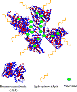
In this study, we evaluated the feasibility of targeted delivery of vincristine to Molt-4 cells (T-cell line, ALL) using HSA as a carrier and sgc8c aptamer as targeting ligand (Fig. 1). To the best of our knowledge, it is for the first time that aptamer–HSA conjugate has been used for targeted delivery of a hydrophobic drug like vincristine.
 |
| Fig. 1 Schematic structure of Vincristine–HSA–Apt complex. | |
2. Materials and methods
2.1. Materials
The sgc8c aptamer (Apt), 5′-ATCTAACTGCTGCGCCGCCGGGAAAATACTGTACGGTTAGA-thiol-3′, and FAM-labeled sgc8c aptamer, 5′-FAM-ATCTAACTGCTGCGCCGCCGGGAAAATACTGTACGGTTAGA-thiol-3′ were purchased from Bioneer (South Korea). Vincristine and human serum albumin (HSA) were ordered from Sigma-Aldrich (USA). DAPI, sulfosuccinimidyl 4-(N-maleimidomethyl)cyclohexane-1-carboxylate (Sulfo-SMCC), salmon sperm DNA and WGA-Alexa 488 were provided by Thermo Fisher Scientific (USA). 10 K centrifugal device was purchased from PALL (USA).
2.2. Cell culture
U266 (C151, B lymphocyte, human myeloma) and Molt-4 (C149, T-cell line, human ALL) cells were obtained from Pasteur Institute of Iran and cultured in RPMI 1640 (Gibco) containing 10% fetal bovine serum (FBS, heat inactivated, Gibco) and 100 units per mL penicillin–streptomycin (Sigma-Aldrich).
2.3. Preparation of Vincristine–HSA conjugate
Vincristine–HSA conjugate was prepared by vincristine-induced self-assembly of HSA proteins. 2 mg HSA was mixed with 0.3 mg vincristine in 1 mL phosphate buffer saline (PBS, pH 7.4), followed by centrifugation with a 10 K centrifugal device to discard the unbound vincristine. The amount of free vincristine was calculated by measuring absorbance at 299 nm. The obtained Vincristine–HSA conjugate was resuspended in PBS (pH 7.4) and stored at 4 °C. The formation of Vincristine–HSA conjugate was evaluated by particle size analyzer (Malvern, UK).
2.4. Preparation of Vincristine–HSA–Apt complex
1 mL PBS (pH 7.4) containing 2 mg Vincristine–HSA conjugate was incubated with 0.25 mg Sulfo-SMCC for 2 h at room temperature. A 10 K centrifugal device was used to remove the excess Sulfo-SMCC. Then, 10 nmol thiol-modified Apt was added to the 0.1 mg Sulfo-SMCC activated HSA in PBS (pH 7.4) to give a final volume of 1 mL. After incubation for 24 h at 4 °C, the free thiol-modified Apt was removed using a 10 K centrifugal device. The obtained Vincristine–HSA–Apt complex was resuspended in 1 mL PBS (pH 7.4) and stored at 4 °C. The formation of Vincristine–HSA–Apt complex was assessed by 2.5% agarose gel electrophoresis, transmission electron microscopy (TEM) (CM120, Philips, Holland) and particle size analyzer.
2.5. Flow cytometry analysis
Molt-4 and U266 cells were seeded in 12-well plates (3 × 10⁵ cells per well) and incubated with Vincristine–HSA–FAM-labeled Apt complex (1 μM FAM-labeled Apt final concentration) for 3 h. Then, the cells were centrifuged at 400 × g for 5 min, followed by treatment with trypsin–EDTA for 7 min. Next, cells were resuspended in PBS for fluorescence analysis by a BD Accuri C6 flow cytometer (BD Biosciences, US). The data were analyzed by FlowJo 7.6.1 software.
In the next step, Molt-4 cells were seeded in 12-well plates (2 × 10⁵ cells per well) and treated with sgc8c aptamer (2 μM final concentration) for 1 h. Then, cells were incubated with Vincristine–HSA–FAM-labeled Apt complex (1 μM FAM-labeled Apt final concentration) for 3 h. Thereafter, Molt-4 cells were treated with trypsin–EDTA for 7 min, followed by resuspension in PBS for fluorescence analysis.
2.6. Confocal imaging
The internalization of Vincristine–HSA–FAM-labeled Apt complex was evaluated using confocal microscopy. U266 and Molt-4 cells were seeded in 6-well plates (confluency between 55–70%) and rinsed with PBS, followed by incubation with Vincristine–HSA–FAM-labeled Apt complex (1 μM FAM-labeled Apt final concentration) and salmon sperm DNA (0.5 mg mL−1) for 3 h. After that, the cells were rinsed with PBS, fixed and stained with WGA-Alexa 488 (plasma membrane staining) and DAPI (nuclei staining). The cellular fluorescent images were obtained by a confocal microscopy (FV500-IX81, Olympus America Inc., Melville, NY, USA).
2.7. MTT assay
Escalating-dose studies of vincristine (0–12 nM), showed that IC50 for Molt-4 and U266 cells were 6 and 4.5 nM, respectively. To analyze cell viability, Molt-4 and U266 cells were seeded in 96-well plates (1 × 10⁴ cells per well) and treated with vincristine (based on the results of escalating-dose assay), HSA–Apt conjugate, Vincristine–HSA conjugate (with the same amount of vincristine) and Vincristine–HSA–Apt complex (with the same amount of vincristine) for 3 h. Thereafter, the cells were centrifuged at 400 × g for 5 min and the supernatants were removed, followed by addition of fresh culture medium to each well. After incubation for 72 h at 37 °C, 10 μL MTT solution was added to each well and incubated for 3.5 h. Finally, 100 μL DMSO was added to each well and A545 was measured by a microplate reader (BioTek, USA).
2.8. Statistical analysis
Statistical tests were done by the Student's t-test. Data were mean ± SD, n = 3 independent treatments. P < 0.05 was considered as significant and P > 0.05 means data were not significantly different.
3. Results
3.1. Characterization of Vincristine–HSA conjugate and Vincristine–HSA–Apt complex
Particle size of HSA, Vincristine–HSA conjugate and Vincristine–HSA–Apt complex were 8.2 ± 0.6 nm, 31.6 ± 1.6 nm and 36.1 ± 3 nm, respectively. Zeta-potential of HSA, Vincristine–HSA conjugate and Vincristine–HSA–Apt complex were −29.6 ± 2.2 mV, −84.8 ± 4.6 mV and −88.3 ± 6.9 mV, respectively. Based on TEM image, the size of HSA and Vincristine–HSA–Apt complex were around 7 and 40 nm (Fig. 2(a) and (b)), which were in agreement with the results collected from particle size analyzer.
 |
| Fig. 2 (a) TEM image of HSA. (b) TEM image of Vincristine–HSA–Apt complex. (c) Agarose gel electrophoresis of Vincristine–HSA–Apt complex. Lane 1: Apt, lane 2: Vincristine–HSA–Apt complex, lane 3: ladder. | |
The formation of Vincristine–HSA–Apt complex was also confirmed by gel retardation assay. As indicated in Fig. 2(c), the band of Vincristine–HSA–Apt complex was retarded relative to the band of free Apt.
Vincristine loading into HSA was calculated through optical absorption. According to absorbance data, the vincristine payload was 0.17 mg.
3.2. Internalization assay
The fluorescence histogram of U266 and Molt-4 cells after treatment with Vincristine–HSA–FAM-labeled Apt complex have been displayed in Fig. 3(a) and (b). FL2 log intensity for Molt-4 cells after no treatment and treatment with Vincristine–HSA–FAM-labeled Apt complex were 204 ± 16 and 853 ± 56, respectively. FL2 log intensity for U266 cells after no treatment and treatment with Vincristine–HSA–FAM-labeled Apt complex were 215 ± 11 and 356 ± 32, respectively.
 |
| Fig. 3 (a) Flow cytometry histogram of U266 cells after treatment with Vincristine–HSA–FAM-labeled Apt complex (green) and nontreated cells (red). (b) Flow cytometry histogram of Molt-4 cells after treatment with Vincristine–HSA–FAM-labeled Apt complex (green) and nontreated cells (red). (c) Flow cytometry histogram of Molt-4 cells (green) and sgc8c aptamer-treated Molt-4 cells (blue) after incubation with Vincristine–HSA–FAM-labeled Apt complex. | |
FL2 log intensity for Molt-4 cells and sgc8c aptamer-treated Molt-4 cells after incubation with Vincristine–HSA–FAM-labeled Apt complex were 887 ± 36 and 334 ± 23, respectively (Fig. 3(c)).
The cellular fluorescent images after incubation with Vincristine–HSA–FAM-labeled Apt complex have been indicated in Fig. 4. The images showed well internalization of Vincristine–HSA–FAM-labeled Apt complex into Molt-4 cells (target), while much less internalization was detected for nontarget cells (U266).
 |
| Fig. 4 (a) Confocal images of Molt-4 cells treated with Vincristine–HSA–FAM-labeled Apt complex stained with WGA-Alexa 488 (green) and DAPI (blue) and the image merged to indicate internalization of Vincristine–HSA–FAM-labeled Apt complex. (b) Confocal images of U266 cells treated with Vincristine–HSA–FAM-labeled Apt complex stained with WGA-Alexa 488 (green) and DAPI (blue) and the image merged to show internalization of Vincristine–HSA–FAM-labeled Apt complex. Red arrows show Vincristine–HSA–FAM-labeled Apt complex. | |
3.3. Cell viability
MTT method was performed to record the viability of Molt-4 and U266 cells (Fig. 5). Molt-4 cell viability after treatment with vincristine, HSA–Apt conjugate, Vincristine–HSA conjugate and Vincristine–HSA–Apt complex were 51.6 ± 2.4%, 98.7 ± 6.8%, 36.7 ± 3.1% and 33.4 ± 3.2%, respectively. U266 cell viability after treatment with vincristine, HSA–Apt conjugate, Vincristine–HSA conjugate and Vincristine–HSA–Apt complex were 48.3 ± 2.8%, 97.1 ± 7.6%, 36.1 ± 2% and 74.5 ± 5.3%, respectively.
 |
| Fig. 5 Effects of vincristine, HSA–Apt conjugate, Vincristine–HSA conjugate and Vincristine–HSA–Apt complex on control and target cells viability (MTT assay). Both cell lines were treated with vincristine, HSA–Apt conjugate, Vincristine–HSA conjugate and Vincristine–HSA–Apt complex for 3 h. After 72 h post-treatment, viability of the cells was analyzed using MTT assay. | |
4. Discussion
Chemotherapy is one of the leading protocols in treatment of cancer. Chemotherapeutic agents are associated with severe toxicity, owing to their poor specificity. Tumor-targeted drug delivery could significantly increase therapeutic efficacy of cytotoxic agents and diminish their serious side effects through delivery of high amounts of cytotoxic drugs into cancer tissues.26,27
High accumulation in tumor site and biocompatibility of HSA make it an appropriate carrier for anticancer drug delivery.23,24 However, HSA could not inherently recognize target cells. Application of aptamer as targeting ligand could overcome this drawback.
Vincristine, as a hydrophobic chemotherapeutic agent, binds to HSA by hydrophobic interactions between the hydrophobic domain of HSA and vincristine. Based on the results of particle size analyzer, upon the addition of vincristine, the size of HSA was significantly increased (about 4-fold), which could be attributed to the loading of drug and self-assembly of HSA that is induced by vincristine. The drug loading efficiency in the designed formulation was measured to be 8.5%, which is a good amount of drug loading. The drug payload of the developed targeted delivery system was much more than the payload that was already reported for using aptamer as both targeting agent and carrier to deliver intercalating agents to cells28 or using super paramagnetic iron oxide nanoparticle-aptamer bioconjugate for targeted delivery of Epi to cancer cells.29 Also compared to aptamer-wrapped carbon nanotubes delivery system, there is no controversial about the safety and biocompatibility of HSA as a nonacarrier.30
Flow cytometry analysis showed the fluorescence intensity of U266 cells (nontarget) treated with Vincristine–HSA–FAM-labeled Apt complex was significantly lower compared to Vincristine–HSA–FAM-labeled Apt complex-treated Molt-4 cells (target, Fig. 3) (P < 0.05). These results confirmed sgc8c aptamers, located on the surface of Vincristine–HSA–Apt complex, could efficiently differentiate between nontarget and target cells. Less entry of Vincristine–HSA–Apt complex into U266 cells was due to the lack of target site of sgc8c aptamer, PTK-7, on the surface of U266 cells. Receptor mediated endocytosis (RME) and albumin-induced delivery are assumed as the internalization mechanisms for Vincristine–HSA–Apt complex because sgc8c aptamer is internalized into target cells via RME.30
Also, fluorescence intensity in sgc8c aptamer-treated Molt-4 cells after incubation with Vincristine–HSA–FAM-labeled Apt complex was significantly lower than Vincristine–HSA–FAM-labeled Apt complex-treated Molt-4 cells (P < 0.05), confirming the internalization of complex into target cells via RME (Fig. 3(c)).
The results of confocal imaging were compatible with the results of flow cytometry analysis and verified more internalization of the designed complex into Molt-4 cells compared to U266 cells (Fig. 4).
MTT assay also verified the internalization data (Fig. 5). Treatment of Molt-4 and U266 cells with HSA–Apt conjugate did not significantly change their cell viability (P > 0.05). Treatment of U266 and Molt-4 cells with Vincristine–HSA conjugate for 3 h, significantly reduced their cell viability (about 36%) compared to vincristine alone (about 50%) (P < 0.05). This could be attributed to the higher accumulation of Vincristine–HSA conjugate in cancer cells because of high rate of metabolism in tumor cells and therefore consumption of HSA as a source of energy. Treatment of U266 cells (nontarget) with Vincristine–HSA–Apt complex significantly increase cell viability in comparison with vincristine and Vincristine–HSA conjugate (P < 0.05) because of the presence of sgc8c aptamers as smart ligands on the surface of HSA.
5. Conclusion
In summary, a Vincristine–HSA–Apt complex was designed for targeted delivery of vincristine to Molt-4 cells (target). This complex inherits characteristics of efficient drug loading, biocompatibility and cancer targeting. Due to these features, the fabricated drug delivery system could reduce side effects of vincristine in nontarget cells (U266 cells). So, these properties make HSA–Apt complex an ideal and applicable delivery system for clinical application.
Conflict of interest
There is no conflict of interest about this article.
Acknowledgements
Financial support of this study was provided by Mashhad University of Medical Sciences.
References
- U. Ali, M. Naveed, A. Ullah, K. Ali, S. A. Shah, S. Fahad and A. S. Mumtaz, Eur. J. Pharmacol., 2016, 771, 199–210 CrossRef CAS PubMed.
- N. M. Danesh, P. Lavaee, M. Ramezani, K. Abnous and S. M. Taghdisi, Int. J. Pharm., 2015, 489, 311–317 CrossRef CAS PubMed.
- H. Goto, Pediatr. Int., 2015, 57, 1059–1066 CrossRef PubMed.
- S. M. Taghdisi, N. M. Danesh, P. Lavaee, A. S. Emrani, K. Y. Hassanabad, M. Ramezani and K. Abnous, Mater. Sci. Eng., Proc. Conf., 2016, 61, 753–761 CrossRef CAS PubMed.
- V. Thakur, P. Kush, R. S. Pandey, U. K. Jain, R. Chandra and J. Madan, Mater. Sci. Eng., Proc. Conf., 2016, 61, 113–122 CrossRef CAS PubMed.
- B. Diouf, K. R. Crews, G. Lew, D. Pei, C. Cheng, J. Bao, J. J. Zheng, W. Yang, Y. Fan, H. E. Wheeler, C. Wing, S. M. Delaney, M. Komatsu, S. W. Paugh, J. R. McCorkle, X. Lu, N. J. Winick, W. L. Carroll, M. L. Loh, S. P. Hunger, M. Devidas, C. H. Pui, M. E. Dolan, M. V. Relling and W. E. Evans, JAMA, J. Am. Med. Assoc., 2015, 313, 815–823 CrossRef CAS PubMed.
- M. W. Chao, M. J. Lai, J. P. Liou, Y. L. Chang, J. C. Wang, S. L. Pan and C. M. Teng, J. Hematol. Oncol., 2015, 8, 1–15 CrossRef CAS PubMed.
- D. Böhme and A. G. Beck-Sickinger, J. Pept. Sci., 2015, 21, 186–200 CrossRef PubMed.
- G. Casi and D. Neri, Mol. Pharm., 2015, 12, 1880–1884 CrossRef CAS PubMed.
- X. Lin, K. H. Leung, L. Lin, S. Lin, C. H. Leung, D. L. Ma and J. M. Lin, Biosens. Bioelectron., 2016, 79, 41–47 CrossRef CAS PubMed.
- Y. Huo, L. Qi, X. J. Lv, T. Lai, J. Zhang and Z. Q. Zhang, Biosens. Bioelectron., 2016, 78, 315–320 CrossRef CAS PubMed.
- C. Reinemann, U. Freiin von Fritsch, S. Rudolph and B. Strehlitz, Biosens. Bioelectron., 2016, 77, 1039–1047 CrossRef CAS PubMed.
- F. Yang, P. Wang, R. Wang, Y. Zhou, X. Su, Y. He, L. Shi and D. Yao, Biosens. Bioelectron., 2016, 77, 347–352 CrossRef CAS PubMed.
- H. Li, Y. Qiao, J. Li, H. Fang, D. Fan and W. Wang, Biosens. Bioelectron., 2016, 77, 378–384 CrossRef CAS PubMed.
- B. Wang, Y. Chen, Y. Wu, B. Weng, Y. Liu, Z. Lu, C. M. Li and C. Yu, Biosens. Bioelectron., 2016, 78, 23–30 CrossRef CAS PubMed.
- E. J. Jo, H. Mun, S. J. Kim, W. B. Shim and M. G. Kim, Food Chem., 2016, 194, 1102–1107 CrossRef CAS PubMed.
- N. Mohammad Danesh, M. Ramezani, A. Sarreshtehdar Emrani, K. Abnous and S. M. Taghdisi, Biosens. Bioelectron., 2016, 75, 123–128 CrossRef CAS PubMed.
- D. Xiang, C. Zheng, S. F. Zhou, S. Qiao, P. H. Tran, C. Pu, Y. Li, L. Kong, A. Z. Kouzani, J. Lin, K. Liu, L. Li, S. Shigdar and W. Duan, Theranostics, 2015, 5, 1083–1097 CrossRef CAS PubMed.
- D. Xiang, S. Shigdar, G. Qiao, T. Wang, A. Z. Kouzani, S. F. Zhou, L. Kong, Y. Li, C. Pu and W. Duan, Theranostics, 2015, 5, 23–42 CrossRef PubMed.
- X. Wu, Z. Zhao, H. Bai, T. Fu, C. Yang, X. Hu, Q. Liu, C. Champanhac, I. T. Teng, M. Ye and W. Tan, Theranostics, 2015, 5, 985–994 CrossRef CAS PubMed.
- Y. F. Huang, D. Shangguan, H. Liu, J. A. Phillips, X. Zhang, Y. Chen and W. Tan, ChemBioChem, 2009, 10, 862–868 CrossRef CAS PubMed.
- G. Jiang, M. Zhang, B. Yue, M. Yang, C. Carter, S. Z. Al-Quran, B. Li and Y. Li, Leuk. Res., 2012, 36, 1347–1353 CrossRef CAS PubMed.
- F. Abedin, M. R. Anwar, R. Asmatulu and S. Y. Yang, J. Biomater. Appl., 2015, 30, 38–49 CrossRef CAS PubMed.
- Q. Chen, X. Wang, C. Wang, L. Feng, Y. Li and Z. Liu, ACS Nano, 2015, 9, 5223–5233 CrossRef CAS PubMed.
- A. Wunder, U. Müller-Ladner, E. H. K. Stelzer, J. Funk, E. Neumann, G. Stehle, T. Pap, H. Sinn, S. Gay and C. Fiehn, J. Immunol., 2003, 170, 4793–4801 CrossRef CAS.
- X. Li, Q. Zhao and L. Qiu, J. Controlled Release, 2013, 171, 152–162 CrossRef CAS PubMed.
- J. K. Vasir and V. Labhasetwar, Technol. Cancer Res. Treat., 2005, 4, 363–374 CrossRef CAS PubMed.
- S. M. Taghdisi, K. Abnous, F. Mosaffa and J. Behravan, J. Drug Targeting, 2010, 18, 277–281 CrossRef CAS PubMed.
- S. H. Jalalian, S. M. Taghdisi, N. Shahidi Hamedani, S. A. Kalat, P. Lavaee, M. Zandkarimi, N. Ghows, M. R. Jaafari, S. Naghibi, N. M. Danesh, M. Ramezani and K. Abnous, Eur. J. Pharm. Sci., 2013, 50, 191–197 CrossRef CAS PubMed.
- S. M. Taghdisi, P. Lavaee, M. Ramezani and K. Abnous, Eur. J. Pharm. Biopharm., 2011, 77, 200–206 CrossRef CAS PubMed.
Footnote |
† These authors contributed equally to the work. |
|
This journal is © The Royal Society of Chemistry 2016 |
Click here to see how this site uses Cookies. View our privacy policy here.