DOI:
10.1039/C6RA08410A
(Paper)
RSC Adv., 2016,
6, 46226-46230
Facile synthesis of core–shell magnetic-fluorescent nanoparticles for cell imaging†
Received
1st April 2016
, Accepted 29th April 2016
First published on 3rd May 2016
Abstract
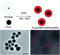
In this work, we present a novel type of magnetic-fluorescent bifunctional nanoparticle (NP). Fe3O4 nanocrystals and cationic fluorescent star polymer perylene diimide–poly(2-aminoethyl methacrylate) (PDI–PAEMA) were simultaneously encapsulated into a silica matrix by a one-pot method. The morphology and fluorescence properties of Fe3O4/PDI–PAEMA@SiO2 core–shell NPs were investigated by ultraviolet-visible (UV-vis) spectrometry, fluorescence spectrometry and high resolution transmission electron microscopy (HRTEM). The analysis of in vitro intracellular uptake and cell viability revealed that the bifunctional NPs possessed favourable biocompatibility. Taken together, the Fe3O4/PDI–PAEMA@SiO2 NPs could be a promising candidate for bioimaging due to their stable red emission and favourable biocompatibility.
Introduction
In recent years, the path for cancer diagnosis and treatment has been opened up by the advent of modern imaging techniques, such as magnetic resonance imaging (MRI),1 computed tomography (CT),2 positron emission tomography (PET),3 photoacoustic tomography (PAT)4 and fluorescence imaging.5 Owing to the unique advantages of high sensitivity, simple operation, rapid imaging and deep tissue penetration,6,7 fluorescence imaging has been widely applied in the biomedical field, allowing cancer diagnosis in the early stage.
Perylene diimide (PDI) derivatives are an attractive class of imaging fluorophores because of their excellent chemical, thermal and photochemical properties.8–10 PDI-cored star polymers have been synthesized and applied for cell-nucleus or extracellular matrix staining.11,12 The outer arms of the polymer chains provide the PDI-cored polymers with water solubility and positive or negative charges. In addition, the fluorescence emissions of the PDI-cored chromophores have no overlap with biological backgrounds, resulting in favourable conditions for in vivo imaging.
Silica is one of the most biocompatible substrates being endogenous to most living organisms. Silica-based matrix shows excellent biocompatibility and stability in aqueous solutions, and facile regulation of silica surface enables the substrate conjugation with biomolecules, thereby improving the selectivity and specificity.13–15 It has been reported that fluorophores can be encapsulated into silica network through covalent bonding, electrostatic interactions and spatial constrain.16 The isoelectric point of pure silica is 3.4, indicating that the silica matrix is negatively charged when dispersed in neutral or basic medium.17 Positively charged dyes are expected to be readily doped into silica due to high affinity.
Multifunctional nanoparticles (NPs) have great potential in biological fields by integrating various beneficial features.18,19 In this work, we reported a facile method to prepare magnetic-fluorescent bifunctional NPs in a well controlled manner. Fe3O4 nanocrystal was readily synthesized by chemical co-precipitation. The PDI–poly(2-aminoethyl methacrylate) (PAEMA) star polymer was prepared by atom transfer radical polymerization (ATRP). Positively charged PDI–PAEMA was encapsulated into silica network during NP formation through electrostatic interactions. In addition to facilitating the encapsulation of PDI-cored star polymer within the silica, the PAEMA arms provide a spatial spacer to avoid PDI-cored fluorophore quenching by Fe3O4.20 Our data showed that the Fe3O4/PDI–PAEMA@SiO2 NPs possessed stable red emission and favourable biocompatibility, making it a promising candidate for bioimaging.
Experimental section
Materials
Ferric chloride hexahydrate (FeCl3·6H2O, Tianjin Fuchen Chemical Reagent Factory), ferrous sulfate heptahydrate (FeSO4·7H2O, Xilong Chemical Co., Ltd.), oleic acid (OA, Tianjin Fuchen Chemical Reagent Factory), tetraethyl orthosilicate (TEOS, Xilong Chemical Co., Ltd.), triton X-100 (Beijing Chemical Reagent Company). Ammonia aqueous solutions (NH3·H2O, 25%), sodium chloride, cyclohexane, hexanol and ethanol were purchased from Beijing Chemical Plant. All chemicals were used directly without any further purification.
Synthesis of Fe3O4 nanocrystals
Fe3O4 nanocrystals were synthesized using chemical co-precipitation as reported previously.21 For a typical reaction, 4.1 g of FeCl3·6H2O and 2.35 g FeSO4·7H2O were dissolved in 100 mL deionized water in a 250 mL three-neck flask. The mixture was mechanical stirred under a flow of nitrogen for 30 min, followed by adding 25 mL NH3·H2O at room temperature. The color of solution immediately changed from orange to black. After the mixture was heated to 80 °C, 1 mL OA was added dropwisely into the dispersion during 1 h. Under a blanket of nitrogen, the mixture was further reacted for 1 h. The black-colored solution was cooled to room temperature by removing the heat source. After small amount of sodium chloride was added into the system, the above solution was mixed with cyclohexane in an extractor. Under the protection of OA, Fe3O4 NPs were extracted from water to organic phase and dispersed well in cyclohexane.
Synthesis of Fe3O4/PDI–PAEMA@SiO2 NPs
The magnetic-fluorescent Fe3O4/PDI–PAEMA@SiO2 core–shell NPs were prepared using one-pot method. In a brief procedure, 1.2 mL of as-synthesized Fe3O4 cyclohexane solution (5 mg mL−1), 37 mL cyclohexane, 8 mL hexanol, 10 g triton X-100, 1.7 mL of PDI–PAEMA aqueous solution (0.6 mg mL−1) were added into a 250 mL three-neck flask, which was sonicated to form a uniform microemulsion. At room temperature, 0.2 mL TEOS were added into the flask. After 4 h of mechanical stirring, 0.6 mL NH3·H2O was introduced to activate the TEOS hydrolysis. The mixture was continued to react at room temperature for 24 h under constant stirring. After this, ethanol was added to the flask resulted in flocculent precipitates. The final brown yellow nanoparticles were collected through magnetic separation. Centrifugation and ultrasonication were applied to remove unreacted reactants for several times.
Characterization
UV-visible absorption was recorded on a spectrometer (Cintra 20, GBC, Australia). Fluorescence spectrum was performed on a fluorescence spectrophotometer (Horiba Jobin Yvon FluoroMax-4 NIR, NJ, USA) at room temperature (25 °C). The quantum yield of nanoparticle was evaluated with Edinburgh Instruments FLS 980 spectrofluorimeter at room temperature. JEOL JEM-3010 high resolution transmission electron microscope (HRTEM) was used to observe morphology of Fe3O4/PDI–PAEMA@SiO2 at an accelerating voltage of 200 kV. Samples were processed by placing a drop of suspension in ethanol on a copper grid, and then evaporating at room temperature. X-ray diffraction (XRD) patterns were measured on D/max2500 VB2+/PC X-ray diffractometer (Rigaku) using Cu Kα radiation in the 2θ range 5–90°. Zeta-potential were performed on Brookhaven 90 Plus/BI-MAS particle size analyzer with dispersed particles in water at 25 °C. Magnetic characterization was manipulated on a vibrating sample magnetometer (VSM, Jilin University JDM-13 VSM).
Dye leakage of Fe3O4/PDI–PAEMA@SiO2 NPs
The leakage of organic dyes from the silica matrix was determined by the change of fluorescence intensity. 1 mg of Fe3O4/PDI–PAEMA@SiO2 NPs was suspended in 2 mL deionized water, and the emission intensity of the solution was measured. Then the sample was centrifuged at 12
000 rpm for 10 min. After discarding the supernate, the Fe3O4/PDI–PAEMA@SiO2 NPs were re-suspended in 2 mL deionized water and the emission of solution was measured again.
Cytotoxicity assay
Cytotoxicity of magnetic-fluorescence NPs was conducted by using Tali™ viability kit-Dead Cell Green (Invitrogen, Catalog A10787) according to a standard protocol. The procedure was manipulated at 48 h post-incubation of Fe3O4/PDI–PAEMA@SiO2 NPs. After 48 h incubation, replace the fresh cell medium and add 1 μL dead cell green into 100 μL cell medium for 0.5 h incubation.
Cellular uptake
The used cell line was HeLa cell. In brief, 7.5 × 10⁴ cells were plated in a 35 mm Petri dish for 7 h to allow the live cells to attach. Cells were washed with PBS and incubated with cell culture medium containing Fe3O4/PDI–PAEMA@SiO2 NPs (50 μg mL−1) for 24 h at 37 °C. After incubation, the cells were washed several times with PBS to remove remaining particles and dead cells, and then observed under a fluorescent microscope.
Results and discussion
Synthesis of Fe3O4/PDI–PAEMA@SiO2 NPs
The PDI-cored star polymer (PDI–PAEMA) was synthesized through ATRP strategy as previously described.22 The repeat units of each arm in PDI–PAEMA were well controlled (r.u. = 50) and determined by 1H NMR. Silica is the most commonly used inorganic shelling material as it has numerous advantages in biological applications. The synthesized polycations can readily bind with silica through electrostatic interactions. Therefore, we designed and prepared the magnetic-fluorescent Fe3O4/PDI–PAEMA@SiO2 NPs. Scheme 1 presents the synthesis procedure of the hybrid NPs. In this framework, Fe3O4 nanocrystal and silica matrix formed core–shell structure, characterized by HRTEM. Simultaneously, the fluorescent polymer PDI–PAEMA was homogeneously dispersed in this structure.
 |
| Scheme 1 Schematic illustration of one-pot synthesis of Fe3O4/PDI–PAEMA@SiO2 NPs. | |
Morphology of NPs
The morphology of the NPs was characterized by HRTEM. Fig. 1A reveals that Fe3O4 nanocrystals possessed an average size of 10 nm. The Fe3O4/PDI–PAEMA@SiO2 NPs were spherical and had a quite uniform size about 60 nm (Fig. 1B), and almost every NP contained one Fe3O4 nanocrystal. As evident from the different electron penetration of silica and Fe3O4, the hybrid NPs displayed a typical core–shell structure. The increased particle size (from 10 nm to 60 nm) indicated the successful coating of silica. Monodispersed Fe3O4/PDI–PAEMA@SiO2 NPs with appropriate size (<200 nm) were suitable for cell uptaking.23
 |
| Fig. 1 HRTEM images of (A) Fe3O4 and (B) Fe3O4/PDI–PAEMA@SiO2 NPs. | |
Optical property assays
Fig. 2 shows the optical spectra of PDI–PAEMA and Fe3O4/PDI–PAEMA@SiO2 NPs. PDI–PAEMA exhibited an emission peak at 609 nm in water (λ = 540 nm). Upon excitation at 540 nm, the Fe3O4/PDI–PAEMA@SiO2 exhibited the same characteristic emission peak as PDI–PAEMA, which was far from cell autofluorescence. The fluorescence quantum yield (Φf) of the Fe3O4/PDI–PAEMA@SiO2 NPs was 15 ± 0.2% in water at room temperature, which was at the equal level of PDI–PAEMA. The fluorescence spectra and quantum yield suggested the well dispersion of PDI–PAEMA in the complex NPs, and the optical property of PDI–PAEMA was not affected by the Fe3O4 nanocrystal because of the spacer provided by the polymer arms of PAEMA.
 |
| Fig. 2 Fluorescence spectra of PDI–PAEMA and Fe3O4/PDI–PAEMA@SiO2 NPs in water. | |
PDI–PAEMA is a pH-sensitive polymer, and the pH response of its eight-armed analogue has been discussed in previous work.24 At low pH values, PDI–PAEMA has a relatively weak fluorescence intensity caused by the volume phase transition (Fig. S2†). Therefore, the exposure of PDI-cored polymer to physiological environment often leads to pH-dependent and unstable optical property, resulting in inhibited fluorophore application for imaging probes. As PDI–PAEMA was encapsulated into silica, Fe3O4/PDI–PAEMA@SiO2 performed stable fluorescence properties under variable pH conditions (Fig. S3†). The final Fe3O4/PDI–PAEMA@SiO2 NPs with enhanced photostability took advantages in biomedical imaging.
Magnetic property of Fe3O4/PDI–PAEMA@SiO2
To evaluate the magnetic properties of Fe3O4/PDI–PAEMA@SiO2 NPs, we applied the magnetization curves on vibrating sample magnetometer (VSM) with a magnetic field cycle between −20 and +20 kOe. Fig. 3 shows that the complex NPs displayed a typical superparamagnetic behaviour at room temperature. After Fe3O4 was encapsulated within silica matrix, the saturation magnetization of Fe3O4 NPs was decreased from 64.21 emu g−1 (Fig. S4†) to 6.67 emu g−1 since less Fe3O4 existed in the Fe3O4/PDI–PAEMA@SiO2 nanostructure compared with the same mass of pure Fe3O4 NPs. However, the magnetic-fluorescent NPs could still be effectively separated by external magnet under the magnetic guidance (Fig. 4).
 |
| Fig. 3 Field-dependent magnetization of Fe3O4/PDI–PAEMA@SiO2 NPs. | |
 |
| Fig. 4 (A) The dispersion of Fe3O4/PDI–PAEMA@SiO2 NPs; (B) Fe3O4/PDI–PAEMA@SiO2 NPs separated by external magnet. | |
Zeta potential
We verified electrostatic interaction capability between PDI–PAEMA and silica matrix by zeta potential. Results showed that the PDI–PAEMA polymer had a positive charge of +26.5 mV, while the Fe3O4@SiO2 had a negative charge of −31.9 mV. After PDI–PAEMA was incorporated into the Fe3O4@SiO2 matrix, the zeta potential of the Fe3O4/PDI–PAEMA@SiO2 NPs was increased from −31.9 mV to −22.9 mV. High affinity between positively charged fluorophore and negatively charged silica resulted in the successful encapsulation of PDI–PAEMA.
Dye leakage assay of Fe3O4/PDI–PAEMA@SiO2
In the present study, we determined leakage of PDI–PAEMA out of the silica according to the change of fluorescence intensity of the Fe3O4/PDI–PAEMA@SiO2 NP suspension. Fig. 5 shows the alteration of Fe3O4/PDI–PAEMA@SiO2 fluorescence intensity. The intensity of the suspension was maintained at 94% as the initial intensity after washing for five times. It was possible that the PDI–PAEMA diffusion was somehow inhibited by the relatively large size of PDI-cored polymer arms as compared with small size of silica pores.
 |
| Fig. 5 Dye leakage of Fe3O4/PDI–PAEMA@SiO2 NPs. | |
Cytotoxicity of Fe3O4/PDI–PAEMA@SiO2
Biocompatibility is an essential requirement for nanomaterials in biological application. To clarify the biocompatibility of final NPs, the cytotoxicity of Fe3O4/PDI–PAEMA@SiO2 NPs was examined using Tali cell viability assay. Fig. 6 shows the effect of concentration of Fe3O4/PDI–PAEMA@SiO2 NPs on cell viability at variable concentrations ranging from 50.0 mg L−1 to 200.0 mg L−1. Within 48 h, the Fe3O4/PDI–PAEMA@SiO2 NPs had low cytotoxicity over a broad range of concentrations, and the cell viability remained as high as 80% even at a high concentration of 200 mg L−1. The biocompatible nature of silica could be potentially used in biological applications.
 |
| Fig. 6 Cell viability of assays of Fe3O4/PDI–PAEMA@SiO2 NPs. | |
Cellular uptake
Herein, we investigated cellular uptake using HeLa cells to test the ability of Fe3O4/PDI–PAEMA@SiO2 entering living cells. After incubation with composite NPs for 24 h, the cells were imaged by fluorescence microscopy. Strong red fluorescence could be observed inside the cells (Fig. 7), suggesting that Fe3O4/PDI–PAEMA@SiO2 was internalized into living cells via endocytosis.25 The real-time monitoring of fluorescence intensity (Fig. 8A) showed that the fluorescence was detected within 0.5 h, and the fluorescence intensity was increased along with incubation time. After incubation for 48 h, the culture medium was replaced without Fe3O4/PDI–PAEMA@SiO2. Although the emission intensity was decreased, the red fluorescence could still be observed for 60 h and 72 h, indicating that the NPs were still stable when internalized into cells (Fig. 8B). The results of cellular uptake assays and cytotoxicity test demonstrated that the Fe3O4/PDI–PAEMA@SiO2 NPs had a favourable biocompatibility.
 |
| Fig. 7 Fluorescence images of HeLa cells incubated with Fe3O4/PDI–PAEMA@SiO2 NPs for 24 h. (A) Bright-field, (B) fluorescence and (C) overlapping images. | |
 |
| Fig. 8 Quantified fluorescence intensities of cellular distributed NPs. (A) Cells were treated with 50 μg mL−1 Fe3O4/PDI–PAEMA@SiO2 NPs for different incubation time. (B) Cells were incubated in culture medium without Fe3O4/PDI–PAEMA@SiO2. | |
Conclusions
In summary, the Fe3O4/PDI–PAEMA@SiO2 NPs were fabricated by facile one-pot method in our present study. Fe3O4 nanocrystal and silica formed well controlled core–shell structure. Simultaneously, the fluorescent polymer PDI–PAEMA was homogeneously dispersed in this nanostructure. The bifunctional NPs exhibited stable red fluorescence and retained magnetic response. Cellular uptake and cytotoxicity assays demonstrated that these hybrid NPs had high intracellular penetration and favourable biocompatibility. Taken together, the magnetic-fluorescent Fe3O4/PDI–PAEMA@SiO2 NPs possessed the potential to be applied in biomedical fields, such as multimode imaging and drug delivery, due to their stable optical property and good internalization capacity.
Acknowledgements
This work was supported by the National Natural Science Foundation of China (51221002 and 21574009), the Beijing Natural Science Foundation (2142026), and the Innovation and Promotion Project of Beijing University of Chemical Technology.
References
- J. Yu, C. Yang, J. Li, Y. Ding, L. Zhang, M. Z. Yousaf, J. Lin, R. Pang, L. Wei and L. Xu, Adv. Mater., 2014, 26, 4114–4120 CrossRef CAS PubMed.
- V. Cnudde, B. Masschaele, M. Dierick, J. Vlassenbroeck, L. Van Hoorebeke and P. Jacobs, Appl. Geochem., 2006, 21, 826–832 CrossRef CAS.
- K. Chen and P. S. Conti, Adv. Drug Delivery Rev., 2010, 62, 1005–1022 CrossRef CAS PubMed.
- L. V. Wang and S. Hu, Science, 2012, 335, 1458–1462 CrossRef CAS PubMed.
- J. Kim, H. S. Kim, N. Lee, T. Kim, H. Kim, T. Yu, I. C. Song, W. K. Moon and T. Hyeon, Angew. Chem., Int. Ed., 2008, 47, 8438–8441 CrossRef CAS PubMed.
- M. Chen and M. Yin, Prog. Polym. Sci., 2014, 39, 365–395 CrossRef CAS.
- J. V. Frangioni, Curr. Opin. Chem. Biol., 2003, 7, 626–634 CrossRef CAS PubMed.
- S. K. Yang, X. Shi, S. Park, S. Doganay, T. Ha and S. C. Zimmerman, J. Am. Chem. Soc., 2011, 133, 9964–9967 CrossRef CAS PubMed.
- C. Kohl, T. Weil, J. Qu and K. Müllen, Chem.–Eur. J., 2004, 10, 5297–5310 CrossRef CAS PubMed.
- S. K. Yang and S. C. Zimmerman, Adv. Funct. Mater., 2012, 22, 3023–3028 CrossRef CAS PubMed.
- M. Yin, J. Shen, R. Gropeanu, G. O. Pflugfelder, T. Weil and K. Müllen, Small, 2008, 4, 894–898 CrossRef CAS PubMed.
- M. Yin, J. Shen, G. O. Pflugfelder and K. Müllen, J. Am. Chem. Soc., 2008, 130, 7806–7807 CrossRef CAS PubMed.
- J. H. Lee, Y. w. Jun, S. I. Yeon, J. S. Shin and J. Cheon, Angew. Chem., 2006, 118, 8340–8342 CrossRef.
- L. Sun, Y. Li, M. Sun, H. Wang, S. Xu, C. Zhang and Q. Yang, New J. Chem., 2011, 35, 2697–2704 RSC.
- J. Kobler, K. Möller and T. Bein, ACS Nano, 2008, 2, 791–799 CrossRef CAS PubMed.
- D. Ma, A. J. Kell, S. Tan, Z. J. Jakubek and B. Simard, J. Phys. Chem. C, 2009, 113, 15974–15981 CAS.
- X. Song and L. Gao, J. Phys. Chem. C, 2007, 111, 8180–8187 CAS.
- Y. Deng, Y. Cai, Z. Sun, J. Liu, C. Liu, J. Wei, W. Li, C. Liu, Y. Wang and D. Zhao, J. Am. Chem. Soc., 2010, 132, 8466–8473 CrossRef CAS PubMed.
- S. Santra, C. Kaittanis, J. Grimm and J. M. Perez, Small, 2009, 5, 1862–1868 CrossRef CAS PubMed.
- E. Dulkeith, A. Morteani, T. Niedereichholz, T. Klar, J. Feldmann, S. Levi, F. Van Veggel, D. Reinhoudt, M. Möller and D. Gittins, Phys. Rev. Lett., 2002, 89, 203002 CrossRef CAS PubMed.
- Y. Sun, X. Ding, Z. Zheng, X. Cheng, X. Hu and Y. Peng, Eur. Polym. J., 2007, 43, 762–772 CrossRef CAS.
- S. You, Q. Cai, Y. Zheng, B. He, J. Shen, W. Yang and M. Yin, ACS Appl. Mater. Interfaces, 2014, 6, 16327–16334 CAS.
- H. Maeda, J. Wu, T. Sawa, Y. Matsumura and K. Hori, J. Controlled Release, 2000, 65, 271–284 CrossRef CAS PubMed.
- S. You, Q. Cai, K. Müllen, W. Yang and M. Yin, Chem. Commun., 2014, 50, 823–825 RSC.
- X. Xing, X. He, J. Peng, K. Wang and W. Tan, J. Nanosci. Nanotechnol., 2005, 5, 1688–1693 CrossRef CAS PubMed.
Footnote |
† Electronic supplementary information (ESI) available. See DOI: 10.1039/c6ra08410a |
|
This journal is © The Royal Society of Chemistry 2016 |
Click here to see how this site uses Cookies. View our privacy policy here.