Lina Zhouab,
Qiuxiang Yinab,
Shichao Dua,
Hongxun Haoab,
Yanfeng Lic,
Mingyan Liu*ab and
Baohong Houab
aSchool of Chemical Engineering and Technology, State Key Laboratory of Chemical Engineering, Tianjin University, Tianjin 300072, China. E-mail: myliu@tju.edu.cn
bCollaborative Innovation Center of Chemistry Science and Engineering, Tianjin 300072, China
cTianjin Pharmaceuticals Research Institute CO. LTD, Tianjin 300000, China
First published on 19th May 2016
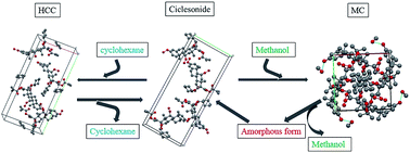
Two novel solvates of ciclesonide were successfully obtained and characterized by various analytical techniques (X-ray powder diffraction, X-ray single-crystal diffraction, differential scanning calorimetry, thermogravimetric analysis and hot-stage microscopy). Crystal structure analysis results indicate that the solvate of ciclesonide formed with cyclohexane (HCC) is a channel solvate and isostructural with ciclesonide, while the solvate formed with methanol (MC) isa discrete position solvate. Thermal analysis results show that HCC could desolvate without destroying the crystal lattice. However, MC would transform into an amorphous form after desolvation and the amorphous form could recrystallize into the crystalline form of ciclesonide. Moreover, the kinetics of the HCC and MC isothermal and non-isothermal desolvation processes is calculated and discussed.
Plenty of solvates have been obtained and studied during the solid form screening of APIs. Researchers mostly focused on the crystal structure and thermal (or moisture) stability analysis of the solvates in the previous literature.6–9 Based on the crystal structure, solvates are divided into two types: discrete position solvates which the solvent molecules occupy isolated position in the crystal structure and channel-type solvates which the solvate molecules exist along channels in the structure.10 There are significant differences in the formation and desolvation processes of the two types of solvates.11–13
The model compound ciclesonide (CAS Registry no. 126544-47-6, Fig. 1) is a novel cortical hormone drug developed by ALTANA company from Germany and is frequently applied to cure asthma.14 In literature, crystalline ciclesonide has been reported and characterized using X-ray diffraction, thermal and spectroscopy analysis methods.15 Due to its poor solubility in water, new solid forms of ciclesonide need to be developed to improve the physicochemical properties. Amorphous state ciclesonide has been developed to improve the solubility, but its poor stability is unfavorable for manufacture and transportation. Therefore, other solid forms such as new polymorphs and new solvates of ciclesonide need to be developed and investigated.
In this work, two kinds of solvates of ciclesonide methanol–ciclesonide (1:
1, ab. MC) and hemi-cyclohexane–ciclesonide (0.5
:
1, ab. HCC) solvates, are developed and fully characterized by X-ray powder diffraction, X-ray single-crystal diffraction, differential scanning calorimetry, thermogravimetric analysis and hot-stage microscopy. Firstly, single crystal structure data was obtained and used to confirm the different incorporating types of solvent molecules and the solute–solvent interactions. Then, the thermal analysis was carried out to investigate the solvent escaping process, the crystal form change and the thermal variation during and after desolvation process. What's more, the desolvation kinetics of HCC and MC in the isothermal and non-isothermal processes was calculated and discussed, respectively.
In order to obtain high-quality single crystals for crystal structure determination, 0.2 g ciclesonide was added into 10 mL cyclohexane and 5 mL methanol, respectively. The solution was heated up until solids completely dissolved to get saturated solution. The saturated solution was filtered through 0.25 μm PTFE filter. The filtrate was gradually cooled to room temperature, and covered with plastic film with a few holes on it for solvent evaporating. After several days, colorless crystals with appropriate size for crystal structure determination were obtained from cyclohexane and methanol, respectively.
Ciclesonide15 | HCC | MC | |
---|---|---|---|
Formula weight | 540.67 | 582.75 | 572.71 |
Crystal system | Orthorhombic | Orthorhombic | Tetragonal |
Crystal habit | Needle | Rectangular | Prismatic |
Space group | P212121 | P21212 | P43 |
a/Å | 6.638 (1) | 14.955 (3) | 11.001 (1) |
b/Å | 14.079 (3) | 32.318 (7) | 11.001 (1) |
c/Å | 32.870 (7) | 6.818 (2) | 25.559 (4) |
α, β, γ/° | α = β = γ = 90° | α = β = γ = 90° | α = β = γ = 90° |
V/Å3 | 3071 (9) | 3295 (2) | 3093 (1) |
Z | 4 | 4 | 4 |
ρ/g cm3 | 1.169 | 1.175 | 1.230 |
θ-Range/° | 1.90–24.39 | 3.0–27.50 | 3.0–27.50 |
F(000) | 1168 | 1264 | 1240 |
Reflections | 4730 | 6119 | 5727 |
R/Rw/% | 4.94/8.96 | 7.42/10.90 | 3.23/10.11 |
![]() | ||
Fig. 2 Crystal structure of (a) ciclesonide, (b) HCC, (c) MC. Several atoms have not been labelled for clarity. |
To further understand the crystal structural changes caused by solvent molecules, the formation of H-bonds from different crystal structure were analyzed. As shown in Table 3 and Fig. 2, although different symmetry elements along c axis lead to different position of adjacent molecules, both ciclesonide and HCC have the same H-bonds type between host molecules, and there is no H-bond between guest and host molecules. On the contrary, MC contains distinct H-bond between guest and host molecules, which causes the variation of host molecules packing. In the arrangement process of host molecules, H-bonds O(2)–H(2A)⋯O(1) is substituted by the new H-bond O(2)–H(2A)⋯O(7) in MC structure. Besides, methanol molecules occupying discrete position are connected with host molecules by H-bond O(8)–H(8B)⋯O(1). Both the inter-molecular and intra-molecular H-bonds play important roles in constructing the MC crystals.
D–H⋯A/Å | d(D–H)/Å | d(H⋯A)/Å | d(D⋯A)/Å | ∠(DHA)/° | |
---|---|---|---|---|---|
a #1: x − 1/2, −y + 1/2, −z; #2: y, −x + 1, z + 1/4; D: donor, A: acceptor. | |||||
Ciclesonide | O(2)–H(2A)⋯O(1) | 0.82 | 1.97 | 2.739 | 156.7 |
HCC | O(2)–H(2A)⋯O(1)#1 | 0.82 | 1.99 | 2.792 | 167.6 |
MC | O(2)–H(2A)⋯O(7)#2 | 0.82 | 2.08 | 2.861 | 158.2 |
O(8)–H(8B)⋯O(1) | 1.00 | 1.79 | 2.784 | 169.0 |
The packing diagrams of two kinds of solvates are displayed along c axis from Fig. 3. It can be seen in Fig. 3a that cyclohexane molecules occupy the cavities in the crystal lattices. Besides, the cyclohexane molecules always appear in the center of plane (100) and plane (010). So they are shared by the adjacent cell with 0.5 contributing rate to every cell and forming two channels parallel along c axis. In addition, the volume of the cavity shown in Fig. 3a was calculated by using PLATON (version 210103) program,18 and the result was 60.1 Å3. Meanwhile, the volume of cyclohexane solvent was also calculated by Materials Studios (version 5.0, Accelrys company), and Smart Minimizer method was used to optimize molecular stability. The cyclohexane molecular volume obtained by simulation was 57.15 Å3, which was smaller than the volume of the cavity. Therefore, it is feasible that the free volume available to the solvent molecules in the cavity is related to strength the host–solvent interactions, and thus cyclohexane molecules can occupy the position of the channels arrangement, forming stable solvates.
![]() | ||
Fig. 3 The packing feature of solvates ((a) HCC, (b) MC) along c axis. Sky blue dashed lines show the H-bond that had been matched completely. |
Experimental TGA traces are shown in Fig. 4. It can be seen that the weight loss of MC/HCC is up to 5.12%/6.74% during desolvation process, which is nearly consistent with the calculated weight loss (5.58%/7.21%) according to stoichiometry of the solvents obtained from the XRSD analysis. The small differences between the experimental results and the theoretical results might be caused by the experiment operation. Fig. 4 also shows that the desolvation temperature of MC is much higher than that of HCC. Besides, there is no more weight loss after solvent escaping until 250 °C for both HCC and MC, illustrating that no decomposition of ciclesonide happens in this temperature range.
The DSC curves are also displayed in Fig. 5. Considering both DSC and TGA results, the first endothermic peaks A of curve (b) (HCC) and (c) (MC) in Fig. 5 are regarded as desolvation peaks and the second endothermic peaks B are considered as melting peaks by comparing with the DSC curve of non-solvated ciclesonide. Detailed thermal data of DSC are obtained and displayed in Table 4. It shows that the onset values of the desolvation peaks are 80.82 °C and 122.28 °C for HCC and MC respectively. As we know, the boiling temperatures of cyclohexane and methanol are 80.7 °C and 64.7 °C under atmospheric pressure. From that, the desolvation temperature of HCC is almost consistent with the boiling temperature of cyclohexane. As a channel-type solvate, cyclohexane molecule can be constrained only by van der Waals force, so it can escape from the cavities of crystal lattice without higher temperature. Conversely, the desolvation temperature of MC is much higher than the boiling temperature of solvent. According to the crystal structure information of MC, the methanol molecules have strong H-bonds interaction with ciclesonide molecules. Therefore higher temperature is needed to break the H-bonds and facilitate the escape of methanol molecules.
Solvate | Tonset, A/°C | ΔHA/kJ mol−1 | Tonset, B/°C | ΔHB/kJ mol−1 |
---|---|---|---|---|
HCC | 80.82 | 20.53 | 207.45 | 30.49 |
MC | 122.28 | 11.73 | 207.22 | 30.49 |
In addition, there is an exothermic peak at about 160 °C in the DSC curve of MC, and no weight change is found from TGA analysis at the same temperature. This kind of exothermic peak might correspond to a phase transformation process, but it cannot be confirmed only by DSC and TGA analysis.
![]() | ||
Fig. 6 Comparison of XRPD patterns: (a) ciclesonide, (b) HCC, (c) MC, (d) simulated HCC, (e) simulated MC. |
Then the XRPD was used to determine the crystal form of the solvates during the heating process and the spectrums were displayed in Fig. 7. The experimental temperature of HCC was elevated from room temperature to 120 °C. As shown in Fig. 7a, there is no obvious change of peak position except the variation of relative peak intensity. The intensity of peak 1 increases gradually and peak 2 weakens at the same time. The relative intensity changes might be caused by the density change of cyclohexane in channels. Furthermore, the XRPD patterns of HCC during the desolvation are always very similar to ciclesonide. It can be concluded that the escaping of cyclohexane molecules does not destroy the crystal lattice and has little influence on the crystal structure.
For Fig. 7b, as the temperature increases, the identified peaks of MC gradually weaken until completely disappear and an amorphous phase appears at 140 °C. This temperature range is in accordance with the desolvation process. During the desolvation process, the H-bonds between ciclesonide and methanol are broken to facilitate the escape of methanol molecules. Then the crystal lattice of MC is destroyed and it transformed into amorphous phase. Interestingly, when temperature was further elevated to 160 °C, a new crystalline phase turned up. Its XRPD pattern is identical with ciclesonide, indicating that the amorphous phase will recrystallize into crystalline as the temperature increases. This phase transformation process of MC could be used to explain the exothermic peak in the DSC data of MC.
In Fig. 8b, MC crystal became opaque from 120 °C because of the escape of methanol molecules from crystal lattice. The opaque region moved from the edge toward the middle due to that desolvation reaction was much easier to take place on the edge of crystals. As methanol molecules escaping from MC crystals, the crystalline structure of solvate was destroyed, and MC transformed from crystalline to amorphous form. When temperature was persistently heated up to 160 °C, the color of the crystals began to turn transparent again. Combing TGA, DSC and XRPD results, the rearrangement of ciclesonide molecules might occur in the process. When temperature was elevated to 213 °C, the crystal was in half-fusion state and some un-fused tiny crystals were left. In order to identify crystal form of these tiny crystals that might come from rearrangement product of amorphous, the hot-state temperature was fast reduced to 201 °C to promote the growth of tiny crystals. After that, the needle crystals were obtained and detected in microscope, whose shape was similar to ciclesonide reported before. The crystal form of needle crystals was measured by XRPD, whose results are compared with ciclesonide in Fig. 9, verifying again that the needle crystals are crystalline non-solvated ciclesonide.
As → Bs + Cg |
Based on the above reaction equation, the rate of an isothermal solid-state reaction can be described by20
![]() | (1) |
![]() | (2) |
For non-isothermal reaction rate, a solid-state reaction can be described as eqn (3):
![]() | (3) |
![]() | (4) |
![]() | (5) |
Both f(α) and g(α) have different expressions according to different reaction models which are shown in Table 5.19,21–23
Model | Rate form f(α) | Integral form g(α) |
---|---|---|
Nucleation models | ||
Power law (P2) | 2α1/2 | α1/2 |
Power law (P3) | 3α2/3 | α1/3 |
Power law (P4) | 4α3/4 | α1/4 |
Avrami–Erofe'ev (A2) | 2(1 − α)[−ln(1 − α)]1/2 | [−ln(1 − α)]1/2 |
Avrami–Erofe'ev (A3) | 3(1 − α)[−ln(1 − α)]2/3 | [−ln(1 − α)]1/3 |
Avrami–Erofe'ev (A4) | 4(1 − α)[−ln(1 − α)]3/4 | [−ln(1 − α)]1/4 |
Prout–Tompkins (B1) | α(1 − α) | ln[α/(1 − α)] + ca |
![]() |
||
Geometrical contraction models | ||
Contracting area (R2) | 2(1 − α)1/2 | 1 − (1 − α)1/2 |
Contracting volume (R3) | 3(1 − α)2/3 | 1 − (1 − α)1/3 |
![]() |
||
Diffusion models | ||
1-D diffusion (D1) | 1/(2α) | α2 |
1-D diffusion (D1) | −[1/ln(1 − α)] | ((1 − α)ln(1 − α)) + α |
3-D diffusion-Jander (D3) | [3(1 − α)2/3]/[2(1 − (1 − α)1/3)] | (1 − (1 − α)1/3)2 |
Ginstling–Brounshtein (D4) | 3/[2((1 − α)−1/3 − 1)] | 1 − (2/3)α − (1 − α)2/3 |
![]() |
||
Reaction-order models | ||
Zero-order (F0/R1) | 1 | α |
First-order (F1) | (1 − α) | −ln(1 − α) |
Second-order (F2) | (1 − α)2 | [1/(1 − α)] − 1 |
Third-order (F3) | (1 − α)3 | (1/2)[(1 − α) − 2 − 1] |
Plots of conversion fraction α against temperature T in transition process from solvates to the corresponding non-solvates at certain heating rates are shown in Fig. 10. Fig. 11 presents the relation between conversion fraction α and time t in isothermal desolvation process.
![]() | ||
Fig. 10 Conversion fraction α versus temperature plots for non-isothermal desolvation of HCC (a) and MC (b) at different heating rates. |
![]() | ||
Fig. 11 Conversion fraction α versus time plots for isothermal desolvation of HCC (a) and MC (b) at different temperatures. |
In this work, both isothermal and non-isothermal kinetic data (α) in the range of 10% to 90% conversion fraction derived from TGA are fitted to all the mathematical models in Table 5, using Microsoft Excel software. R2, defined as eqn (6) was employed to evaluate the applicability of different models. The fitting results are summarized in Table S1† (non-isothermal), Table S2† (non-isothermal) and Table S3† (isothermal).
![]() | (6) |
In general, a particular solid-state reaction should belong to the same model. But from Tables S1† and 2, it can be concluded that the best fitting model changes with the heating rates for non-isothermal process. Table 3 states that the best fitting model for isothermal process is consistent. It also indicates that the variation rate of temperature will influence the choice of the reaction kinetics models.
For HCC solvate, the F0 model is suitable when heating rate is 1 K min−1 and 2 K min−1 while the F1 model is suitable for 5 K min−1 and F2 model is optimal when the heating rate was elevated to 10 K min−1.
Based on the mechanism of established model, F0, F1 and F2 are reaction-order models. When heating rate is slow, desolvation process of HCC solvate belongs to the zero order reaction, but when heating rate is increased to 10 K min−1, the desolvation process is regarded as two-step reaction.
Considering the kinetics models and the crystal structure of HCC, desolvation process may be proposed as two steps: (a) dissociation of cyclohexane molecules from the crystal lattice and (b) the release of solvent molecules by diffusion through the channels. For isothermal desolvation process, R3 model always gives the best fitting results in the temperature range of 80 °C to 90 °C. The R3 model is a three-dimensional phase boundary model, which means that desolvation process can simultaneously happen in three directions under the isothermal condition.
F2 and F3 models are appropriate for non-isothermal desolvation process of MC. H-Bond interactions existing between methanol and ciclesonide molecules account for an additional step for breaking H-bond compared with HCC in the desolvation process of MC. R2 model has excellent performance in fitting the α–time curves in isothermal desolvation process, which could testify that MC desolvation process belongs to two-dimensional phase boundary reaction.
The activation energy Ea and preexponential factor A calculated from best fitting model, are shown in Table 6. It can be seen that the kinetic parameters Ea and A obtained from non-isothermal and isothermal process are not consistent. Vyazovkin and Wight22 explained the reason from two aspects: (1) in non-isothermal process both T and α vary simultaneously. But it is difficult to achieve a thoroughly separation between T and f(α). As a result, any f(α) can satisfactorily fit data at the cost of drastic variations in the Arrhenius parameters, which compensate for the difference between the assumed form of f(α) and the true but unknown reaction model. (2) The temperature regions are different between isothermal and non-isothermal process. If a desolvation process contains not only one step and every step might have different activation energy, the contribution of every step to overall conversion rate of desolvation process will vary with both temperature and time. It means that the activation energy determined from thermal analysis experiments will also be a function of these two variables. These kinetic parameters obtained from the best-fitting model are an average of all steps, so they don't reflect the changes of parameters values in desolvation process.
HCC | MC | |||
---|---|---|---|---|
Non-isothermal | Isothermal | Non-isothermal | Isothermal | |
A (s−1) | 1.2218 × 1029 | 1.4741 × 10²¹ | 2.4798 × 1030 | 3.1758 × 10²³ |
Ea (kJ mol−1) | 87.656 | 103.843 | 123.283 | 157.341 |
Footnote |
† Electronic supplementary information (ESI) available: The values of R2 for fitting models. CCDC 1450418 and 1450419. For ESI and crystallographic data in CIF or other electronic format see DOI: 10.1039/c6ra08351j |
This journal is © The Royal Society of Chemistry 2016 |