Nanosilver rainbow: a rapid and facile method to tune different colours of nanosilver through the controlled synthesis of stable spherical silver nanoparticles

Pramujitha Mendisa, Rohini M. de Silva*a, K. M. Nalin de Silvaab, Lahiru A. Wijenayakab, Kalana Jayawardanacd and Mingdi Yancd
aDepartment of Chemistry, University of Colombo, Colombo 03, Sri Lanka. E-mail: rohini@chem.cmb.ac.lk
bSri Lanka Institute of Nanotechnology (SLINTEC), Nanotechnology and Science Park, Pitipana, Homagama, Sri Lanka
cDepartment of Chemistry, University of Massachusetts, Lowell, Massachusetts, USA
dDepartment of Chemistry, Portland State University, Portland, Oregon, USA

Received 31st March 2016 , Accepted 4th May 2016

First published on 5th May 2016


Abstract

A rapid and simple one-pot reaction to synthesize stable, spherically shaped silver nanoparticles (AgNps) of different sizes producing distinct optical properties in aqueous solution at ambient temperature has been developed. Each system contains various sizes of silver nanoparticles showing rainbow colours with the peak wavelength of the absorption spectra ranging from 400 to 750 nm. Seven different colours of nano silver were developed through the controlled synthesis of spherical silver nanoparticles using silver nitrate as the metal precursor. Sodium borohydride was used as the main reducing agent and trisodium citrate and hydrazine sulphate were used as the stabilizing and auxiliary reducing agents respectively. The colour of the solution was controlled by varying the concentrations of reagents and the optimum conditions for all the colours are reported. Characterization of silver nanoparticles was carried out using UV-visible spectrophotometry and Transmission Electron Microscopy (TEM). Factors affecting the formation of different sizes of silver nanoparticles, such as silver nitrate concentration, reducing agent concentrations, reaction temperature and reaction pH are also reported. The stability of these coloured silver colloidal solutions was also investigated at different temperatures and the most stable temperature was found to be 4 °C, while the optimum pH to synthesize distinctively coloured silver nanoparticles was found to be in the range of 7–8. The outlined procedure provides a rapid, facile and reproducible synthetic route to spherical AgNps of varying size and ensuing optical properties. Thus, this method is certain to find value in the many applications where size tunability of AgNps is desired.
Introduction

Noble metal nanoparticles show size and shape dependent optical properties. Controlled synthesis of nanoparticles of various noble metals and their alloys has been strongly motivated by the necessity to harness their potential applications in various fields. Silver for example is used in an array of applications ranging from optoelectronics, catalysis, spectroscopy, etc. Most of these applications require a specific shape and size of silver nanoparticles for the desired applicability and efficiency to be achieved. Nevertheless, the intrinsic physical and chemical properties of the nanoparticles would in turn be related to the size and shape of the synthesized nanoparticles. Thus, controlled synthesis of silver nanoparticles is an overarching challenge in all applications of silver nanoparticles.

The first synthesis of noble metal nanoparticles was reported in China and Egypt, and dates back to the 5th or 4th century BC.1 These noble metal nanoparticles were exploited for improving the aesthetic properties of various artefacts due to their attractive colours. One of the most fascinating objects created using this technology is the famous Lycurgus cup which is displayed in the British Museum in London. This object was a result of craftsmanship of Romans in the 4th century BC and features an astonishing property of changing colour depending on the light falling on it. It has already been proved that the presence of mixed silver and gold particles of approximately 70 nm in the vase within the glass matrix resulted in a bright red colour.2,3

Noble metal nanoparticles and in particular, silver nanoparticles with superior thermal, electrical and optical properties have become the focus of current research in view of their potential applications in various areas such as catalysis,4,5 surface enhanced Raman scattering,6–10 optoelectronics,11–15 and surface enhanced fluorescence.16,17 Additionally, silver nanoparticles have received considerable attention due to their capacity to interact with viruses,18 as well as their antifungal and antibacterial properties.19–27 The control of size, shape and stability of these nanoparticles is significant as many intrinsic properties are governed by these factors. Over the past few years there was an emphasis towards fine tuning of properties through the control of shape of silver nanoparticles. Typically, synthesized silver nanoparticles are yellow in colour as yellow silver nanoparticles seem to be the most stable. Various methods have been developed to prepare numerous shapes of silver nanoparticles including rods,28–33 disks,34–37 plates,38 prisms,39–44 wires,29,30,33,45–47 and cubes.48–52 However, there are limitations associated with many methods reported in literature. The major disadvantage of these methods is the reaction time which varies from hours44,53 to days in some cases.41,54–56 Further, the stability of nanoparticles has been a key issue as the optical properties of silver nanoparticles change with time while less attention has been paid on stability regardless of its perceived importance.

Unlike gold nanoparticles, silver colloidal solutions of varying optical properties have rarely been realised using spherical particles. The first comprehensive study in obtaining silver nanoparticles of different colour was reported by Ledwith et al.57 This methodology was based on the reduction of silver ions by ascorbic acid in the presence of citrate stabilized silver seeds, trisodium citrate and polyvinylpyrrolidone. Later Xu et al.58 have also reported the synthesis of coloured colloidal silver nanoparticles using silver nitrate, sodium borohydride, polyvinylpyrrolidone, hydrogen peroxide and sodium citrate. They synthesized a range of colours using different sizes and shapes of silver nanoparticles suggesting the possibility of tuning optical properties without emphasizing to probe the shape and size of the nanoparticle.

The origin of the colour of noble metal nanoparticles was not understood until 1857 when Michael Faraday59 recognized and explained the role of noble metal nanoparticles as a colorant in general terms. He explained that the mere variation in the size of the particles gave rise to a variety of resultant colours. In 1908 Gustav Mie60,61 provided a theoretical explanation by solving Maxwell's equations for the absorption and scattering of electromagnetic radiation by very small metallic particles. The Surface Plasmon Band (SPB)62–65 is a very strong and broad band observed in the UV-visible spectra of silver nanoparticles bigger than 2 nm. The very strong extinction coefficient of nanosilver is due to Surface Plasmon Resonance (SPR) effects meaning that very little nanosilver is required to provide an intense colour. Apart from the applications mentioned, coloured nanosilver can be used as a colorant and a functional component in wool to manufacture high value functional textiles.66

Solution phase synthesis techniques that are based on various modifications of Turkevich method which were introduced in early 1950s have a host of examples in which different silver salts and reducing agents were used.67,68 The Turkevich method could be used to make AgNps but the particles formed were larger, less uniform and had a greater degree of polydispersity. This has been attributed to the fact that fewer seeds are formed of larger nanocrystals of varying shape and size. Despite this or perhaps even because of it, this type of colloid is a popular substrate for Surface Enhanced Raman Scattering (SERS).57 Another successful procedure is to use sodium borohydride; as this is a strong reducing agent, a larger number of nuclei are formed when it is used to reduce metal salts and therefore the resulting particles formed are typically smaller.69 These particles can be used as seeds or nucleation centres for the preparation of nanoparticles with larger sizes and different shapes. Seed particles have acted as catalysts for the reduction of silver ions by weak reducing agents such as ascorbic acid.57 Spherical silver nanoparticles could mainly be synthesised using sodium borohydride at room temperature70 while hydrazine and hydroxylamine have also been used as reducing agents.71

An average size of 16 nm of AgNps has been reported using ethanol.73 Various other reagents such as sodium dodecyl sulphate,72 polyvinylpyrrolidone,57,74 formaldehyde,75 cetyltrimethyl ammonium bromide76 have been used to control the size and stability of these silver nanoparticles.

The method we report here has made significant progress and addressed various issues regarding the synthesis and stability of silver nanoparticles of various colours. In this study our objective therefore was to develop simple, cost effective and quick procedures to synthesize silver nanoparticles of different colours, i.e. spherical particles of varying diameter. The method reported here is very convenient compared to the seed mediated methods reported in literature. More emphasis was placed on controlling the colour of the system rather than achieving monodispersity, while the colour of silver nanoparticles was mainly obtained through spherical nanoparticles as opposed to the previous reports where colour was obtained through non-spherical morphologies.38,57,58 This is important as many systematic studies have been done to evaluate the stability, toxicity, and biological compatibility of spherical nanoparticles as opposed to non-spherical geometries, allowing spherical nanoparticles to be readily applied in a variety of applications.

Further, another reason for having few reports on various colours other than yellow is the stability of silver nanoparticles of other colours. Therefore in our study we have investigated the effect of temperature on the stability of silver nanoparticles of all the colours including violet, purple, blue, green, yellow, orange and red. In addition, factors such as pH, concentration of metal salt and concentrations of reducing agents have also been investigated.

Materials and methods

Materials

Silver nitrate (99%), sodium borohydride (99%), trisodium citrate (99%) and hydrazinium sulphate (99%) were purchased from BDH Chemicals. All chemicals were used as received without further purification. All other reagents were of analytical grade. Glassware were cleaned with concentrated nitric acid and washed thoroughly with water followed by freshly prepared deionized double distilled water before use.

Procedures

The general procedure for the synthesis of different colours of silver nanoparticles involves the following method. Sodium borohydride, hydrazine sulphate and trisodium citrate were added into a beaker and stirred for 10 minutes. After 10 minutes AgNO3 was added into the solution while stirring. The reagents were added according to the amounts given in Table 1. In all procedures freshly prepared sodium borohydride was used as it plays a major role in the resultant colour of the final solution.
Table 1 The optimum concentrations required for the preparation of distinctly coloured AgNps
Colour of AgNps Concentration of reagents (M)
AgNO3 (5 mL) NaBH4 (20 mL) Citrate (30 mL) Hydrazine (20 mL)
Red (RAgNp) 1 × 10⁻³ 4 × 10⁻³ 1 × 10⁻² 2 × 10⁻³
Orange (OAgNp) 1 × 10⁻³ 4 × 10⁻³ 2 × 10⁻² 2 × 10⁻³
Yellow (YAgNp) 1 × 10⁻³ 3 × 10⁻³ 1 × 10⁻²
Green (GAgNp) 1 × 10⁻³ 3 × 10⁻³ 1 × 10⁻² 2 × 10⁻³
Purple (PAgNp) 2 × 10⁻³ 4 × 10⁻³ 1 × 10⁻² 2 × 10⁻³
Blue (BAgNp) 3 × 10⁻³ 4 × 10⁻³ 3 × 10⁻² 2 × 10⁻³
Violet (VAgNp) 3 × 10⁻³ 4 × 10⁻³ 1 × 10⁻² 2 × 10⁻³


Understanding the factors affecting AgNp formation

It has been observed that many factors affect the formation of AgNps. Therefore in our study we investigated the effect of temperature, pH, metal salt concentration, and reducing agent concentrations on the formation of green colour silver nanoparticles (GAgNps). Note the choice of GAgNps for the above investigation was arbitrary.

First, the synthesis of GAgNps was repeated to investigate the effect of temperature on the formation of GAgNps. Here, the reaction temperature was maintained at room temperature, 50 °C, 70 °C, or 100 °C using a water bath while keeping the other factors constant. The absorbance and the wavelengths of the resulting solutions were measured spectrophotometrically. Using the spectra it was identified that room temperature maximizes the yield and thus all the following reactions were performed at an ambient temperature (28–31 °C) (ESI).

Similarly, the synthesis of GAgNps was repeated with buffer solutions of varying pH to investigate the effect of reaction pH. The pH was adjusted by using acetic acid/sodium acetate buffer for pH 4, phosphate buffer for pH 7 and sodium bicarbonate/sodium carbonate buffer for pH 10. The pH in the solution mixtures were monitored using a pH meter (ESI). It was observed that the yield was maximized when basic conditions were used for the synthesis and thus a pH of 10 was used for all the following synthesis reactions discussed.

Then, the procedure for the synthesis of GAgNps was repeated to investigate the effect of silver nitrate concentration at the optimized temperature and pH. The reaction was monitored using different concentrations of silver nitrate (1 × 10⁻³, 2 × 10⁻³, 3 × 10⁻³, 4 × 10⁻³, 5 × 10⁻³ M) keeping the other factors constant. The extinction spectra of the resulting solutions were measured spectrophotometrically (ESI).

Above procedure was also repeated to investigate the effect of varying the concentration of each reducing agent at the optimized temperature and pH. Thus, the reducing agent concentrations were varied between 1 and 5, 10–50, and 0.2–20 mM for sodium borohydride, trisodium citrate, and hydrazine sulphate respectively, keeping the other factors constant in each case. The absorbance of the resulting solutions were measured spectrophotometrically while the pH variations in the solution mixtures were monitored using a pH meter where appropriate (ESI).

Optimization of storage temperature of AgNps

Silver nanoparticle solutions of red, orange, green, yellow, blue, purple and violet colour were kept in the freezer at −20 °C, refrigerator at 4 °C, and under room temperature at 30 °C. Samples that were kept at room temperature were studied over an hour at 6 minute intervals using ultraviolet-visible (UV/Vis) spectroscopy. Samples which were refrigerated at 4 °C were monitored after 1 week, 1 month and 3 months using UV/Vis spectroscopy. Samples that were kept in the freezer were observed after 24 hours using UV/Vis spectroscopy. By analysing the results obtained, it was found that the AgNps preserved at 4 °C were stable for more than 3 months in the refrigerator in the colloidal form with no further aggregation.

Characterization

UV/Vis spectroscopy was used to characterize the optical properties using a 1 cm quartz cuvette in a JASCO V560 UV/Vis spectrophotometer with a resolution of 1 nm between 350 and 900 nm at a scanning speed of 400 nm min−1. Transmission Electron Microscopy (TEM) was performed with FEI Tecnai F-20 and Philips EM400.

Results and discussion

UV/Vis spectra and transmission electron microscopic analysis of silver nanoparticles

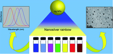
The spectrum of colours of silver sols obtained using the above procedure is given in Fig. 1 and 2 shows the extinction spectra of individual as-synthesized coloured silver nanoparticles. The corresponding extinction values at the extinction maximum wavelengths are summarized in Table 2.
image file: c6ra08336f-f1.tif
Fig. 1 Photograph of (a) violet (b) blue (c) purple (d) green (e) yellow (f) orange and (g) red AgNps.

image file: c6ra08336f-f2.tif
Fig. 2 Extinction spectra of (a) violet (b) blue (c) purple (d) green (e) yellow (f) orange and (g) red AgNps.
Table 2 Extinction maximum wavelengths and the extinction thereof of the synthesized AgNps
Colour of AgNps Peak 1 Peak 2
λ (nm) Extinction λ (nm) Extinction
Orange 402.0 Weak shoulder 482.5 0.476
Red 402.0 0.630 509.0 0.559
Yellow 402.5 0.521
Purple 402.5 0.494 535.5 0.378
Violet 403.0 0.590 572.5 0.529
Blue 398.0 1.226 599.5 0.827
Green 409.0 0.839 719.0 0.245


It is clear from the photograph that the procedures developed have given rise to a spectrum of colours of silver sols. Up to now, there are only few reports published about silver nano colloids of different colours.38,57,58

The different colours of silver sols are the result of the extinction of selected portions of visible light as shown in Fig. 2. All samples, with the exception of the yellow coloured sample indicate two distinct absorption features, one centered between 450 and 700 nm and another at ∼400 nm. The spectra indicate good agreement to the colour in which the solutions are observed. For example, the absorption spectrum of red colour AgNps shows two peaks at 402 nm and 509 nm. The peak at 509 nm corresponds to the bluish green region. The particles in the red sol absorb the bluish green portion of the visible light, and the complementary colour which transmits from the solution has to be red as we have observed. Accordingly, as λmax of peak 2 is red shifted, the particles change their colour from red to blue as seen in Fig. 1 and the corresponding spectra in Fig. 2.

The colours of silver and gold nano sols are attributed to surface plasmon resonance.62,77–80 In metal nanoparticles such as silver, the conduction band and valence band lie very close to each other in which electrons move freely.81 These free electrons give rise to a surface plasmon resonance (SPR) absorption62,82,83 due to the collective oscillation of electrons of silver nanoparticles in resonance with the incident light wave.84 Classically, the electric field of an incoming wave induces a polarization of the electrons with respect to the much heavier ionic core of AgNps. As a result a net charge difference occurs and this in turn acts as a restoring force. This creates an in-phase dipolar oscillation of all the electrons. When the frequency of the electromagnetic field becomes resonant with the coherent electron motion, a strong absorption takes place, which is the origin of the observed colour.

The wavelength of the plasmon absorption maximum in a given solvent can be used to estimate particle size.83,84,87 For instance, small spherical nanoparticles (<20 nm) exhibit a single surface plasmon band near 400 nm.83 However, instead here the exact particle size and distribution of each coloured silver colloidal solution was determined using transmission electron microscopy. TEM images of orange, yellow, green, blue, red, purple and violet AgNps are given in Fig. 3. It can be seen that the AgNps are spherical in shape with a smooth surface morphology in all samples. The average sizes for different colours are analyzed and found to be in the range between 10 and 50 nm. Closer look at the TEM images shows that the resulted particles indicate a bimodal distribution of sizes in each sample, with the exception of the yellow AgNps (vide infra), as can be seen from Fig. 4. This bimodal nature is clearly visible in Fig. 4a–d. However, it should be noted that Fig. 4e and f does not clearly demonstrate this behavior as the sample size in the TEM image is insufficient to result in an accurate size distribution.


image file: c6ra08336f-f3.tif
Fig. 3 TEM images of (a) orange (b) yellow (c) green (d) blue (e) red (f) purple and (g) violet AgNps.

image file: c6ra08336f-f4.tif
Fig. 4 Size distributions of (a) orange (b) yellow (c) green (d) blue (e) red (f) purple and (g) violet AgNps.

The TEM image obtained for yellow AgNps given in Fig. 3b exhibits an extinction feature at 402.5 nm as previously observed for many yellow silver sols.57,58,78,79,83,85,86 The diameters of these particles are in the 10–30 nm range, however high abundance can be seen between 15 and 20 nm which is in agreement with previously reported data.72,73,88 Interestingly, AgNps of above size range can be clearly seen in each of the other TEM images and the corresponding size distributions, thus justifying the observation of a spectral feature ∼400 nm in each of the coloured solutions. Hence, we attribute the spectral feature at ∼400 nm to the formation of AgNps of 10–30 nm in size (centred ∼20 nm); a result independent of the presence of hydrazine in the reaction medium.

Notably, no nanoparticle in the above size range is seen for the violet coloured AgNps (Fig. 3g and 4g), and it is seen that all particles in that sample have significantly smaller diameters (<5 nm). However, previous reports suggest that smaller AgNps, specifically of the smaller diameters given in Fig. 4g, are yellow in colour,58 thus justifying the peak observed at 403.0 nm for the violet AgNps.

According to Fig. 3a from the TEM image obtained for orange AgNps, it is clear that the particles are spherical in shape. The most abundant particle size is 10 nm. However, some larger sized particles (∼25 nm) can also be seen from the image, in addition to the few agglomerates that can be seen. This behavior is again visible from the size distribution in Fig. 4a. The UV/Vis spectrum of these orange AgNps shows one dominant peak with a small shoulder as described; features attributable to the two different sizes of AgNps present in the system. However, the appearance of a shoulder suggests that the variation of the sizes of the AgNps here should be narrow such that the bimodal size distributions overlap. This behavior is clearly visible in Fig. 4a, hence justifying the above statement.

Further, the presence of one intense peak at 482.5 nm is in good agreement with the reported value for orange color silver nanoparticles.57,58,85 The shoulder around 402 nm is consistent with the smaller AgNps present here (vide supra). The larger AgNps here are formed via the presence of hydrazine in the reaction medium.

Additionally, GAgNps indicate the largest variation in λmax of the two LSPR peaks, suggesting that these should thus have the largest variation in the particle sizes. The size distribution in Fig. 4C confirms the above statement as this shows particles ranging in diameter from ∼10 nm up to ∼65 nm; the largest range observed for the various samples prepared here. Thus, it can be clearly stated that there is a correlation between the surface plasmon band and the size and size distributions of the particles as previously reported,86,90 while the colours observed were typical for AgNps.89

According to previously reported data, the position and the shape of the plasmon absorption depends on the particle size, shape, chemical surrounding and the dielectric constant of the surrounding medium.91–94 In order to clearly understand the observed variations in terms of particle size and optical properties, it is important to investigate the origin of the variation in the particle size. Silver nitrate was used as the only metal precursor. NaBH4 was used as the common reducing agent and this also acts as a stabilizing agent. In addition, trisodium citrate and hydrazine sulphate were used as secondary reducing agents and citrate as the stabilizing agent. The main reaction was the reduction of Ag+ to Ag0 with hydrogen evolution, while Ag0 is combined to make nano sized particles. Hence, any changes observed in terms of the synthesized AgNps can be attributed to the different concentrations of the reducing agents used as outlined in Table 1.

Citrate ion is a commonly used reducing agent in metal colloid synthesis. It undergoes strong surface interaction with silver nano crystallites.67,68 Although hydrazine sulphate is known to be a reducing agent, it has never been tested as a stabilizing agent for AgNps. In order to gain some insight into how AgNps of different colours were obtained, the concentrations and amounts of AgNO3, NaBH4, and other agents used were examined (Table 1). When the experiment was carried out only with hydrazine sulphate and without trisodium citrate only yellow coloured silver sols were obtained. Similarly, in the presence of trisodium citrate but without hydrazine sulphate, the result was the same yellow and no other colours could be obtained. Therefore it is clear that in order to obtain a range of sizes, and hence a range of colours the presence of NaBH4, hydrazine sulphate and trisodium citrate are necessary in the synthesis of AgNps. This explains why only a single plasmonic feature is observed when the synthesis was conducted in the absence of hydrazine whereas two plasmonic features are visible on the spectra where both hydrazine sulphate and trisodium citrate were used as reducing agents.

Similar multiple resonance behaviours have been previously observed with AgNps and have been attributed to the effect of reducing agents leading to distinct variations in the resulted nanoparticle morphology.57 Interestingly, in this study we accomplish variations in optical properties, while maintaining spherical morphologies as visible from the TEM images in Fig. 3; a significant result owing to the well-known stability, lower toxicity, and widespread applicability of spherical nanoparticle morphologies as opposed to its anisotropic counterparts.

The TEM image of OAgNps indicates an average particle size of 10 nm. Red AgNps are uniformly distributed with an average size of 18 nm. During the synthesis of orange and red coloured AgNps, the only variation was the concentration of trisodium citrate. Since the silver colloidal particles possessed a negative charge due to the adsorbed citrate ions, a repulsive force would work along the particles and prevent aggregation.95 The citrate layer is clearly visible in red silver nanoparticles as shown in Fig. 3e. Citrate ions influence particle growth at the early stages by complexing with positively charged Ag2+ dimers.95 When the concentration is doubled, the solution gets more citrate ions where it can process smaller particles as seen with orange AgNps.87 As the particles increase in size, the absorption peak usually shifts towards the red wavelengths as seen in Fig. 2 for orange and red sols. The average sizes of red and orange AgNps as shown in the TEM images confirm this fact.

When only hydrazine sulfate and NaBH4 are present, the pH of the sample is near 7.28, but with the addition of citrate the pH is raised. The pH range of 7 to 8.5 is required in order to produce AgNps. The concentration of AgNO3 was another major factor to be considered when synthesizing AgNps. When synthesizing purple and violet AgNps the only difference was the concentration of AgNO3. When the concentration of AgNO3 is increased, more Ag+ ions are present in the medium. Since the stabilizing citrate ions are constant for both the procedures, with the increase in concentration of silver nitrate, the number of AgNps increase. This result is mainly attributed to the fact that the rate of spontaneous nucleation increases significantly more than the growth rate of silver nano crystals where a larger number of nuclei are formed during the nucleation burst.96,97 This is visible in the TEM images of violet and purple AgNps as number of VAgNps are greater than that of PAgNps.97 Due to this, VAgNps are more prone for aggregation.

In summary, this study outlines a rapid, facile and reproducible method of preparing silver nanoparticles of a range of colours primarily due to the size of the spherical nanoparticles produced and the method suggests the possibility of tuning deferent colours through combination of sizes. The combination of various sizes results in different plasmon resonance bands and their resultant plasmon band produces the colours which can be tuned by changing the amount of reactants in the medium.

These developed procedures can be used in preparing AgNps of various sizes and these AgNps can be applied in a variety of commercial applications such as medicine, textiles, coatings, water purification and food industry. For example, size and ensuing plasmonic tunability is an important consideration in spectroscopic applications of AgNps. In surface-enhanced Raman spectroscopy (SERS) where AgNps are widely used, it is important that the plasmonic features of the nanoparticle substrates are located close in energy to the excitation source, which may vary between different visible wavelengths based on the excitation laser source. Plasmonic tuning of AgNps achieved here would thus allow one to synthesize and utilize AgNps according to the optimal conditions required for a specific spectroscopic analysis. Similarly, the above tunability of particle size and the ensuing optical properties is certain to find value in the many other applications of nanoparticles where structure–property tunability is desired.

Conclusions

A new method to synthesize silver sols of various colours has been developed. It is a simple aqueous phase method obviating the need for any extra surfactant to stabilize the particles. The complexation of citrate ions with silver plays an important role in dictating the size and shape of the AgNps. When compared with the previous studies, it has been proven that a combination of reducing agents trisodium citrate, hydrazine sulphate and sodium borohydride were suitable for making distinctively coloured spherically shaped AgNps with an average size of 10–50 nm range, where good correlation was seen between the size distributions obtained for the AgNps of each colour and the corresponding optical properties. Nanoparticles were identified as violet, blue, purple, green, yellow, orange and red AgNps. Hydrazine sulphate was recognized as a supportive reducing agent as well as an agent that controls the effective pH of the medium. Sodium borohydride was identified as the most powerful reducing agent of the three. Trisodium citrate was identified as the most effective stabilizing agent rather than a reducing agent. According to UV/Vis observations, as the concentration of AgNO3 increases, larger number of bigger sized AgNps are formed, as the increased concentration of sodium borohydride favoured smaller sized particles. Among the concentrations of hydrazine sulphate which were employed in this study, 2 × 10⁻³ M has been identified as the best concentration to make distinctively coloured AgNps. Room temperature was identified as the best reaction temperature to produce stable and reproducible distinctive coloured AgNps. It has been identified that the best pH to make these coloured AgNps is 10. As far as the stability is concerned, it was found that the AgNps preserved at 4 °C are stable for more than 3 months in the refrigerator in the colloidal form with no further aggregation.

Acknowledgements

We thank the Department of Chemistry, University of Colombo, Sri Lanka for providing the facilities. We are grateful to Mr Ananda Hettiarachchy of P & E Consultants for the continuous support.

References

  1. M. C. Daniel and D. Astruc, Chem. Rev., 2004, 104, 293 CrossRef CAS PubMed.
  2. D. J. Barber and I. C. Frestone, Archaeometry, 1990, 32, 33 CrossRef.
  3. G. L. Hornyak, C. J. Patrissi, E. B. Oberhauser, C. R. Martin, J. C. Valmalette, L. Lemaire and H. Hofmann, Nanostruct. Mater., 1997, 9, 571 CrossRef CAS.
  4. K. Watanabe, D. Menzel, N. Nilius and H. J. Freund, Chem. Rev., 2006, 106, 4301–4320 CrossRef CAS PubMed.
  5. L.-S. Wang, J.-C. Deng, F. Yanga and T. Chen, Mater. Chem. Phys., 2008, 108, 165–169 CrossRef CAS.
  6. S. Nie and S. R. Emory, Science, 1997, 275, 1102–1106 CrossRef CAS PubMed.
  7. L. Lu, A. Kobayashi, K. Tawa and Y. Ozaki, Chem. Mater., 2006, 18, 4894–4901 CrossRef CAS.
  8. J. M. McLellan, A. Siekkinen, J. Chen and Y. Xia, Chem. Phys. Lett., 2006, 427, 122–126 CrossRef CAS.
  9. D. Graham, K. Faulds and W. E. Smith, Chem. Commun., 2006, 4363–4371 RSC.
  10. S. E. J. Bell and N. M. S. Sirimuthu, J. Am. Chem. Soc., 2006, 128, 15580–15581 CrossRef CAS PubMed.
  11. M. S. Hu, H. L. Chen, C. H. Shen, L. S. Hong, B. R. Huang, K. H. Chen and L. C. Chen, Nat. Mater., 2006, 5, 102–106 CrossRef CAS PubMed.
  12. S. V. Karpov, V. S. Gerasimov, I. L. Isaev and V. A. Markel, Phys. Rev. B: Condens. Matter Mater. Phys., 2005, 72, 205425 CrossRef.
  13. A. M. Whelan, M. E. Brenan, W. J. Blau and J. M. Kelly, J. Nanosci. Nanotechnol., 2004, 4, 66–68 CrossRef CAS PubMed.
  14. A. Balamurugan, K.-C. Ho and S.-M. Chen, Synth. Met., 2009, 159, 2544–2549 CrossRef CAS.
  15. S.-J. Park, S.-W. Lee, K.-J. Lee, J.-H. Lee, K.-D. Kim, J.-H. Jeong and J.-H. Choi, Nanoscale Res. Lett., 2010, 5, 1570–1577 CrossRef CAS PubMed.
  16. R. L. Stoermer and C. D. Keating, J. Am. Chem. Soc., 2006, 128, 13243–13254 CrossRef CAS PubMed.
  17. K. Aslan, J. R. Lokowicz and C. D. Geddes, J. Phys. Chem. B, 2005, 109, 6247–6251 CrossRef CAS PubMed.
  18. J. L. Elechiguerra, J. L. Burt, J. R. Morones, A. Camacho-Bragado, X. Gao, H. H. Lara and M. J. Yacaman, J. Nanobiotechnol., 2005, 3, 6 CrossRef PubMed.
  19. J. R. Morones, J. L. Elechiguerra, A. Camacho, K. Holt, J. B. Kouri, J. Tapia Ramirez and M. J. Yacaman, Nanotechnology, 2005, 16, 2346–2353 CrossRef CAS PubMed.
  20. M. Rai, A. Yadev and A. Gade, Biotechnol. Adv., 2009, 27, 76–83 CrossRef CAS PubMed.
  21. B. Despax, C. Saulou, P. Raynaud, L. Dates and M. Mercier-Bonin, Nanotechnology, 2011, 22(175101), 1 Search PubMed.
  22. J. S. Kim, E. Kuk, K. N. Yu, J. H. Kim, S. J. Park, H. J. Lee, S. H. Kim, Y. K. Park, Y. H. Park, C. Y. Hwang, Y. K. Kim, Y. S. Lee, D. H. Jeong and M. H. Cho, Nanomedicine, 2007, 3, 95 CAS.
  23. C. N. Lok, C. M. Ho, R. Chen, Q. Y. He, W. Y. Yu, H. Sun, P. K. H. Tam, J. F. Chiu and C. M. Che, JBIC, J. Biol. Inorg. Chem., 2007, 12, 527 CrossRef CAS PubMed.
  24. S. Pal, Y. K. Tak and J. M. Song, Appl. Environ. Microbiol., 2007, 12, 527 Search PubMed.
  25. O. Choi, K. K. Deng, N. J. Kim, L. Ross Jr, R. Y. Surampalli and Z. Hu, Water Res., 2008, 42, 3066–3074 CrossRef CAS PubMed.
  26. A. Bankar, B. Joshi, A. R. Kumar and S. Zinjarde, Colloids Surf., A, 2010, 368, 58–63 CrossRef CAS.
  27. Z.-M. Xiu, Q.-B. Zhang, H. L. Puppala, V. L. Colvin and P. J. J. Alvarez, Nano Lett., 2012, 12(8), 4271–4275 CrossRef CAS PubMed.
  28. X. Gu, C. Nie, Y. Lai and C. Lin, Mater. Chem. Phys., 2006, 96, 217–222 CrossRef CAS.
  29. G. Wei, H. Zhou, Z. Liu, Y. Song, L. Wang, L. Sun and Z. Li, J. Phys. Chem. B, 2005, 109, 8738–8743 CrossRef CAS PubMed.
  30. G.-J. Lee, S.-I. Shin, Y.-C. Kim and S.-G. Oh, Mater. Chem. Phys., 2004, 84(2–3), 197–204 CrossRef CAS.
  31. C. Ni, P. A. Hassan and E. W. Kaler, Langmuir, 2005, 21, 3334–3337 CrossRef CAS PubMed.
  32. N. R. Jana, L. Gearheart and C. J. Murphy, Chem. Commun., 2001, 617–618 RSC.
  33. B. Pietrobon, M. McEachran and V. Kitaev, ACS Nano, 2009, 3(1), 21–26 CrossRef CAS PubMed.
  34. E. Hao, K. L. Kelly, J. T. Hupp and G. C. Schatz, J. Am. Chem. Soc., 2002, 124, 15182–15183 CrossRef CAS PubMed.
  35. M. Maillard, S. Giorgio and M.-P. Pileni, Adv. Mater., 2002, 14, 1084–1086 CrossRef CAS.
  36. S. Chen, Z. Fan and D. L. Carroll, J. Phys. Chem. B, 2002, 106, 10777–10781 CrossRef CAS.
  37. M. Maillard, S. Giorgio and M.-P. Pileni, J. Phys. Chem. B, 2003, 107, 2466–2470 CrossRef CAS.
  38. Q. Zhang, N. Li, J. Goeble, Z. Lu and Y. Yin, J. Am. Chem. Soc., 2011, 133, 18931–18939 CrossRef CAS PubMed.
  39. A. J. Frank, N. Cathcart, K. E. Maly and V. Kitaev, J. Chem. Educ., 2010, 87, 1098–1101 CrossRef CAS.
  40. X. Dong, X. Ji, J. Jing, M. Li, J. Li and W. Yang, J. Phys. Chem. C, 2010, 114(5), 2070–2074 CAS.
  41. V. Bastys, I. Pastoriza-Santos, B. Rodriguez-Gonzalez, R. Vaisnoras and L. M. Liz-Marzan, Adv. Funct. Mater., 2006, 16, 766–773 CrossRef CAS.
  42. G. S. Metraux and C. A. Mirkin, Adv. Mater., 2005, 17, 412–414 CrossRef CAS.
  43. A. Sarkar, S. Kapoor and T. Mukherjee, J. Colloid Interface Sci., 2005, 287, 496–500 CrossRef CAS PubMed.
  44. I. Pastoriza-Santos and L. M. Liz-Marzan, Nano Lett., 2002, 2, 903–905 CrossRef CAS.
  45. Y. Sun, Y. Yin, B. T. Mayers, T. Herricks and Y. Xia, Chem. Mater., 2002, 14, 4736–4745 CrossRef CAS.
  46. J.-Q. Hu, Q. Chen, Z.-X. Xie, G.-B. Han, R.-H. Wang, B. Ren, Y. Zhang, Z.-L. Yang and Z.-Q. Tian, Adv. Funct. Mater., 2004, 14, 183–189 CrossRef CAS.
  47. Y. Sun, B. Mayers, T. Herricks and Y. Xia, Nano Lett., 2003, 3, 955–960 CrossRef CAS.
  48. S. H. Im, Y. T. Lee, B. Wiley and Y. Xia, Angew. Chem., Int. Ed., 2005, 44, 2154–2157 CrossRef CAS PubMed.
  49. D. Yu and V. W. Yam, J. Am. Chem. Soc., 2004, 126, 13200–13201 CrossRef CAS PubMed.
  50. Y. Ma, W. Li, J. Zeng, M. McKiernan, Z. Xie and Y. Xia, J. Mater. Chem., 2010, 20, 3586–3589 RSC.
  51. Y. Sun and Y. Xia, Science, 2002, 298, 2176–2179 CrossRef CAS PubMed.
  52. B. Wiley, Y. Sun, B. Mayers and Y. Xia, Chem.–Eur. J., 2005, 11, 454–463 CrossRef CAS PubMed.
  53. Y. Sun, B. Mayers and Y. Xia, Nano Lett., 2003, 3, 675–679 CrossRef CAS.
  54. Y. Sun and Y. Xia, Adv. Mater., 2003, 15, 695–699 CrossRef CAS.
  55. R. C. Jin, Y. W. Cao, C. A. Mirkin, K. L. Kelly, G. C. Schatz and J. G. Zheng, Science, 2001, 294, 1901–1903 CrossRef CAS PubMed.
  56. X. L. Tian, K. Chen and G. Y. Cao, Mater. Lett., 2006, 60, 828–830 CrossRef CAS.
  57. D. M. Ledwith, A. M. Whelan and J. M. Kelly, J. Mater. Chem., 2007, 17, 2459–2464 RSC.
  58. T. Huang and X.-H. Nancy Xu, J. Mater. Chem., 2010, 20, 9867–9876 RSC.
  59. M. Faraday, Philos. Trans. R. Soc. London, 1857, 147, 145 CrossRef.
  60. G. Mie, Ann. Phys., 1908, 25, 377 CrossRef CAS.
  61. U. Kreibig and M. Vollmer, Optical Properties of Metal Clusters, Springer, Berlin, 1995 Search PubMed.
  62. A. Moores and F. Goettmann, New J. Chem., 2006, 30, 1121–1132 RSC.
  63. K. L. Kelly, E. Coronado, L. L. Zhao and G. C. Schatz, J. Phys. Chem. B, 2003, 107, 668 CrossRef CAS.
  64. U. Kreibig and L. Genzel, Surf. Sci., 1985, 156, 678 CrossRef CAS.
  65. E. Hutter and J. H. Fendler, Chem. Mater., 2004, 16, 1685 CAS.
  66. J. H. Johnston, K. A. Burridge, F. M. Kelly and A. C. Small, NSTI-Nanotech 2010, 2010, vol. 1, http://www.nsti.org, ISBN 978-1-4398-3401-5 Search PubMed.
  67. J. Kimling, M. Maier, B. Okenve, V. Kotaidis, H. Ballot and A. Plech, J. Phys. Chem. B, 2006, 110, 15700–15707 CrossRef CAS PubMed.
  68. M. P. Mallin and C. J. Murphy, Nano Lett., 2002, 2, 1235–1237 CrossRef CAS.
  69. X. Dong, X. Ji, J. Jing, M. Li, J. Li and W. Yang, J. Phys. Chem. C, 2010, 114, 2070–2074 CAS.
  70. S. Sedaghat and A. Nasseri, Int. Nano Lett., 2011, 1, 22–24 CAS.
  71. M. G. Guzmán, J. Dille and S. Godet, Int. J. Chem. Biol. Eng., 2009, 2, 104–111 Search PubMed.
  72. K. C. Song, S. M. Lee, T. S. Park and B. S. Lee Korean, J. Chem. Eng., 2009, 26, 153–155 CAS.
  73. R. Das, S. S. Nath, D. Chakdar, G. Gope and R. Bhattacharjee, J. Nanotechnol., 2009, 5, 1–6 Search PubMed.
  74. Z. Jiang, Y. Y. Chen, A. H. Liang, H. L. Tao, N. L. Tang and F. X. Zhong, Sci. China, Ser. B: Chem., 2007, 3, 345–350 CrossRef.
  75. H. Y. Song, K. K. Ko, I. H. Oh and B. T. Lee European, Cells Mater., 2006, 11, 58–61 Search PubMed.
  76. S. Chen and D. L. Carroll, Nano Lett., 2002, 2, 1003–1007 CrossRef CAS.
  77. S. D. Solomon, M. Bahadory, A. V. Jeyarajasingam, S. A. Rutkowsky and C. Boritz, J. Chem. Educ., 2007, 84(2), 322 CrossRef CAS.
  78. S. K. Ghosh and T. Pal, Chem. Rev., 2007, 107, 4797–4862 CrossRef CAS PubMed.
  79. M. L. Rao and I. Bell, Mater. Lett., 2008, 62, 1487–1490 CrossRef CAS PubMed.
  80. C. Dahmen and G. von Plessen, Aust. J. Chem., 2007, 60, 447–456 CrossRef CAS.
  81. E. Hutter and J. S. Fendler, Adv. Mater., 2004, 16(19), 1685–1706 CrossRef CAS.
  82. A. Taleb, C. Petit and M. P. Pileni, J. Phys. Chem. B, 1998, 102, 2214–2222 CrossRef CAS.
  83. R. Das, S. S. Nath, D. Chakdar, G. Gope and R. Bhattacharjee, J. Nanotechnol., 2009, 5, 1–6 Search PubMed.
  84. S. D. Solomon, M. Bahadory, A. V. Jeyarajasingam, S. A. Rutkowsky and C. Boritz, J. Chem. Educ., 2007, 84(2), 322 CrossRef CAS.
  85. N. Cathcart, A. J. Frank and V. Kitaev, Chem. Commun., 2009, 7170–7172 RSC.
  86. K. G. Stamplecoskie and J. C. Scaiano, J. Am. Chem. Soc., 2010, 132, 1825–1827 CrossRef CAS PubMed.
  87. M. G. Guzmán, J. Dille and S. Godet, Int. J. Chem. Biol. Eng., 2009, 2(3), 104–111 Search PubMed.
  88. S. Egger, R. P. Lehmann, M. J. Height, M. J. Loessner and M. Schuppler, Appl. Environ. Microbiol., 2009, 75(9), 2973–2976 CrossRef CAS PubMed.
  89. R. Jin, Y. W. Cao, C. A. Mirkin, K. L. Kelly, G. C. Schatz and J. G. Zheng, Science, 2001, 294, 1901–1903 CrossRef CAS PubMed.
  90. C. S. Seney, B. M. Gutzman and R. H. Goddard, J. Phys. Chem. C, 2009, 113, 74–80 CAS.
  91. A. Moores and F. Goettmann, New J. Chem., 2006, 30, 1121–1132 RSC.
  92. L. M. Liz-Marzán, M. Giersig and P. Mulvaney, Langmuir, 1996, 12, 4329–4335 CrossRef.
  93. L. Sironi, Nanoparticles for in vitro and in vivo biosensing and imaging, Ciclo XXIII, 2008–2010 Search PubMed.
  94. S. Link and M. A. El-Sayed, Annu. Rev. Phys. Chem., 2003, 54, 331–366 CrossRef CAS PubMed.
  95. Z. S. Pillai and P. V. Kamat, J. Phys. Chem. B, 2004, 108, 945–951 CrossRef CAS.
  96. D. Chen, X. Qiao, X. Qiu and J. Chen, J. Mater. Sci., 2009, 44, 1076–1081 CrossRef CAS.
  97. Y. Xia and N. J. Halas, MRS Bull., 2005, 30, 338–343 CrossRef CAS.

Footnote

Electronic supplementary information (ESI) available. See DOI: 10.1039/c6ra08336f

This journal is © The Royal Society of Chemistry 2016
Click here to see how this site uses Cookies. View our privacy policy here.