Bioinformatical analysis and preliminary study of the role of lipase in lipid metabolism in Mucor circinelloides

Xinyi Zana, Xin Tanga, Lina Zhaoa, Linfang Chua, Haiqin Chenac, Wei Chenac, Yong Q. Chenac and Yuanda Song*ab
aState Key Laboratory of Food Science and Technology, School of Food Science and Technology, Jiangnan University, Wuxi, P. R. China. E-mail: ysong@jiangnan.edu.cn
bColin Ratledge Center for Microbial Lipids, School of Agriculture Engineering and Food Science, Shandong University of Technology, Zibo, P. R. China
cSynergistic Innovation Center for Food Safety and Nutrition, Wuxi, P. R. China

Received 31st March 2016 , Accepted 14th June 2016

First published on 15th June 2016


Abstract

The filamentous fungus Mucor circinelloides has been widely used as a model organism to investigate the mechanisms of lipid accumulation. Although a lot of work has been done to analyze and explore many of the essential enzymes/genes related to lipid accumulation in M. circinelloides, the function of lipase in this fungus has not been studied at all. In this study, we report some important characteristics of all 30 lipases, and especially 4 lipases Lip6, Lip10, Lip19 and Lip24 with functional analyses in Mucor circinelloides CBS 277.49 based on the genome sequences. Transcriptional analyses revealed that the Lip6 or Lip10 gene expression increased significantly during 24 h to 72 h of lipid accumulation while Lip19 and Lip24 genes were down-regulated along with the increase in lipid accumulation. Over-expression of either lipase Lip6 or Lip10 led to a slight increase in cell dry weight, but no significant effect on lipid accumulation. More interestingly over-expression of Lip6 resulted in a 17% increase in C18:3 content of the lipids. However, over-expression of lipase Lip19 and Lip24 decreased the cell dry weight by 9.08–18.8% and lipid content by 37.86–41.42%, respectively. The fatty acid profiles of strains with Lip19 and Lip24 over-expression were also significantly changed as compared to control. Analysis of lipase genes revealed that the sequences of Lip6 and Lip10 not only contained the typical lipase motif (G/A) XSXG, but also the consensus sequence motif HXXXXD (a typical of acyltransferase motif). These results suggest that Lip6 or Lip10 may control acyltransferase activity in Mucor circienlloides and play a role in fatty acid reconstruction of TAG, while Lip19 or Lip24 may work as a TAG lipase involved in TAG metabolism/degradation.

1. Introduction

The pressure of depleting petroleum reserve and environmental problems has led to an increasing interest in alternative renewable fuel materials.1,2 Microbial lipids can be used as precursors to produce biodiesel because of the similar fatty acid composition with plant oils and animal fats, high productivity and low cost.3–5 Additionally, some oleaginous microorganisms can convert lignocellulosic sugars6–8 or low-value hydrophobic substrates such as waste oils9 to several nutritionally-important polyunsaturated fatty acids (PUFAs)10,11 such as γ-linolenic acid (GLA),12 arachidonic acid (AA),13 eicosapentaenoic acid (EPA),14 and docosahexaenoic acid (DHA).15 These high-value products could counteract the production cost of microbial lipids.

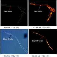
Oleaginous microorganisms, including bacteria, microalgae, yeast and some fungi, could generally accumulate cellular lipid up to 20–80% of cell dry weight.16 Lipids in these species are stored, mainly as triacylglycerols (TAGs), in a specialized globular organelle named lipid particle (LP, Fig. 1).17 A wide range of proteins are specifically targeted to the LP surface, where they can regulate lipid synthesis and degradation.18,19


image file: c6ra08285h-f1.tif
Fig. 1 Lipid droplets in the oleaginous fungus Mucor circinelloides and yeast Yarrowia lipolytica. (A) represents a low lipid-producing strain M. circinelloides CBS 277.49 and (B) represents a high lipid-producing strain M. circinelloides WJ11 which are stored in our lab. (A1) and (B1) were the photos of lipid droplets at white light, and fluorescence at (A2) and (B2) were obtained by staining cells with Nile red (10 mg mL−1) for 5 min.

Lipid-metabolizing enzymes, especially lipases and hydrolases, were the major classes of LP enzymes. Peer researches have revealed that intracellular lipases/hydrolases play an important role in the lipid metabolism including lipid homeostasis, phospholipids synthesis, regulation of fatty acid composition, and the viabilities and spore formation of cells.20–23 For example, Tgl3p, Tgl4p and Tgl5p are the major lipases of yeast S. cerevisiae and located on the surface of LP. Compared with wild type, deletion of Tgl3p or Tgl4p resulted in an increase of TAG level by 138.95% and 72.67% respectively, and also altered fatty acid compositions. More interestingly, Tgl3p, Tgl4p and Tgl5p not only acted as lipases but also exhibited lysophospholipid acyltransferase activities. Over-expression of Tgl3p induced a rise of glycerophospholipids, which was essential for phospholipids synthesis and efficient sporulation of yeast cells.21–23 Furthermore, three genes ptl1, ptl2 and ptl3 in the fission yeast Schizosaccharomyces pombe have high homology to lipase genes Tgl3p and Tgl5p in S. cerevisiae. Deletion of each gene increased TAG content by approximately 1.7-fold compared to control, and their triple deletion mutant led to higher TAG accumulation (2.7-fold) in S. pombe when the glucose (2%, w/v) was taken as sole carbon source.24 In addition, transcriptomic analyses of the oleaginous fungus Mortierella alpina suggested that down-regulation of triacylglycerol lipase (EC 3.1.1.3) might be involved in the lipid accumulation.13 However, transcriptomic analyses of Y. lipolytica revealed that a lipase encoding gene Lip15 was over-expressed during lipid accumulation, while the exact function of Lip15 has not been fully clarified.25

As an oleaginous microorganism, the filamentous fungus M. circinelloides, can accumulate 10–36% lipid of cell dry weight.26 Especially, it produces high levels of GLA, which has beneficial effects on the prevention or treatment of inflammatory disorders, diabetes, cardiovascular disorders, cancers, and some other diseases.27 M. circinelloides has been used as a model organism to investigate the mechanisms of lipid accumulation. A lot of work has been done to analyze and explore those essential enzymes and/or genes related to lipid accumulation in M. circinelloides such as malic enzyme, glucose 6-phosphate dehydrogenase and 6-phosphogluconate dehydrogenase.28–30 However, the function of lipase in this fungus has not been studied at all. Moreover, it is plausible that alteration of lipase activity could enhance lipid accumulation and even remodel the fatty acid composition in oleaginous microorganisms. Thus, in the present study we searched all genes encoding lipase in M. circinelloides CBS 277.49 based on its genome sequence reported by Joint Genome Institute (JGI). Then bioinformatics and transcriptional analyses of lipase genes during lipid accumulation were preformed. The results of transcriptional analyses revealed that lipase genes Lip6 and Lip10 were over-expressed during lipid accumulation while lipase genes Lip19 and Lip24 were inhibited notably. Furthermore, we investigated the effects of over-expression of these four lipase genes on fatty acid profiles of the fungus. To our knowledge, this is the first report of the roles of lipase in lipid accumulation in M. circinelloides.

2. Materials and methods

2.1 Microorganisms and cultivation

M. circinelloides CBS 277.49 strain was used in this study. 100 μL spore suspension (approx. 107 spores per mL) was inoculated into 150 mL K&R medium31 held in a 1 L flask equipped with baffles to increase aeration. Cultures were incubated at 30 °C for 24 h with shaking at 150 rpm and used as a seed culture. A 2 L fermentor containing 1.5 L modified K&R medium (2 g diammonium tartrate, 80 g glucose per liter plus inorganic salts) was used for fermentation of M. circinelloides mycelia. 10% (v/v) of the seed culture was inoculated with aeration at 0.5 v/v min−1 and stirring at 700 rpm. pH was maintained at 6.0 by auto-addition of sterilized 4 M KOH or 2 M H2SO4.

2.2 Determination of cell dry weight and lipid content

Mycelia were sampled at 6 h, 9 h, 12 h, 24 h, 48 h, 72 h and 96 h and collected on a dried and weighed filter paper by filtration through a Buchner funnel under reduced pressure. Then collections were washed three times with distilled water, frozen overnight at −80 °C, and lyophilized. Cell dry weight was determined gravimetrically. Lipid extraction and analysis were using the method described by Tang et al.12

2.3 Determination of glucose and ammonium

Glucose concentration in the culture was measured using a glucose oxidase Perid-test kit (Shanghai Rongsheng Biotech Co., Ltd.). Ammonium concentration in the culture filtrate was determined using the indophenols test.32

2.4 RNA extractions and transcriptional analyses of lipase genes by qRT-PCR

The genome sequence of M. circinelloides CBS 277.49 showed that it might have 30 lipase genes. Based on the characteristic lipid accumulation in M. circinelloides CBS 277.49, mycelia at 6, 24 and 72 h were harvested and used for transcriptional analysis of lipase genes. Total RNA was extracted by an RNAiso Plus kit after grinding under liquid N2 and reverse-transcribed using the Prime ScriptRT reagent kit (Takara, Japan) according to the manufacturer's instructions. Real-time quantitative PCR was performed in BioRad CFX96 (BioRad, CA, USA) using the iTaq™ Universal SYBR® Green Supermix (BioRad, CA, USA). Relative quantification was based on the 2−ΔΔCt method using 18S rRNA of M. circinelloides as a housekeeping gene.33 The thermal cycling conditions for the amplification reaction were as follows: 95 °C 30 s, 51/53/55/58 °C 30 s (40 cycles). Three replicates, prepared from independent biological samples, were analyzed. The primer sequences used for amplification of lipase genes are listed in Table S1.

2.5 Strains, plasmid construction and transformation

The uridine auxotroph, pleu-MU402, was used as recipient strain in transformation experiments to over-express lipase genes of lipases. MU402 is an auxotrophic strain for leucine and uridine. Plasmid pLEU4, containing the wild-type allele of leuA gene, was transformed into MU402 to generate pleu-MU402 strain. Cultures were grown at 30 °C in YPG media containing 3 g L−1 yeast extract, 10 g L−1 tryptone, and 20 g L−1 glucose at pH 4.5. The media were supplemented with uridine (200 μg mL−1) when required.

Plasmid pMAT1552 (Fig. 2a), which contains the M. circinelloides pyrG gene surrounded up- and down-stream by 1 kb of CarRP sequences, was used for construction of four lipases-overexpressing plasmids. These four lipases genes were isolated by PCR amplification from the genome of M. circinelloides CBS 277.49 with corresponding primers listed in Table S2. They contain 30 bp sequences homologous as in up- and down-stream of XhoI restriction sites in pMAT1552. The PCR fragment was then cloned into plasmid pMAT1552 using XhoI to generate plasmids pMAT1552-Lip6, pMAT1552-Lip10, pMAT1552-Lip19 and pMAT1552-Lip24 (Fig. 2a) (One step cloning kit, Takara). These plasmids were used for transformation using the method described by Zhao et al.30


image file: c6ra08285h-f2.tif
Fig. 2 (a) Structure of plasmid pMAT1552-gene for target genes over-expressing in M. circinelloides. Target genes represent gene Lip6, Lip10, Lip19 and Lip24. (b) PCR amplification of genome of the over-expressing strains and the control strain Mc-1552 with the primers 1552 F/R. M, GeneRuler DNA Ladder Mix. Sizes in kb of the relevant maker fragments are indicated. The numbers of 1–5 represent strain Mc-Lip6, Mc-Lip10, Mc-Lip19, Mc-Lip24 and Mc-1552, respectively.

Transformants, contained plasmids pMAT1552, pMAT1552-Lip6, pMAT1552-Lip10, pMAT1552-Lip19 and pMAT1552-Lip24, were named as Mc-1552, Mc-Lip6, Mc-Lip10, Mc-Lip19 and Mc-Lip24, respectively. Mc-1552 was used as the control strain. Transformants were initiated by inoculation of 2.5 × 10⁵ spores per mL into 200 mL modified K & R medium and held in 1 L flasks equipped with baffles to improve aeration. The cultures were incubated at 30 °C with shaking at 150 rpm for 72 h. Cell dry weight and lipid accumulation of transformants were analyzed by the method described in 2.2 and 2.3.

2.6 Analyses of expression levels of Lip6, Lip10, Lip19 and Lip24 in transformants

For reverse transcription-quantitative PCR (qRT-PCR) analysis, Mc-1552, Mc-Lip6, Mc-Lip10, Mc-Lip19 and Mc-Lip24 were grown in 1 L baffled flasks with modified K & R medium, and the mycelium were harvested during lipid accumulation phase (24, 48 and 72 h). The method for total RNA extraction and qPCR were described in 2.4.

2.7 Bioinformatics analysis

The multiple alignments of the primary sequences of lipases Lip6, Lip10, Lip19 and Lip24 were analyzed by using Clustal Omega (http://www.ebi.ac.uk/Tools/msa/clustalo/). Phylogenic analyses of these four lipases and homology analysis were performed in MEGA 6.06 and the Pairwise Sequence Alignment program (http://ebi.ac.uk/Tools/psa/emboss_needle/), respectively. The transmembrane spanning region of Lip10 and Lip24 were predicted in TMHMM (http://www.cbs.dtu.dk/service/TMHMM). TargetP1.1 Server (http://www.cbs.dtu.dk/services/TargetP/) and SignalP 4.1 Server (http://www.cbs.dtu.dk/services/SignalP/) program were used to predict the sub-cellular location, and the presence and location of signal peptide of lipases from M. circinelloides CBS 277.49.

3. Results and discussion

3.1 Analysis of lipase genes in M. circinelloides CBS 277.49

In this study, criteria for the identification of lipase genes in M. circinelloides include gene annotations and the presence of typical lipase conserved domain GXSXG. Based on the published genomic sequences of M. circinelloides CBS 277.49, 30 potential genes of lipase were retrieved. The protein ID, amino acid number, molecular weight, motif sequence and domain of lipases were shown in Table 1. All lipases contain the consensus sequence motif (G/A) XSXG (where X stands for any amino acid), which is typical character of serine hydrolases. Among them, 16 lipases (protein ID: 116027, 114978, 161426, 104484, 185587, 184709, 115761, 107413, 119940, 166875, 35076, 155817, 111734, 111320, 114445 and 110553) contain not only the typical lipase motif (G/A) XSXG, but also the consensus sequence motif HXXXXD (a typical of acyltransferase motif). The sequence variation within the motif (G/A) XSXG is one of the factors to distinguish various lipolytic families.34 The domains of lipase were mainly divided into four types including α/β-hydrolase_1, α/β-hydrolase_3, lipase class 3 and lipase_GDSL. For example, lipolytic enzymes are called true lipases conserve the GHS (H/Q) G sequence, whereas the GDSLS motif is characteristic of GDSL family. If another enzyme harbors a motif which is closely related to GDSL families, they may share similar broad substrate specificity toward triglycerides or lysophospholipids. The predicted results of sub-cellular location and the signal peptide were shown in Table S3. Three lipases (protein ID: 116027, 106404 and 170034) are most likely belonging to secretary lipases with a signal peptide based on the online TargetP1.1 Server (RC value > 0.8) and SignalP 4.1 Server program (the predicted result is “Yes”). For the remained lipases, the information of sub-cellular location is limited.
Table 1 Amino acid number, molecular weight, motif sequence and domain of lipases in Mucor circinelloides CBS 277.49
Protein ID Amino acid number Molecular weight Motif sequence Domain
Lipase Acyltransferase
116027 384 42 610 GASVG HDLRVD Lipase_GDSL
114978 368 40 638 GYSKG HDLRMD Lipase_GDSL
72954 364 39 935 GISYG Lipase_GDSL
34010 387 44 170 GFSQG α/β-Hydrolase_1
143450 420 48 064 GFSQG   α/β-Hydrolase_1
115264 294 33 086 GDSAG α/β-Hydrolase_3
81713 341 37 137 GDSAG α/β-Hydrolase_3
113168 403 45 420 GDSAG α/β-Hydrolase_3
161426 444 49 758 GDSAG HLYLDD α/β-Hydrolase_3
104484 620 68 763 GDSAG HGKMTD α/β-Hydrolase_3
167388 455 50 054 GDSAG α/β-Hydrolase_3
185587 370 40 714 GDSAG HWIVKD α/β-Hydrolase_3
184709 624 69 275 GHSLG HHHHQD Class 3
39585 336 37 016 GTSAG Class 3
130354 304 33 853 GTSAG Class 3
115761 621 70 554 GLSHG HHHSLD Class 3
107413 592 67 385 GHSLG HKGFWD Class 3
106404 344 38 092 GHSYG Class 3
119940 233 26 283 GHSFG HTALYD Class 3
154931 218 23 142 GHSLG Class 3
166875 862 95 666 GHSLG HDKAED Class 3
35076 352 37 956 GHSLG HLSYYD Class 3
155817 384 41 143 GHSLG HFSYYD Class 3
170034 410 45 254 GHSLG Class 3
111734 695 77 919 GHSLG HSKAWD Class 3
155202 395 43 422 GHSLG Class 3
111320 934 106 245 GHSLG HKFNPD Class 3
114445 948 107 579 GHSLG HNQLFD Class 3
110553 815 90 965 GHSLG HNQLFD Class 3
167246 839 93 584 GHSLG Class 3


3.2 Cell growth, lipid content, and transcription level of lipase genes in M. circinelloides CBS 277.49

In oleaginous microorganisms, the initiation of lipid accumulation during lipid synthesis is caused by the exhaustion of nitrogen in the cultural medium. So in this study, we first grew the fungus in the N-limited medium, and then analyzed the lipid accumulation and the transcription of lipase genes during the fermentation process.

The concentrations of ammonium and glucose in the culture medium, cell dry weight and lipid accumulation of M. circinelloides CBS 277.49 during growth are shown in Fig. 3. Glucose was sufficient during the entire fermentation period, but ammonium was used up at approx. 9 h. Cell dry weight initially increased rapidly during the balance growth phase from 0 h to 9 h, and then slowed down after nitrogen exhaustion. But the fungus started to accumulate lipid rapidly immediately after nitrogen depletion in the growth medium; from 9 h to 48 h, the total fatty acids (TFAs) content in strain CBS 277.49 increased rapidly from 3.1% to 11.7% of cell dry weight and then slowed down.


image file: c6ra08285h-f3.tif
Fig. 3 Cell growth and lipid accumulation of M. circinelloides CBS 277.49. Glucose and ammonium concentration in the growth media, cell dry weight (CDW) and total fatty acids (TFAs) content (w/w, CDW) of strain CBS 277.49 in the modified K & R medium in 2 L fermenter. ◇ stands for glucose concentration; □ stands for ammonium concentration; △ stands for CDW; ○ stands for TFAs/CDW. Values were measured as mean of three biological replicates. Error bars represent the standard error of the mean.

For transcriptional analysis, M. circinelloides CBS 277.49 cells were collected at 6 h (N rich i.e. balanced growth stage), 24 h (after N depletion i.e. fast lipid accumulation stage) and 72 h (after N depletion i.e. slow lipid accumulation stage), and qRT-PCR was carried out to analyze the transcriptional levels of lipase genes. The results revealed that eight lipase genes (no. 3, 6, 7, 8, 10, 14, 16, 21 and 30) were significantly up-regulated by 1.49–2.87 folds at fast lipid accumulation stage (24 h), while nine lipase genes (no. 2, 5, 9, 11, 12, 13, 17, 18, 19, and 29) were significantly down-regulated by 0.1–0.78 fold (Table 2). At slow lipid accumulation stage, three lipase genes (no. 6, 7 and 10) were up-regulated by 1.85–3.32 folds, but 13 lipase genes (no. 2, 3, 5, 9, 12, 14, 15, 17, 19, 23, 24, 29 and 30) were down-regulated by 0.05–0.79 fold. Compared with other lipase genes, the transcription of two lipase genes (no. 6 and 10) continued to increase most significantly (>1.5-fold) during the lipid accumulation from 24 h to 72 h, and the expression of other two lipase genes (no. 19 and 24) were down-regulated most sharply along with the increase of lipid accumulation. Interestingly, a lipase encoding gene in Y. lipolytica, Lip15, was also up-regulated during lipid accumulation, while related genes of triacylglycerol lipase were down-regulated during lipid accumulation in the fungus M. alpina.13,25 However, the essential roles of Lip15 in Y. lipolytica and relevant triacylglycerol lipases in M. alpina have not been investigated. Therefore, in this study, these four lipase genes (no. 6, no. 10, no. 19 and no. 24) were chosen to further investigate the effects of lipase on lipid accumulation (Table 2). Thus, these four lipase genes were named as Lip6, Lip10, Lip19 and Lip24, respectively.

Table 2 The transcriptional levels of 30 lipase genes were analyzed by qRT-PCR at 6, 24 and 72 h. Error bars represent the standard error of the mean. Values which do not share common superscripts were significantly different to each other. a: p-value < 0.05, b: p-value < 0.01
No. ID The relative transcriptional level
6 h 24 h 72 h
1 116027 1.00 1.16 ± 0.14a 1.08 ± 0.02a
2 114978 1.00 0.14 ± 0.05b 0.26 ± 0.14b
3 81713 1.00 1.64 ± 0.23b 0.05 ± 0.03b
4 104484 1.00 1.21 ± 0.04b 1.18 ± 0.16a
5 115264 1.00 0.56 ± 0.21b 0.44 ± 0.12b
6 161426 1.00 2.11 ± 0.34b 3.32 ± 0.35b
7 167388 1.00 1.58 ± 0.21b 1.85 ± 0.33b
8 72954 1.00 2.25 ± 0.34b 1.01 ± 0.23a
9 185587 1.00 0.23 ± 0.13b 0.14 ± 0.15b
10 115761 1.00 1.13 ± 0.04b 1.98 ± 0.21b
11 167246 1.00 1.14 ± 0.17a 1.10 ± 0.22a
12 170034 1.00 0.71 ± 0.21b 0.73 ± 0.11b
13 184709 1.00 0.57 ± 0.35b 1.29 ± 0.41a
14 154931 1.00 1.73 ± 0.25b 0.54 ± 0.21b
15 35076 1.00 1.12 ± 0.24a 0.56 ± 0.23b
16 155817 1.00 1.92 ± 0.41b 0.91 ± 0.21a
17 155202 1.00 0.10 ± 0.03b 0.15 ± 0.07b
18 114445 1.00 0.54 ± 0.21b 0.72 ± 0.19b
19 143450 1.00 0.78 ± 0.08b 0.29 ± 0.11b
20 113168 1.00 1.24 ± 0.21a 1.28 ± 0.14b
21 34010 1.00 2.87 ± 0.56b 1.36 ± 0.21b
22 106404 1.00 0.82 ± 0.13b 0.87 ± 0.21a
23 11320 1.00 0.94 ± 0.31a 0.79 ± 0.27b
24 107413 1.00 0.86 ± 0.02b 0.42 ± 0.04b
25 166875 1.00 0.91 ± 0.12a 0.86 ± 0.21a
26 39585 1.00 1.26 ± 0.31a 1.12 ± 0.25a
27 111734 1.00 1.21 ± 0.22a 1.18 ± 0.19a
28 110553 1.00 0.89 ± 0.22a 0.91 ± 0.17a
29 119940 1.00 0.60 ± 0.11b 0.63 ± 0.13b
30 130354 1.00 1.49 ± 0.32b 0.47 ± 0.24b


3.3 Altered cell growth and lipid accumulation associated with the over-expression of lipase genes

The effects of the over-expression of four lipases on cell dry weight and the lipid accumulation were analyzed. As our preliminary data showed that the lipid contents of three transformants of each mutant strain were not significantly different (data not given), so only one transformant for each mutant strain was selected for cell dry weight and lipid content analysis (Fig. 4).
image file: c6ra08285h-f4.tif
Fig. 4 Cell dry weight (A), lipid content (B) and fatty acid composition (C) in the transformants grown in 1 L baffled flasks with 200 mL modified K & R medium. These results are mean values ± S.D. from three independent biological replicates. Values which do not share common superscripts were significantly different to each other (P < 0.05).

After 24 h of cultivation, cell dry weights of the transformants were around 4 g L−1 with no significant difference. Cell dry weights of strains Mc-Lip6 and Mc-Lip10 were slightly higher than the control at 48 and 72 h, whereas cell dry weights of strains Mc-Lip19 and Mc-Lip24 decreased significantly at 48 and 72 h compared to control (Fig. 4A).

Fig. 4B and C showed the altered lipid content and fatty acid composition of the transformants compared with wild type. Though over-expression of Lip6 had no significant effect on lipid content, a 7.34% decrease in the content of C18:1 and a 17% increase in the content of C18:3 were observed. This effect could be mainly attributed to the increased conversion of C18:1 to C18:3. The lipid content of strain Mc-Lip10 did not change significantly before 48 h, but decreased slightly by 5.39% along with 15.63% reduction in C18:0 content. Similarly, a shift from C18:0 to C18:1 was observed in the triple mutants S. cerevisiae with only the acyltransferases activity of DGA1.35 Analysis of lipase genes revealed that the sequences of Lip6 and Lip10 also contained the typical of acyltransferase motif HXXXXD. Therefore, we proposed that Lip6 and Lip10 mainly function as an acyltransferase activity in M. circienlloides, and may play a role in fatty acid reconstruction of TAG.

In contrast, lipid contents of strains Mc-Lip19 and Mc-Lip24 were decreased markedly by 37.86% and 41.42%, respectively. It had been proved that alteration of lipase activity could affect lipid accumulation. For example, deletion of ptl1, ptl2 or ptl3 in S. pombe increased TAG content by approximately 1.7-fold compared to control, and the level of TAG in their triple deletion mutant was up to 2.7-fold of that in wild type.24 The amount of C14:0 and C18:0 in the lipids of strain Mc-Lip19 was higher, but amounts of C16:0, C18:1 and C18:3 were lower than the control. In strain Mc-Lip24, the content of C16:0, C16:1, C18:0 and C18:2 were enhanced by 11.7%, 29.45%, 42.81% and 8.76%, while a 21.19% dip in the content of C18:3 was also observed compared to control. These variations of fatty acid composition might be due to the acyl-chain selectivity of lipases. Relevant reports have suggested that yeast S. cerevisiae Tgl5p and Tgl3p exhibited a clear preference for oleoyl-CoA (18:1) over palmitoyl-CoA (16:0).21 These results indicated that Lip19 and Lip24 work as a TAG lipase involved in TAG metabolism/degradation.

3.4 Expression levels of lipase genes Lip6, Lip10, Lip19 and Lip24 in the transformants

The transcriptional levels of Lip6, Lip10, Lip19 and Lip24 in transformants were analyzed by qRT-PCR. As shown in Fig. 5, transcriptional levels of Lip6 and Lip10 were enhanced notably during the lipid accumulation from 24 to 72 h. However, no significant lipid degradation was observed in strains Mc-Lip6 and Mc-Lip10 except the altered contents of C18:1, C18:3 and C18:0. This result also supported our previous hypothesis that Lip6 and Lip10 may not be involved in lipid metabolism/degradation, but play a role in fatty acid reconstruction of TAG as an acyltransferase in M. circienlloides. Though the levels of Lip19 and Lip24 genes in the transformants were up-regulated by approximately 1.70-fold at fast lipid accumulation stage (24 h), Lip19 and Lip24 genes were still inhibited during lipid accumulation. This result is consistent with that in the fungus M. alpina which was also reported that the expressions of some triacylglycerol lipase genes were down-regulated during the lipid accumulation.13 These results further confirmed that lip19 and lip24 were involved in lipid degradation.
image file: c6ra08285h-f5.tif
Fig. 5 qRT-PCR analyses of the transcription level of Lip6, Lip10, Lip19 and Lip24 in the transformants. Values are means of three biological replicates. Error bars represent the standard error of the mean. Values which do not share common superscripts were significantly different to each other (P < 0.05).

3.5 Bioinformatics analysis of four lipases

The multiple alignments of the primary sequences of lipases Lip6, Lip10, Lip19 and Lip24 were analyzed using Clustal Omega (Fig. 6). These results revealed that these four lipases harbored the consensus motif GXSXG (where X stands for any amino acid), which is characteristic of alpha/beta hydrolases. Additionally, Lipases Lip6, Lip10 and Lip24 contained the consensus sequence motif HXXXXD, which is typical of acyltransferase motif. Similarly it has been reported that Lipases Tgl3p, Tgl4p and Tgl5p from the yeast S. cerevisiae not only act as lipases but also have lysophospholipid acyltransferase activities.22,23 Furthermore, over-expression of Tgl3p led to an increase of glycerophospholipids.23
image file: c6ra08285h-f6.tif
Fig. 6 Multiple sequence alignment for of the primary sequences of lipases Lip6, Lip10, Lip19 and Lip24.

To investigate the evolutionary relationship of lipase genes, a neighbor-joining tree was generated by aligning protein sequences of 30 lipases in M. circinelloides CBS 277.49. The phylogram was clustered into three major groups (Fig. 7A). Interestingly, the same type of lipases annotated by the genome was not strictly clustered into the same group. The α/β-hydrolase_1 family, α/β-hydrolase_3 family and class_3 family of M. circinelloides mainly conserve the GFSQG, GDSAG and GHSLG sequence, respectively, whereas the motif (G/A) XSXG of GDSL family is diverse. The class_3 family showed a closer relationship to the α/β-hydrolase_3 family. This result indicated that class_3 family and α/β-hydrolase_3 family are the conserved lipase family in the fungus M. circinelloides. Catalytic triads formed by Ser His and Asp residues were observed in the amino acid sequences of 30 lipases in M. circinelloides CBS 277.49.


image file: c6ra08285h-f7.tif
Fig. 7 (A) Phylogenetic analysis of 30 lipases in M. circinelloides. (B) Phylogenetic analysis of four lipases from M. circinelloides, S. cerevisiae lipases Tgl3p, Tgl4p and Tgl5p, and Y. lipolytica TGL3 and TGL4. (C) Phylogenetic analysis of Lip6 and Lip10 from M. circinelloides and lysophospholipid acyltransferases (LPAT) from M. circinelloides.

Phylogenic analysis of these four lipases from M. circinelloides, lipases Tgl3p, Tgl4p and Tgl5p from S. cerevisiae, and lipases TGL3 and TGL4 from Y. lipolytica, were shown in Fig. 7B. Lip10 revealed a closer relationship with Lip24, with 41% identical sequences. It was conspicuously interesting that effect of Lip10 and Lip24 on cell growth and lipid accumulation was significantly different. Lip6 from M. circinelloides might be closely related to Tgl4p of S. cerevisiae, TGL3 of Y. lipolytica and Tgl5p of S. cerevisiae. When homology between Lip6, Tgl4p, TGL3 and Tgl5p were analyzed, the amino acid sequence identify was 6.9% (between Lip6 and Tgl4p), 10.3% (between Lip6 and TGL3), and 11.5% (between Lip6 and Tgl5p). The low homologies between these lipases suggest that Lip6 from M. circinelloides was possibly a novel lipolytic enzyme.

Because Tgl3p, Tgl4p and Tgl5p of yeast S. cerevisiae exhibited lysophospholipid acyltransferase activities and the activities of acyltransferase might contribute to the lipid accumulation,22,23 phylogenic analysis and the homologies between Lip6/Lip10 and lysophospholipid acyltransferases (LPAT) from M. circinelloides were further investigated (Fig. 7C). The amino acid sequence identity was 10.8% (between Lip6 and LPAT-156700), 12.2% (between Lip10 and LPAT-77297), and 6.2% (between Lip10 and LPAT-148013). The lower homology between Lip6/Lip10 and LPAT indicated that Lip6 and Lip10 in M. circinelloides could be functionally different from Tgl3p, Tgl4p and Tgl5p in S. cerevisiae.

Using TMHMM, Lip10 and Lip24 were predicted to contain three transmembrane spanning regions at their N-terminus (data not given), but not in Lip6 and Lip19. In the yeast S. cerevisiae, Tgl3p and Tgl4p did not contain any transmembrane spanning region, but lipases TGL3 and TGL4 from Y. lipolytica were predicted to contain one transmembrane spanning region.36

4. Conclusion

The aim of this study was to explore the potential role of lipases in cell metabolism in the oleaginous fungus Mucor circinelloides. We found that the oleaginous fungus M. circinelloides CBS 277.49 possesses 30 lipases and all of them contain the consensus sequence motif (G/A) XSXG, while among which 16 lipases also harbor the acyltransferase motif HXXXXD. Additionally, three lipases (protein ID: 116027, 106404 and 170034) are very likely belonging to secretary lipases.

The transcription of two lipase genes Lip6 and Lip10 in wild type continued to increase significantly during the lipid accumulation. However, over-expression of Lip6 and Lip10 had no significant effect on lipid content except the altered contents of C18:1, C18:3 or C18:0. Though transcriptional levels of Lip6 and Lip10 in the transformants were enhanced notably during the lipid accumulation, no significant effect on lipid degradation was observed in strains Mc-Lip6 and Mc-Lip10. In addition, the sequences of Lip6 and Lip10 contained the typical of acyltransferase motif HXXXXD. These results suggest that Lip6 and Lip10 may control acyltransferase activity in M. circienlloides and play a role in fatty acid reconstruction of TAG. Moreover, the lower homology between Lip6/Lip10 and LPAT indicated that Lip6 and Lip10 in M. circinelloides are functionally different from Tgl3p, Tgl4p and Tgl5p in S. cerevisiae.

The expression of lipase genes Lip19 and Lip24 were down-regulated along with the increase of lipid accumulation in wild type. Over-expression of Lip19 or Lip24 decreased lipid contents markedly by 37.86% and 41.42%, respectively. Though the levels of Lip19 and Lip24 genes in the transformants were up-regulated at fast lipid accumulation stage, the expression of Lip19 and Lip24 genes were still inhibited during lipid accumulation. These suggested that role of lip19 and lip24 in M. circinelloides is involved in lipid degradation. To clearly understand the role of these important lipases in M. circienlloides, further experiments of deletion and site-directed mutagenesis of these genes are required to explore their effect on cellular metabolism.

Acknowledgements

This research was supported by the National Natural Science Foundation of China (31271812 and 21276108), the National High Technology Research and Development Program of China (2012AA022105C), the Program for Changjiang Scholars and Innovative Research Team in University (IRT1249), the Program for New Century Excellent Talents (NCET-13-0831).

References

  1. X. Yang, G. Jin, Z. Gong, H. Shen, F. Bai and Z. K. Zhao, Bioresour. Technol., 2014, 175, 91–96 CrossRef PubMed.
  2. F. Donot, A. Fontana, J. C. Baccou, C. Strub and S. Schorr-Galindo, Biomass Bioenergy, 2014, 68, 135–150 CrossRef CAS.
  3. A. Beopoulos, J. M. Nicaud and C. Gaillardin, Appl. Microbiol. Biotechnol., 2011, 90, 1193–1206 CrossRef CAS PubMed.
  4. M. Chao, M. Chakraborty, D. Tao, X. Yu, Z. Chi and S. Chen, Bioresour. Technol., 2014, 164, 106–112 CrossRef PubMed.
  5. I. R. Sitepu, L. A. Garay, R. Sestric, D. Levin, D. E. Block, J. B. German and K. L. Boundy-Mills, Biotechnol. Adv., 2014, 32, 1336–1360 CrossRef CAS PubMed.
  6. X. Yu, Y. Zheng, X. Xiong and S. Chen, Biomass Bioenergy, 2014, 71, 340–349 CrossRef CAS.
  7. Z. Yubin, Y. Xiaochen, L. Tingting, X. Xiaochao and C. Shulin, Biotechnol. Biofuels, 2014, 7, 1–8 CrossRef PubMed.
  8. R. L. Wang, J. C. Wang, R. H. Xu, Z. Fang and A. Z. Liu, BioResources, 2014, 9, 7027–7040 Search PubMed.
  9. A. Najjar, S. Robert, C. Guérin, M. Violet-Asther and F. Carrière, Appl. Microbiol. Biotechnol., 2010, 89, 1947–1962 CrossRef PubMed.
  10. C. Ratledge, Biochimie, 2004, 86, 807–815 CrossRef CAS PubMed.
  11. O. P. Ward and A. Singh, Process Biochem., 2005, 40, 3627–3652 CrossRef CAS.
  12. T. Xin, C. Haiqin, Y. Q. Chen, C. Wei, G. Victoriano, S. Yuanda and R. Colin, PLoS One, 2015, 10(6), e0128396 CrossRef PubMed.
  13. H. Chen, G. Hao, L. Wang, H. Wang, Z. Gu, L. Liu, H. Zhang, W. Chen and Y. Q. Chen, Sci. Rep., 2014, 5 DOI:10.1038/srep11247.
  14. A. J. Klok, P. P. Lamers, D. E. Martens, R. B. Draaisma and R. H. Wijffels, Trends Biotechnol., 2014, 32, 521–528 CrossRef CAS PubMed.
  15. Y. Taoka, N. Nagano, Y. Okita, H. Izumida, S. Sugimoto and M. Hayashi, J. Biosci. Bioeng., 2011, 111, 420–424 CrossRef CAS PubMed.
  16. C. Ratledge, Acta Biotechnol., 1991, 11, 429–438 CrossRef CAS.
  17. L. A. Garay, K. L. Boundy-Mills and G. J. Bruce, J. Agric. Food Chem., 2014, 62, 2709–2727 CrossRef CAS PubMed.
  18. K. Grillitsch, M. Connerth, H. Köfeler, T. N. Arrey, B. Rietschel, B. Wagner, M. Karas and G. Daum, Biochim. Biophys. Acta, 2011, 1811, 1165–1176 CrossRef CAS PubMed.
  19. V. A. Ivashov, K. Grillitsch, H. Koefeler, E. Leitner, D. Baeumlisberger, M. Karas and G. Daum, Biochim. Biophys. Acta, 2012, 1831, 282–290 CrossRef PubMed.
  20. M. O. Debelyy, S. Thoms, M. Connerth, G. Daum and R. Erdmann, Eukaryotic Cell, 2011, 10, 776–781 CrossRef CAS PubMed.
  21. S. Rajakumari and G. Daum, Mol. Biol. Cell, 2009, 21, 501–510 CrossRef PubMed.
  22. R. Sona and D. Günther, J. Biol. Chem., 2010, 285, 15769–15776 CrossRef PubMed.
  23. S. Claudia, A. Karin, K. Barbara, P. Birgit and D. Günther, J. Biol. Chem., 2013, 288, 19939–19948 CrossRef PubMed.
  24. H. Yazawa, H. Kumagai and H. Uemura, Appl. Microbiol. Biotechnol., 2012, 96, 981–991 CrossRef CAS PubMed.
  25. J. C. Nicolas Morin, A. Beopoulos, G. Lelandais, V. Le Berre, J.-L. Uribelarrea, C. Molina-Jouve and J.-M. Nicaud, J. Cell Sci., 2011, 124, 1726–1738 CrossRef PubMed.
  26. X. Tang, L. Zhao, H. Chen, Y. Q. Chen, W. Chen, Y. Song and C. Ratledge, PLoS One, 2014, 10(9), e0137543 CrossRef PubMed.
  27. A. K. F. Carvalho, J. D. Rivaldi, J. C. Barbosa and H. F. D. Castro, Bioresour. Technol., 2015, 181, 47–53 CrossRef CAS PubMed.
  28. Z. Ying, I. P. Adams and R. Colin, Microbiology, 2007, 153, 2013–2025 CrossRef PubMed.
  29. R. F. Rosa Amarilis, G. Adrián, T. M. Santiago and G. Victoriano, Appl. Microbiol. Biotechnol., 2012, 97, 3063–3072 Search PubMed.
  30. L. N. Zhao, X. Tang, X. Luan, H. Q. Chen, Y. Q. Chen, W. Chen and Y. D. Song, RSC Adv., 2015, 5(118), 97658–97664 RSC.
  31. A. Kendrick and C. Ratledge, Eur. J. Biochem., 1992, 209, 667–673 CrossRef CAS PubMed.
  32. A. L. Chaney and E. P. Marbach, Clin. Chem., 1962, 8, 130–132 CAS.
  33. K. J. Livak and T. D. Schmittgen, Methods, 2001, 25, 402–408 CrossRef CAS PubMed.
  34. J. L. Arpigny and K. E. Jaeger, Biochem. J., 1999, 343(1), 177–183 CrossRef CAS PubMed.
  35. C. Tibor, W. Andrea, Z. Dagmar, L. Karl, L. Erich, I. Elisabeth and D. Günther, J. Biol. Chem., 2008, 283, 17065–17074 CrossRef PubMed.
  36. T. Dulermo, B. Tréton, A. Beopoulos, A. P. K. Gnankon, R. Haddouche and J. M. Nicaud, Biochim. Biophys. Acta, 2013, 1831, 1486–1495 CrossRef CAS PubMed.

Footnote

Electronic supplementary information (ESI) available: Table S1 sequence of primers used in quantitative RT-PCR. Table S2 sequence of primers used in gene over-expression of Lip6 (161426), Lip10 (115761), Lip19 (143450) and Lip24 (107413). Table S3 the sub-cellular location, and the presence and location of signal peptide of lipases from M. circinelloides CBS 277.49. See DOI: 10.1039/c6ra08285h

This journal is © The Royal Society of Chemistry 2016