Synthesis and characterization of an injectable and self-curing poly(methyl methacrylate) cement functionalized with a biomimetic chitosan–poly(vinyl alcohol)/nano-sized hydroxyapatite/silver hydrogel

Man Wanga, Xiaowei Fenga, Tianfeng Wanga, Yixue Gaoa, Yining Wangab, Yue Sa*ab and Tao Jiang*ab
aThe State Key Laboratory Breeding Base of Basic Science of Stomatology (Hubei-MOST) & Key Laboratory of Oral Biomedicine Ministry of Education, School & Hospital of Stomatology, Wuhan University, 237 Luoyu Road, Wuhan 430079, PR China. E-mail: jiangtao2006@whu.edu.cn; sayue@whu.edu.cn; Fax: +86 27 87873260; Tel: +86 27 87686318
bDepartment of Prosthodontics, Hospital of Stomatology, Wuhan University, 237 Luoyu Road, Wuhan 430079, PR China

Received 30th March 2016 , Accepted 13th June 2016

First published on 13th June 2016


Abstract

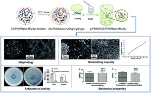
Injected bone substitutes (IBSs) have become increasingly attractive in the field of tissue engineering due to their patient convenience, easy administration as well as minimally invasive procedure for tissue repair. To develop a novel and smart IBS with desirable properties for future in vivo bone regenerative, nano-sized hydroxyapatite (Nano-HA) or antibacterial Ag+, or a combination of them, was initially loaded into a chitosan–poly(vinyl alcohol) (CS–PVA) thermo-sensitive hydrogel. Then the functionalized hydrogel was mixed with PMMA to optimize the final bulk behaviors of the PMMA cement. Finally, the physicochemical properties, anti-bacterial activity, biomineralization ability, and mechanical property changes under simulated physiological conditions of the cements were tested by a type K thermocouple, scanning electron microscopy (SEM), X-ray diffraction (XRD), micro-computed tomography (μ-CT), X-ray photoelectron spectroscopy (XPS), calcium ion test kit and mechanical compression tests. The results showed that the CS–PVA thermo-sensitive hydrogel decreased the Tmax, prolonged the working time, created irregular pores and led to appropriate mechanical properties of the cements. Nano-HA particles induced better mineralization capacity of the cements without acting negatively on the mechanical properties. Ag+ incorporation remarkably enhanced the anti-bacterial activity of the cements for prevention of post-operative infection. Ultimately, such results suggested the injectable and multi-functional cement with p-PMMA/CS–PVA/Nano-HA/Ag+ combination would hold strong promise for future bone reconstruction applications.
Introduction

Although bone tissue possesses the capacity for regenerative growth and remodelling, every year millions of patients worldwide sustain bone defects stemming from pathological events such as inflammation, trauma or tumor resection.1 In this context, it is critical to develop implant technologies to increase or promote bone healing. Despite that autografts are still considered as the gold standard by many orthopedic surgeons for treating bone defects due to the osteogenic, osteoconductive and osteoinductive capacity,1–3 the disadvantages such as donor limitations and reimplant injury4 have resulted in the development of artificial bone substitution biomaterials. Among available materials, injected bone substitutes (IBSs) have been attractive as they could bring significant benefits in several clinical situations, for instance, when defects are complex and irregular, not easily accessible, or when minimally invasive surgery is desired to minimize patient discomfort.5–8

Poly(methyl methacrylate) (PMMA) cements, due to their favorable properties such as easy operating, moldability, robustness, low-price and FDA-approval,9,10 remains the most common injectable material used for filling bone defects, particularly for arthroplasty, vertebroplasty and screw augmentation.11,12 There are, however, several documented shortcomings of PMMA cements,13–16 which include: (1) thermal necrosis caused by a highly exothermic setting reaction; (2) aseptic loosening, the most common cause of arthroplasties due to the inappropriate stiff properties and poor bioactivity of PMMA; (3) infection, another common complication that necessitate prosthesis removal. Given the fact that these drawbacks significantly restrict the application of plain PMMA, several strategies have been employed to modify PMMA cements over the last few decades.17

Incorporating osteoconductive materials or drugs into plain PMMA was regarded as the effective strategy to improve the performance of PMMA cement in the early days.17,18 Previous studies revealed that enriching with calcium phosphate or bioglass could indeed promote the bonding between bone and cement, but did not introduce bone ingrowth into the bone cement and improve the inappropriate stiff properties.19 Moreover, the loaded drugs were enwrapped within the dense PMMA structure and the results of antibacterial efficacy were also not as ideal as expected.20,21 To further overcome these shortcomings, introducing open porosity into the dense PMMA was believed to establish the anchorage at bone/PMMA interface, balance the mechanical properties of bulk PMMA cement, and facilitate drug release against local infection.22–24

Studies performed by de Wijn25 and Shi et al.26 have shown that a porous PMMA (p-PMMA) structure can be produced by mixing the hydrophobic PMMA phase with a hydrophilic hydrogel. This porosity was shown to have beneficial effects on the bone ingrowth, stiffness and polymerization temperature of the PMMA bone cement.27–30 To further improve the performance of p-PMMA, osteoconductive CaP particles were also added into the hydrogel. It is hypothesized that CaP mineral and extracellular matrices (ECMs)-like hydrogel composites could mimic the structure and composition of bone in local sites and also facilitate the mineralization process to obstruct the formation of unfavorable fibrous capsules.

To test this hypothesis, our group have focused on the in vitro and in vivo behaviors of PMMA/CMC hydrogel/CaP composites in recent years.11,19 Although these modified composites were biocompatible and supported bone ingrowth and material fixation, the addition of CaP particles did not increase the amount of bone formation and ingrowth.19 That suggested the way by which the CaP particles were included into the PMMA matrix previously restricted the osteoconductive advantage of the cement. Additionally, antibiotics had not been added into the cement to prevent the possible infection in previous studies. Therefore, a more ideal combination of PMMA/hydrogel porogen/CaP/antibiotic should be established and tested to further optimize the behaviors of composites.

Chitosan (CS), a unique polysaccharide derived from naturally abundant chitin, offers remarkable features for biomedical applications due to its good biocompatibility, biodegradability, bioactivity and antibacterial properties.31 Poly(vinyl alcohol) (PVA), a widely used polymer, is also receiving great interest for its biocompatibility, biodegradability, good mechanical properties and ability to support protein adsorption for healthy cellular growth.32,33 Recently, injectable CS–PVA based thermosensitive hydrogels have attracted increasing attention in the field of biomedical applications.34 Such hydrogels could undergo sol–gel transitions at a temperature close to body temperature (37 °C) under controllable proceeding time without requiring organic solvents, copolymerization agents or externally applied triggers for gelation.35 Additionally, they can approximate the viscoelastic properties of native tissue. Excellent injectability, biocompatibility and ability to deliver drugs or bioactive materials indicates such hydrogels as promising artificial ECM for use in regenerative medicine. Therefore, CS–PVA thermo-sensitive hydrogels could be regarded as ideal porogens to fabricate p-PMMA cements. To further improve the performance of injectable p-PMMA based cements at the bone-material interface, nano-sized hydroxyapatite (Nano-HA), which mimics the composition and structure of bone in local sites and antibacterial Ag+, which has high bacteriostatic and bactericidal activity toward a broad spectrum of bacteria, viruses and other eukaryotic micro-organisms,36,37 could be enriched in CS–PVA thermo-sensitive hydrogels to establish a multifunctional injectable p-PMMA-based cement.

To this end, nano-sized hydroxyapatite (Nano-HA) or antibacterial Ag+, or a combination of them, was initially loaded into a CS–PVA thermo-sensitive hydrogel. Then the functionalized hydrogel was mixed with PMMA to optimize the final bulk behaviors of the PMMA cement. It was hypothesized that the combination of injectable p-PMMA/CS–PVA/Nano-HA/Ag+ could serve as a promising candidate with easy handling properties, suitable mechanical performance, favorable biomineralization capacity and effective infection-resistance ability for future clinical bone reconstruction.

Experimental

Materials

A two-component (powder and liquid) PMMA kit (self-curing PMMA, type II) was purchased from Shanghai New Century Dental Materials Co. (Shanghai, China). The solid component consisted of PMMA, dibenzoyl peroxide (BPO), silicon dioxide and barium sulfate powders. The liquid part was a mixture of methyl methacrylate (MMA) solution and N,N-dimethyl-p-toluidine (DMPT) solution. BPO was used as initiator and DMPT was used as accelerator for the polymerization reaction. Silicon dioxide was used as reinforcer and barium sulfate was used as radiopaque agent.

Chitosan (CS) (Mw = 179.17 kDa; N-deacetylation rate of ≥95%; viscosity = 100–200 mPa s) and poly(vinyl alcohol) (PVA) (Mw = 44.05 g mol−1; alcoholysis degree = 99.8–100%) were purchased from Aladdin Co., Ltd. (Shanghai, China). AgNO3 (Mw = 169.87 g mol−1, purity ≥ 99.8%) was purchased from Sinopharm Chemical Reagent Co., Ltd. (Shanghai, China). Ca(NO3)2·4H2O, (NH4)2HPO4, NH3 (aq) and NaHCO3 were purchased from Aladdin Co., Ltd. (Shanghai, China). All other chemical reagents were of analytical grade or better.

Synthesis and characterization of Nano-HA crystals

Nano-HA was synthesized according to our previous study.38 Briefly, 20 ml of 1.08 M Ca(NO3)2·4H2O solution (pH = 10, adjusted by NH3 (aq)) was heated to 90 °C. Subsequently, 20 ml of 0.65 M (NH4)2HPO4 solution was added dropwise under stirring. The precipitate was maintained in contact with the reaction solution for 5 h at 90 °C under stirring, and then centrifuged at 1800g for 10 min. After that, the product was repeatedly washed with distilled water (DW), centrifuged six times, and then dried at 37 °C overnight.

The morphologies of the synthesized precipitates were evaluated by SEM (Sigma, Zeiss, Germany) and TEM (JEM-2100, JOEL, Japan). The crystal phase was analyzed by X-ray diffraction (XRD, X’Pert Pro, The Netherlands) with a wavelength of 1.5406 Å at a voltage of 40 kV and a current of 40 mA. The scanning range was 10–80°, with a scanning rate of 25 s per step and a step size of 0.026° (2θ).

Synthesis of thermosensitive hydrogels

CS–PVA thermo-sensitive hydrogel was prepared according to the method by Tang et al.34 with slight modification (Fig. 1). In brief, a clear solution of chitosan was obtained by dissolving chitosan (100 mg) into 0.1 M HCl (5 ml) and then chilled in an ice-bath for 15 min. PVA was added to DW to a concentration of 2 wt% and heated at 80 °C for 1 h. Subsequently, 1 M NaHCO3 (0.5 ml) and 2 wt% PVA solution (5 ml) were mixed and similarly chilled for 15 min. Then the PVA–NaHCO3 solution was added into the chitosan solution dropwise in an ice-bath under stirring for another 10 min. The whole solution was degassed by centrifugation of samples at 3500g for 3 min at 5 °C. Then gel-forming process of the solution was conducted in a thermostat at 37 °C for 10 min and the CS–PVA hydrogel was obtained. To prepare the Ag+ loaded thermo-sensitive hydrogel, 50 mg AgNO3 was initially added into 5 ml PVA solution, then the PVA–Ag+ solution was mixed with NaHCO3 solution as described above. To prepare the Nano-HA loaded hydrogel, 500 mg Nano-HA was initially added into 10.5 ml of homogeneous CS–PVA–NaHCO3 solution. Similarly, to prepare the Nano-HA and Ag+ loaded hydrogel, AgNO3 and Nano-HA were added into the PVA solution and CS–PVA–NaHCO3 solution, respectively. The whole solution was mixed uniformly by using sonication and then transformed into the hydrogel at 37 °C for 5 min (Fig. 2).
image file: c6ra08182g-f1.tif
Fig. 1 Schematic diagram of the synthesis of p-PMMA based cements.

Preparation of the cements

The required raw materials for the preparation of the bone cements are listed in Table 1. Four types of porous cements were fabricated, denoted as p-PMMA/CS–PVA, p-PMMA/CS–PVA/Nano-HA, p-PMMA/CS–PVA/Ag+ and p-PMMA/CS–PVA/Nano-HA/Ag+. The solid PMMA cement was used as a control. To prepare plain PMMA cement, PMMA kit powder part and PMMA kit liquid part were manually blended to uniformity using a solid[thin space (1/6-em)]:[thin space (1/6-em)]liquid mass ratio of 1[thin space (1/6-em)]:[thin space (1/6-em)]1 under ambient conditions at room temperature. To prepare the p-PMMA based cements, the mixed PMMA phase was further blended with the four types of CS-based hydrogel separately, with a volume ratio of 3[thin space (1/6-em)]:[thin space (1/6-em)]4 until a homogeneous paste was obtained. Subsequently, the mixture was injected into a Teflon mould (6 mm in diameter; 12 mm in height) to obtain cylinder samples (Fig. 1). After curing overnight, samples were removed from the molds, washed with DW, freeze-dried and imaged by a Nikon D3100 digital camera.
Table 1 Compositions of the prepared cements
Group PMMA matrix CS–PVA matrix Nano-HA crystals/g AgNO3/g
PMMA/g MMA/ml CS/g PVA/g 1 M NaHCO3/ml DW/ml
Plain PMMA 6 3.18
p-PMMA/CS–PVA 6 3.18 0.1 0.1 0.5 5
p-PMMA/CS–PVA/Nano-HA 6 3.18 0.1 0.1 0.5 5 0.5
p-PMMA/CS–PVA/Ag+ 6 3.18 0.1 0.1 0.5 5 0.05
p-PMMA/CS–PVA/Nano-HA/Ag+ 6 3.18 0.1 0.1 0.5 5 0.5 0.05


image file: c6ra08182g-f2.tif
Fig. 2 Different states of CS–PVA solution at room and body temperature. All the CS–PVA solutions remained liquid at room temperature (24.6 °C) and transformed into semisolid hydrogels at body temperature (37 °C).

Polymerization temperature and working time

The polymerization temperature and working time were monitored simultaneously by recording the temperature of the cement mixture as a function of time. According to ISO5833 for acrylic resin cements,39 the homogeneous mixture was packed into a Teflon cylindrical mold in which a probe of a type K thermocouple (Victory high electronic technology co., China) connected to a data logger (Victor E86, Victory high electronic technology co., China) was positioned at the center of the cement surface to record the temperature every 0.2 s. The maximum temperature (Tmax) was obtained during the polymerization reaction (n = 3). The working time was defined as the interval during which the mixture is safely injectable, and before which the mixture becomes too stiff to inject through the access needle;40 room temperature is 24.6 °C.

Micro-computed tomography (μ-CT) examination

Porometrical properties examination within the PMMA matrix was conducted using a micro-computed tomography set-up (μ-CT 50, Scanco Medical, Basersdorf, Switzerland) at 12 μm intervals, with acquisition conditions of 90 kV and 88 μA in high resolution mode. Then, the images were imported into the software and reconstructed in 3D format.

X-Ray photoelectron spectroscopy analysis

Ag[thin space (1/6-em)]3d XPS was detected by X-ray photoelectron spectroscopy (XPS; ESCALAB 250Xi, Thermo Fisher Scientific, Britain) with an Al-Kα X-ray source (1486.6 eV) to confirm the loading of silver within the cement. The cements were mounted on stubs with conductive carbon tape. Then the survey scans were collected using a fixed pass energy of 100 eV with an energy step size of 1.0 eV, while narrow scans were obtained with a pass energy of 30 eV and an energy step size of 0.1 eV. All XPS spectra were recorded using an aperture slot of 500 × 500 μm. Binding energies were calibrated by using contaminant carbon (C[thin space (1/6-em)]1s = 285.0 eV) as a reference.

Anti-bacterial activity assay

Following guidelines of the Clinical and Laboratory Standards Institute,41 Escherichia coli (E. coli) and Staphylococcus aureus (S. aureus) were used as reference strains for Gram-negative and Gram-positive bacteria to test the antimicrobial efficacy of the CS–PVA based hydrogels as well as PMMA-based cements, respectively.

To investigate the inhibitory effect of the CS–PVA based hydrogels by a disk-diffusion method, all hydrogels were freeze-dried and pressed into discs (diameter = 13 mm). Then 30 μl of suspensions of the microbial cells over the range of 0.05–2 mg ml−1 was spread on Mueller–Hinton agar evenly and the tested discs were separately placed on agar for 16 h incubation at 37 °C. After that, the antimicrobial activity was evaluated by considering the zone of inhibition (absence of viable microbial cells) around the disc samples and optical images were taken for analysis.

To compare the contact bacteriostatic activities, autoclaved PMMA-based cements were prepared and soaked in bacteria suspension (1 × 10³ colony-forming units [CFUs] per ml) of E. coli and S. aureus with mass–volume ratio of 0.2 g ml−1 for incubation at 37 °C, respectively. Bacteria suspension without immersing cements was used as the negative control. After 24 h incubation, the soaked cements of each type were washed with PBS three times, fixed in 2.5% glutaraldehyde for 3 h, dehydrated using Analytical grade ethanol and freeze-dried. Subsequently, SEM (Sigma, Zeiss, Germany) was performed to reveal the contact-killing activity.

To further test the sustained-release bacteriostatic activities for different cements, 100 μl of dilute inoculums which were subjected to immersed PMMA-based cements for 24 h, were plated on Mueller–Hinton agar and incubated at 37 °C for 16 h (n = 3). Finally, CFUs of bacterial colonies were counted for group comparisons.

Simulated body fluid (SBF) immersion

To evaluate the reaction of different PMMA cements in physiological conditions, SBF was prepared according to the protocol of Kokubo et al.42 Sterile cements were separately immersed in SBF (10 ml SBF for each cement) at 37 °C which was refreshed weekly. After 28 days, cements were washed in DW, freeze-dried, and used for the following studies along with the unsoaked cements.

Mineralizing capacity of cements

In order to reveal internal structure variation of cements before and after immersion in SBF, SEM (Sigma, Zeiss, Germany) was performed on the cross section of fractured samples which were mounted on aluminum stubs using conductive carbon tape and previously sputter-coated with gold for 90 s. Morphology images were recorded using an In lens detector at an accelerating voltage of 10 kV and working distance of 4–7 mm.

To further evaluate the mineralization ability, cumulative calcium uptake of cements after immersion was evaluated by assessing the remaining calcium concentration in SBF after 7, 14, 21 and 28 days (n = 3, per time point). The concentration was determined using methyl thymine blue colorimetric assay through a calcium ion test kit (Jiancheng Co. Ltd, Nanjing, China) according to the manufacturer’s instructions. The kit includes calcium standard solution (2.5 mmol L−1), methylthymine blue (MTB) reagent and alkaline solution. The working solution is a mixture of 1 ml MTB reagent and 2 ml alkaline solution. 50 μl SBF samples or standard calcium solution was added to 3 ml working solution and then left to stand for 5 min. Meanwhile, 50 μl DW added into 3 ml working solution was regarded as the blank control. Subsequently, the color intensity of the solution complex was measured at 610 nm using an ultraviolet spectrophotometer, which was proportional to the quantity of calcium present in the sample.

Then the calcium content was calculated:

image file: c6ra08182g-t1.tif
where t is the SBF sample solution, s is the standard calcium solution and c is the blank control. Subsequently, the cumulative calcium uptake was calculated by adding up the reduced calcium content at each time point. Finally, the accumulation curve was drawn.

Mechanical properties

Modulus of elasticity (E) and the compressive yield strength (σy) were measured to evaluate the effect of the CS–PVA gel, Nano-HA and Ag+ on mechanical performance of cements by using a universal testing machine (AGS-J, SHIMADZU, Japan). In accordance with ISO 5833,39 cylindrical samples of cured cements were prepared with diameter of 6 mm and length of 12 mm. Three specimens were measured per cement type, operated at a crosshead speed of 0.5 mm min−1 along the long axis.

Statistical analysis

A one-way analysis of variance (ANOVA) was used to determine the statistical significance, which was followed by post hoc analysis using the Turkey test (SPSS 22.0). Results were considered significantly different when p < 0.05.

Results and discussion

Characterization of Nano-HA

The SEM and TEM micrograph of HA are shown in Fig. 3a and b. The precipitates are needle-like nanocrystals and the average size of Nano-HA was estimated to be about 30 nm in width and 100 nm in length. The X-ray diffraction pattern of the precipitate in Fig. 3c showed intensity peaks corresponding to Joint Committee on Power Diffraction Standard for HA (JCPDS file no. 00-024-0033) which could mimic the mineral component of bone with potential osteoconductive capacity.6
image file: c6ra08182g-f3.tif
Fig. 3 Characterizations of Nano-HA particles. (a) SEM observation, (b) TEM observation and (c) XRD analysis (the yellow lines show the experimental XRD scan of the sample and the blue lines indicate the match with the JCPDS file for HA).

Polymerization temperature and working time

Despite the immiscibility between hydrophobic PMMA and hydrophilic CS–PVA phase, we easily synthesized p-PMMA based cements in vitro by manual mixing. Fig. 4 shows typical temperature curves of the cements during polymerization and the values of the curing parameters are summarized in Table 2. Due to the exothermic nature of the PMMA/MMA polymerization reaction, the pure PMMA cement showed a high Tmax which rapidly increased to 75.3 ± 0.2 °C. The high Tmax limits the types of loaded drugs that can be applied and may cause thermal necrosis to surrounding tissue.15,20 Luckily, the addition of CS–PVA hydrogel not only effectively reduced the relative amount of polymer (relatively less polymer per unit volume) but it also effectively absorbed the heat generated by PMMA during the polymerization reaction,9 so all the CS–PVA based p-PMMA cements brought down the Tmax to the range of 25.5–29.1 °C. Furthermore, Nano-HA or Ag+ did not compromise this beneficial low polymerization temperature, even though Nano-HA exhibited a poor thermal conductivity.
image file: c6ra08182g-f4.tif
Fig. 4 Typical exotherms of PMMA-based cements.
Table 2 Mean values of Tmax and working time of the prepared cements
Group Tmax/°C Working time/s
PMMA 75.3 ± 0.2 501 ± 0.8
p-PMMA/CS–PVA 25.5 ± 0.4 739 ± 1.1
p-PMMA/CS–PVA/Nano-HA 29.1 ± 1.0 787 ± 1.3
p-PMMA/CS–PVA/Ag+ 27.8 ± 0.5 771 ± 0.6
p-PMMA/CS–PVA/Nano-HA/Ag+ 28.4 ± 1.0 804 ± 1.3


Since the amount of released heat is closely linked with the polymerization speed of PMMA, the decreased Tmax resulted in an increased working time in this study. The working time of p-PMMA based cements ranged between 739 and 804 s with no significant difference among cements (p > 0.05), which was significantly longer than that of plain PMMA of around 501 s (p < 0.01). Sufficient working time is essential for appropriate handling, pressurization and the insertion of cements into complex- and irregular-shaped bone defects without additional techniques, such as pre-cooling the cement or decreasing the operation room temperature.

Morphology

Representative images of the cement surface are shown in Fig. 5A. In comparison with the dense polymer structure of plain PMMA, the p-PMMA based samples showed rough and porous surfaces. The aggregation of Nano-HA particles was evident within the porosities for Nano-HA loaded cements, which can form direct chemical bonds with bone for the future bony conjunction.17 Additionally, the Ag+ loaded cements showed a light brown surface while the Ag+-free cements presented a white appearance.
image file: c6ra08182g-f5.tif
Fig. 5 (A) Surface morphologies, (B and C) μ-CT graphs and (D) SEM observations at low magnification (250×, scale bar represents 100 μm) of PMMA-based cements: (I) PMMA; (II) p-PMMA/CS–PVA; (III) p-PMMA/CS–PVA/Nano-HA; (IV) p-PMMA/CS–PVA/Ag+; (V) p-PMMA/CS–PVA/Nano-HA/Ag+.

μ-CT analysis nondestructively yielded 3D reconstruction images and 2D cross section views of cements to reveal their bulk morphologies, internal porous structures and particle distributions (Fig. 5B and C). By visual inspection, p-PMMA based cements showed loose structures with more irregular and extensive porosities compared with plain PMMA cements. Additionally, μ-CT results revealed agglomeration of Nano-HA within the matrix of Nano-HA loaded cements. Moreover, from 2D cross section views, some highly radiopaque agents were found within the Ag+ loaded cements.

SEM observations at low magnification (500×) further displayed the general morphologies of cross-sectional cements (Fig. 5D). The plain PMMA showed spherically shaped MMA particles compacted to present a smooth surface. By contrast, irregular porous structures are seen within the other p-PMMA based cements.

All the morphological results showed that only a few separated pores are contained in plain PMMA caused by the introduction of air during preparation. In contrast, many irregular pore structures were found in p-PMMA based cements, which is attributed to the phase separation of hydrophobic PMMA and hydrophilic CS–PVA gel. In addition to pore structures enabling tissue infiltration, these irregular pores may be harnessed to facilitate the release of therapeutics, such as antibiotics, which can be incorporated into the CS–PVA gel. For Nano-HA loaded cements, both SEM and μ-CT have evidenced that most, if not all, of the needle-like precipitates were agglomerated within porous structures produced by CS–PVA. This result indicated that the Nano-HA and CS–PVA hydrogels could mimic the native HA crystal/ECM combination in the human body, which could further simulate the natural function of bone to activate in vivo mechanisms of tissue regeneration.43 For the μ-CT results of Ag+ loaded cements, materials with radiopaque property were found. The occurrence of this phenomenon may be attributed to the chemical valence state change of Ag+, such as silver reduction caused by chitosan under alkaline condition44 or photochemical decomposition and silver oxidation, caused by hydrolysis reaction.

Antimicrobial activity

Fig. 6A shows the high-resolution spectrum of Ag[thin space (1/6-em)]3d within the p-PMMA/CS–PVA/Ag+ and p-PMMA/CS–PVA/Nano-HA/Ag+ cements which confirmed the successful incorporation of silver. It can be seen that the spectrum consists of two major peaks at 373.6–373.8 and 367.6–367.8 eV, corresponding to Ag[thin space (1/6-em)]3d3/2 and Ag[thin space (1/6-em)]3d5/2 binding energies, respectively.
image file: c6ra08182g-f6.tif
Fig. 6 (A) Ag[thin space (1/6-em)]3d XPS spectra for the Ag+ loaded PMMA cements; (B) antibacterial activity of disc-like samples by zone of inhibition (ZOI) test against E. coli and S. aureus: (a) CS–PVA; (b) CS–PVA/Nano-Ha; (c) CS–PVA/Ag+; (d) CS–PVA/Nano-Ha/Ag+ (translucent zones indicated inhibition of bacterial growth).

The results of disk-diffusion tests against E. coli and S. aureus was demonstrated by the presence of an inhibition zone (Fig. 6B). The comparison of ZOI indicated that all CS based hydrogels exhibited antibacterial activities but that Ag+ enriched cements revealed clearer and greater area translucent inhibition zones. No statistical significance were found between the Ag+ loaded samples for each reference strain. In addition, the CS–PVA hydrogel and CS–PVA/Nano-HA hydrogels showed slightly higher antibacterial activity towards Gram-negative bacteria than Gram-positive bacteria, which could be attributed to the structural and compositional differences of the cell membrane.45 In comparison with Gram-negative bacteria, Gram-positive bacteria usually have thicker peptidoglycan cell membranes. Therefore, it is harder to compromise their integrity, which resulted in a lower antibacterial response.

The colonization of surfaces by bacteria is well known to adversely affect the function of a variety of specific interfaces.46 In order to eliminate or substantially reduce the bacterial attachment, the antibacterial material should not only act by being toxic when in contact with the bacteria, but also have the ability to release an effective antibacterial agent to the site of infection.46,47 So besides ZOI, contact bacteriostatic and the sustained-release bacteriostatic tests of different cements against E. coli and S. aureus were also evaluated to fully reveal the anti-bacterial abilities of the cements. From the SEM results (Fig. 7A), we found that almost no bacteria adhered to the surface of the Ag+ loaded group. Besides, the results of CFU counts also showed less bacteria for the Ag+ loaded cements than that of the other three cements (Fig. 7B and C). No significant difference were found between the two versions of silver-containing cements (p > 0.05). The possible mechanism for the antimicrobial action of Ag+ may be that they could penetrate inside the bacteria to interact with phosphorus-containing DNA and RNA to inhibit their replication abilities48 as well as interact with thiol groups in proteins, which induces the inactivation of the bacterial proteins.45 Therefore, both the SEM and CFUs count results suggested that enriched Ag+ cements showed excellent contact bacteriostatic and sustained-release bacteriostatic properties with E. coli and S. aureus, which would thus avoid biofilm formation, sterilize the surgical site and reduce the incidence of drug resistance.


image file: c6ra08182g-f7.tif
Fig. 7 (A) Contact bacteriostatics and (B and C) sustained-release bacteriostatics of PMMA-based cements towards E. coli and S. aureus: (I) PMMA; (II) p-PMMA/CS–PVA; (III) p-PMMA/CS–PVA/Nano-HA; (IV) p-PMMA/CS–PVA/Ag+; (V) p-PMMA/CS–PVA/Nano-HA/Ag+.

Mineralizing capacity

The in vitro mineral formation abilities of the cements were observed by SEM micrographs at high magnification (20[thin space (1/6-em)]000×) (Fig. 8A). Before immersion in SBF, plain PMMA cement displayed a clear and smooth surface. p-PMMA/CS–PVA and p-PMMA/CS–PVA/Ag+ cements revealed uniformly formed CS/PVA-films covering the PMMA pore surfaces, and p-PMMA/CS–PVA/Nano-HA and p-PMMA/CS–PVA/Nano-HA/Ag+ cements revealed agglomerated Nano-HA crystals lying on the CS–PVA films. After immersion in SBF for 28 days, no obvious change was found in plain PMMA. Some newly formed CaP-like depositions were occasionally distributed on the pore surface of p-PMMA/CS–PVA and p-PMMA/CS–PVA/Ag+ cements. The different behavior may be due to that plain PMMA was bioinert while CS incorporation could induce crystal formation due to its beneficial surface roughness as well as the wettability of PMMA introduced by CS.49 Most excitingly, Nano-HA enriched cements effectively facilitate the formation of a dense ball-like apatite layer on the pore surface of PMMA after immersion in SBF.
image file: c6ra08182g-f8.tif
Fig. 8 (A) SEM images (20[thin space (1/6-em)]000×, scale bar represents 1 μm) of PMMA-based cements before and after immersion in SBF for 28 days and (B) cumulative calcium uptake of PMMA-based cements in SBF at different time points (data show means and standard deviations): (I) PMMA; (II) p-PMMA/CS–PVA; (III) p-PMMA/CS–PVA/Nano-HA; (IV) p-PMMA/CS–PVA/Ag+; (V) p-PMMA/CS–PVA/Nano-HA/Ag+.

Cumulative calcium uptake showed agreement with SEM observations in a quantitative manner (Fig. 8B). In general, all cements showed an increasing trend of cumulative calcium uptake during 28 days immersion. The two Nano-HA loaded cements revealed more dramatic uptake than the other three HA-free cements. After 21 days, the calcium uptake for Nano-HA-free PMMA cements reached a plateau while Nano-HA loaded cements continued to rise sharply. There was no statistical significance between the two Nano-HA loaded cements or among the three Nano-HA-free cements (p > 0.05).

Therefore, both SEM and calcium uptake results proved better mineral forming ability of Nano-HA enriched cements, which evidently improve the potential bioactivity of such cements for future in vivo bone-bonding ability.

Mechanical properties

Mechanical results are displayed in Fig. 9. Before immersion, plain PMMA shows E of 925.93 ± 23.94 MPa and σy of 66.99 ± 2.52 MPa. These values are considerably higher than E and σy of p-PMMA based cements, which range from 109.01 to 128.72 MPa and 4.31 to 4.77 MPa, respectively (p < 0.001). No significant difference of E or σy was found among these p-PMMA based cements (p > 0.05). After 28 days immersion in SBF, no statistically significant differences were found among the various time points for each group regarding both σy and E (p > 0.05).
image file: c6ra08182g-f9.tif
Fig. 9 Mechanical properties of PMMA-based cements before and after immersion in SBF for 28 days: (a) modulus of elasticity (E); (b) compressive yield strength (σy): (I) PMMA; (II) p-PMMA/CS–PVA; (III) p-PMMA/CS–PVA/Nano-HA; (IV) p-PMMA/CS–PVA/Ag+; (V) p-PMMA/CS–PVA/Nano-HA/Ag+ (* shows significant difference between plain PMMA and other cements regarding E or σy, p < 0.05).

Mechanical characteristics are critical in determining the long-term stability of PMMA-based cements since suitable mechanical properties could play a beneficial role in transferring loads between bone and cement.11 In the present study, the creation of a porous structure by the CS–PVA hydrogel led to declined but still acceptable mechanical properties for p-PMMA based cements, in accordance with the typical porosity–mechanical property relationship.9 All the p-PMMA based cements revealed E values and σy values matching with those of cancellous bone (50–800 MPa of E and 4–12 MPa of σy, respectively),23,50,51 which allow better stress distribution in the filled bone defects, thus reducing the risk of fractures adjacent to the operated sites. Moreover, mechanical results demonstrated that loading Nano-HA or Ag+ into porous PMMA cements was feasible, without acting negatively on the mechanical properties.

Conclusions

The current in vitro study suggested that an injectable bone cement with smart combinations of added components could extract advantages from the composite concept. Particularly, PMMA constructed the main part to provide the mechanical support. CS–PVA thermo-sensitive hydrogel decreased the Tmax, prolonged the working time, created irregular pores and led to appropriate mechanical properties of cements. Nano-HA particles induced better mineralization capacity of the cements without acting negatively on mechanical properties. Ag+ incorporation remarkably enhanced the anti-bacterial activity of cements for prevention of postoperative infection. Ultimately, such results suggested injectable and multi-functional cements with p-PMMA/CS–PVA/Nano-HA/Ag+ combination would hold strong promise for future bone reconstruction applications. More importantly, the p-PMMA/porogen/osteoconductive materials/antibacterial agent construct can establish a multifunctional platform to load different components as a combination for meeting different clinical needs for future applications.

Acknowledgements

This work was supported by the National Natural Science Foundation of China (No. 81470771, No. 81500887), and the Natural Science Fund of Hubei Province (No. 2013CFA068).

Notes and references

  1. A. S. Greenwald, S. D. Boden, V. M. Goldberg, Y. Khan, C. T. Laurencin and R. N. Rosier, J. Bone Jt. Surg., Am. Vol., 2001, 83(2), 98–103 CrossRef.
  2. M. A. Merkx, J. C. Maltha, H. P. Freihofer and A. M. Kuijpers-Jagtman, Biomaterials, 1999, 20, 639–645 CrossRef CAS PubMed.
  3. C. G. Finkemeier, J. Bone Jt. Surg., Am. Vol., 2002, 84, 454–464 CrossRef.
  4. D. Apelt, F. Theiss, A. O. El-Warrak, K. Zlinszky, R. Bettschart-Wolfisberger, M. Bohner, S. Matter, J. A. Auer and B. von Rechenberg, Biomaterials, 2004, 25, 1439–1451 CrossRef CAS PubMed.
  5. B. R. Constantz, I. C. Ison, M. T. Fulmer, R. D. Poser, S. T. Smith, M. VanWagoner, J. Ross, S. A. Goldstein, J. B. Jupiter and D. I. Rosenthal, Science, 1995, 267, 1796–1799 CrossRef CAS PubMed.
  6. M. Bongio, J. J. van den Beucken, M. R. Nejadnik, Z. Tahmasebi Birgani, P. Habibovic, L. A. Kinard, F. K. Kasper, A. G. Mikos, S. C. Leeuwenburgh and J. A. Jansen, Acta Biomater., 2013, 9, 5464–5474 CrossRef CAS PubMed.
  7. Y. J. No, S. I. Roohani-Esfahani and H. Zreiqat, Nanomedicine, 2014, 9, 1745–1764 CrossRef CAS PubMed.
  8. M. Bongio, J. J. van den Beucken, S. C. Leeuwenburgh and J. A. Jansen, J. Tissue Eng. Regener. Med., 2015, 9, 191–209 CrossRef CAS PubMed.
  9. M. Shi, J. D. Kretlow, A. Nguyen, S. Young, L. Scott Baggett, M. E. Wong, F. K. Kasper and A. G. Mikos, Biomaterials, 2010, 31, 4146–4156 CrossRef CAS PubMed.
  10. J. C. Webb and R. F. Spencer, J. Bone Jt. Surg., Br. Vol., 2007, 89–B, 851–857 CrossRef PubMed.
  11. M. A. Lopez-Heredia, Y. Sa, P. Salmon, J. R. de Wijn, J. G. Wolke and J. A. Jansen, Acta Biomater., 2012, 8, 3120–3127 CrossRef CAS PubMed.
  12. H. L. Tan, Z. X. Peng, Q. T. Li, X. F. Xu, S. R. Guo and T. T. Tang, Biomaterials, 2012, 33, 365–377 CrossRef CAS PubMed.
  13. K. W. Lye, H. Tideman, M. A. Merkx and J. A. Jansen, Tissue Eng., Part B: Rev., 2009, 15, 485–496 CrossRef CAS PubMed.
  14. Y. Sa, F. Yang, S. C. Leeuwenburgh, J. G. Wolke, G. Ye, J. R. de Wijn, J. A. Jansen and Y. Wang, J. Biomed. Mater. Res., Part B, 2015, 103, 548–555 CrossRef PubMed.
  15. K. W. Lye, S. Lee, H. Tideman, M. A. Merkx and J. A. Jansen, Int. J. Oral Maxillofacial Surg., 2011, 40, 86–93 CrossRef CAS PubMed.
  16. K. W. Lye, H. Tideman, M. A. W. Merkx and J. A. Jansen, Tissue Eng., Part B: Rev., 2009, 15, 485–496 CrossRef CAS PubMed.
  17. Q. He, H. Chen, L. Huang, J. Dong, D. Guo, M. Mao, L. Kong, Y. Li, Z. Wu and W. Lei, PLoS One, 2012, 7, e42525 CrossRef CAS PubMed.
  18. Y. Sa, M. Wang, H. Deng, Y. Wang and T. Jiang, RSC Adv., 2015, 5, 91082–91092 RSC.
  19. K. W. Lye, H. Tideman, J. C. Wolke, M. A. Merkx, F. K. Chin and J. A. Jansen, Clin. Oral Implants Res., 2013, 24, 100–109 CrossRef PubMed.
  20. H. van de Belt, D. Neut, W. Schenk, J. R. van Horn, H. C. van der Mei and H. J. Busscher, Acta Orthopaedica Scandinavica, 2000, 71, 625–629 CrossRef CAS PubMed.
  21. P. P. Spicer, S. R. Shah, A. M. Henslee, B. M. Watson, L. A. Kinard, J. D. Kretlow, K. Bevil, L. Kattchee, G. N. Bennett, N. Demian, K. Mende, C. K. Murray, J. A. Jansen, M. E. Wong, A. G. Mikos and F. K. Kasper, Acta Biomater., 2013, 9, 8832–8839 CrossRef CAS PubMed.
  22. P. J. van Mullem, J. M. Vaandrager, J. P. Nicolai and J. R. de Wijn, Biomaterials, 1990, 11, 299–304 CrossRef CAS PubMed.
  23. S. J. Hollister, Nat. Mater., 2005, 4, 518–524 CrossRef CAS PubMed.
  24. M. Shi, J. D. Kretlow, P. P. Spicer, Y. Tabata, N. Demian, M. E. Wong, F. K. Kasper and A. G. Mikos, J. Controlled Release, 2011, 152, 196–205 CrossRef CAS PubMed.
  25. J. R. de Wijn, PhD thesis, Catholic University, 1982.
  26. M. Shi, J. D. Kretlow, P. P. Spicer, Y. Tabata, N. Demian, M. E. Wong, F. K. Kasper and A. G. Mikos, J. Controlled Release, 2011, 152, 196–205 CrossRef CAS PubMed.
  27. N. L. Simone, T. Dan, J. Shih, S. L. Smith, L. Sciuto, E. Lita, M. E. Lippman, E. Glatstein, S. M. Swain, D. N. Danforth and K. Camphausen, Breast Cancer Res. Treat., 2012, 132, 197–203 CrossRef PubMed.
  28. P. J. van Mullem, J. R. de Wijn and J. M. Vaandrager, Ann. Plast. Surg., 1988, 21, 576–582 CrossRef CAS PubMed.
  29. A. M. Termuhlen, J. M. Tersak, Q. Liu, Y. Yasui, M. Stovall, R. Weathers, M. Deutsch, C. A. Sklar, K. C. Oeffinger, G. Armstrong, L. L. Robison and D. M. Green, Pediatr. Blood Cancer, 2011, 57, 1210–1216 Search PubMed.
  30. M. L. Bruens, H. Pieterman, J. R. de Wijn and J. M. Vaandrager, J. Craniofacial Surg., 2003, 14, 63–68 CrossRef PubMed.
  31. Y. Wan, H. Wu, A. Yu and D. Wen, Biomacromolecules, 2006, 7, 1362–1372 CrossRef CAS PubMed.
  32. S. Y. Lee, B. P. Pereira, N. Yusof, L. Selvaratnam, Z. Yu, A. A. Abbas and T. Kamarul, Acta Biomater., 2009, 5, 1919–1925 CrossRef CAS PubMed.
  33. K. Ishihara, H. Nomura, T. Mihara, K. Kurita, Y. Iwasaki and N. Nakabayashi, J. Biomed. Mater. Res., 1998, 39, 323–330 CrossRef CAS PubMed.
  34. Y.-F. Tang, Y.-M. Du, X.-W. Hu, X.-W. Shi and J. F. Kennedy, Carbohydr. Polym., 2007, 67, 491–499 CrossRef CAS.
  35. B. Jeong, S. W. Kim and Y. H. Bae, Adv. Drug Delivery Rev., 2012, 64, 154–162 CrossRef.
  36. I. Chopra, J. Antimicrob. Chemother., 2007, 59, 587–590 CrossRef CAS PubMed.
  37. P. Gong, H. Li, X. He, K. Wang, J. Hu, W. Tan, S. Zhang and X. Yang, Nanotechnology, 2007, 18 DOI:10.1088/0957-4484/18/28/285604.
  38. T. Jiang, X. Ma, Z. Wang, H. Tong, J. Hu and Y. Wang, J. Dent., 2008, 36, 907–914 CrossRef CAS PubMed.
  39. ISO-5833/1, International Standards organization, 2002 Search PubMed.
  40. T. S. Eckel and W. Olan, Tech. Vasc. Intervent. Radiol., 2009, 12, 44–50 CrossRef PubMed.
  41. I. Wiegand, K. Hilpert and R. E. Hancock, Nat. Protoc., 2008, 3, 163–175 CrossRef CAS PubMed.
  42. T. Kokubo, H. Kushitani, S. Sakka, T. Kitsugi and T. Yamamuro, J. Biomed. Mater. Res., 1990, 24, 721–734 CrossRef CAS PubMed.
  43. J. Venkatesan and S. K. Kim, Mar. Drugs, 2010, 8, 2252–2266 CrossRef CAS PubMed.
  44. A. Murugadoss and A. Chattopadhyay, Nanotechnology, 2008, 19, 015603 CrossRef CAS PubMed.
  45. M. Rai, A. Yadav and A. Gade, Biotechnol. Adv., 2009, 27, 76–83 CrossRef CAS PubMed.
  46. J. Hasan, R. J. Crawford and E. P. Ivanova, Trends Biotechnol., 2013, 31, 295–304 CrossRef CAS PubMed.
  47. E. P. Ivanova, J. Hasan, V. K. Truong, J. Y. Wang, M. Raveggi, C. Fluke and R. J. Crawford, Appl. Microbiol. Biotechnol., 2011, 91, 1149–1157 CrossRef CAS PubMed.
  48. Y. Matsumura, K. Yoshikata, S. Kunisaki and T. Tsuchido, Appl. Environ. Microbiol., 2003, 69, 4278–4281 CrossRef CAS PubMed.
  49. H. Tan, S. Guo, S. Yang, X. Xu and T. Tang, Acta Biomater., 2012, 8, 2166–2174 CrossRef CAS PubMed.
  50. S. Yang, K. F. Leong, Z. Du and C. K. Chua, Tissue Eng., 2001, 7, 679–689 CrossRef CAS PubMed.
  51. A. Boger, M. Bohner, P. Heini, S. Verrier and E. Schneider, J. Biomed. Mater. Res., Part B, 2008, 86B, 474–482 CrossRef CAS PubMed.

This journal is © The Royal Society of Chemistry 2016
Click here to see how this site uses Cookies. View our privacy policy here.