DOI:
10.1039/C6RA08080D
(Paper)
RSC Adv., 2016,
6, 54503-54509
Hydrothermal synthesis of analcime and hydroxycancrinite from K-feldspar in Na2SiO3 solution: characterization and reaction mechanism
Received
29th March 2016
, Accepted 18th May 2016
First published on 19th May 2016
Abstract
Hydroxycancrinite and analcime were generated in the reaction process of K-feldspar and various concentration solutions of Na2SiO3. The effect of the concentration of Na2SiO3 on the formation of the product samples and the reaction mechanism were investigated. The X-ray diffraction (XRD) results indicate that there exists hydroxycancrinite (Na8Al6Si6O24(OH)2·2H2O) with a hexagonal structure at a Na2SiO3 concentration of 2.85–3.9 mol kg−1 and analcime (NaAlSi2O6·H2O) with a cubic structure at 2.34–2.85 mol kg−1 in the reaction of K-feldspar–Na2SiO3–H2O system. Rietveld refinements were utilized to investigate the crystallographic parameters of hydroxycancrinite based on the XRD results, showing that Na1 atoms existed in the form of [Na·H2O]+ cluster and water molecule (O8) are located in the center of those six-membered rings and slightly displace from the plane of the six-membered ring, as well as Na2 atoms and larger anionic group OH−(O7) are seated in the larger channel. Fourier transformation infrared spectrometry (FTIR) results suggest that the existence of the –OH and water molecules makes the absorption bands at 3569 and 3451 cm−1 broaden and overlap with each other as the concentration of Na2SiO3 decreases. The sharp bands at 1634 cm−1 disappear gradually with the decrease of Na2SiO3 concentration owing to the difference of product. It turns out that the reaction process of K-feldspar and Na2SiO3 as well as the synthesis of analcime and hydroxycancrinite is an interface coupled dissolution–reprecipitation process mainly in the presence of Na+, [Al(OH)4]−, [H2SiO4]2− and SiO32− and followed by the repolymerization process of the silicate framework.
1. Introduction
Analcime is a crystalline microporous aluminosilicate mineral, the three-dimensional framework of which consists of a network of pores and cavities built by the corner-sharing of [SiO4] and [AlO4].1,2 Analcime (NaAlSi2O6·H2O) is a scarce zeolite in nature with small irregular channels consisting of 4, 6 and 8 membered distorted rings and some cavities occupied by Na ions generating an octahedral-coordination.3 The unique structure makes analcime have wide applications in catalysis, ion exchange, ion adsorption reactions, and fluoride wastewater treatment.4,5
The synthesis of analcime has been reported in several papers and patents. Generally, analcime is synthesized using chemical reagents sodium silicate and aluminate. Azizi et al.6 used effectively NaOH solution and rice husk ash as a silica source for the synthesis of analcime. Analcime was synthesized using a hydrothermal method using chrysotile and rice husk ash as the sources of silica in the report of Petkowicz et al.7 In addition, there are many aluminosilicate minerals that could be used as silica, such as kaolin,8 coal fly ash9 and perlite.10
As a feldspathoid mineral, cancrinite found in nature with an ideal chemical composition Na6Ca[AlSiO4]6CO3·2H2O is formed by the stacking of layers of six-membered rings in an ABAB sequence and consists of 5-cages and larger twelve-membered channels similar to those of zeolite. In general, the channels of cancrinite are easily blocked by inorganic guest anions which limits the application of the porous structure.11–13 Moreover, the removal of guest anions by annealing of the guest anion causes the destruction of the framework. However, OH− and H2O molecules can be removed under mild conditions, which makes hydroxycancrinite a suitable material to utilize the porous structure as compared with cancrinite.14
K-feldspar is widely distributed in China as an important potassic mineral resource for relieving the shortage of soluble potassic salt if utilized efficiently.15,16 The stable structure of K-feldspar means it cannot be used in soil directly. The reported dissolution methods of K-feldspar have more than 30 categories, including limestone sintering,17,18 molten salt leaching,19 inorganic acid dissolution, hydrothermal alkaline dissolution,20,21 and so on. Hydrothermal alkaline digestion has great potential in industrial applications, and has several advantages, such as a lower reaction temperature, higher heat utilization rate and lower environmental impact. The hydrothermal dissolution of K-feldspar in Ca(OH)2, NaOH and KOH solutions has been studied in our previous work.22–24
However, synthesis of analcime from K-feldspar and Na2SiO3 is seldom reported. Moreover, the insoluble potassium can be extracted from K-feldspar in the formation process of analcime and hydroxycancrinite, which is very significant for China owing to the rare potassium resource. In this work, analcime and hydroxycancrinite were synthesized in the hydrothermal dissolution process of K-feldspar. The effect of the concentration of Na2SiO3 on the formation of different products and the reaction mechanism were discussed. The products were characterized by means of X-ray diffraction (XRD), transmission electron microscopy (TEM), wet chemistry analysis, Fourier transform infrared spectroscopy (FTIR), and scanning electron microscope (SEM).
2. Experimental
2.1. Materials
The K-feldspar (RC15) sample used in this study was collected from Rongcheng County of Shandong Province, China. The chemical composition of the K-feldspar powder is shown in Table 1. The main chemical components of K-feldspar are SiO2 (64.80%) and Al2O3 (17.35%) with a K2O content of about 15.31%. Sodium metasilicate nonahydrate (analytical reagent grade) was supplied by Beijing Modern Eastern Finechemical Co., Ltd. The deionized water produced in the local laboratory was used in the experiments.
Table 1 Chemical composition of potassic feldspar (wB/%)
| Sample |
SiO2 |
TiO2 |
Al2O3 |
Fe2O3 |
FeO |
MgO |
CaO |
Na2O |
K2O |
P2O5 |
Loss |
Total |
| RC15 |
64.8 |
0.00 |
17.35 |
0.078 |
0.29 |
0.17 |
0.40 |
0.66 |
15.31 |
0.007 |
0.55 |
99.62 |
2.2. Experimental
In the hydrothermal experiments, 10 g of K-feldspar powder was ground, passed through a 200-mesh sieve, and mixed with 3.90, 3.19, 2.85, 2.57, or 2.34 mol kg−1 solvent (expressed in molality representing the number of moles of solute divided by the kilograms of solvent) in a 100 mL Teflon liner. After magnetic stirring, the liner was placed in a steel hydrothermal autoclave assembled in a temperature controlled homogenous reactor at 250 °C for 5 h with a rotating speed of 10 rpm. The solid products (named with RC-N-1–RC-N-5) were separated from the solution using filtration and then investigated using X-ray diffraction, scanning electron microscope, transmission electron microscope and Fourier transform infrared spectroscopy. The elemental contents in all the aqueous samples and solid samples were analyzed using wet chemical analysis. The schematic illustration of the reaction is displayed in Fig. 1.
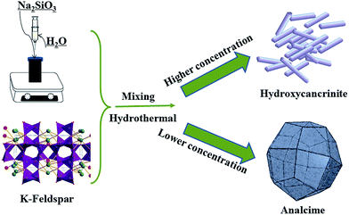 |
| | Fig. 1 Schematic illustration of the synthesis process of analcime and hydroxycancrinite. | |
2.3. Characterization
The XRD patterns of the K-feldspar powder and solid products obtained under different conditions were recorded using a SmartLab (Rigaku) X-ray diffractometer with Cu Kα radiation. The powder diffraction data were analyzed using a computer software General Structure Analysis System (GSAS) package.25 Transmission electron microscopy using a JEM-2100 made in Japan was used to study the microstructure of the as-prepared samples. The morphologies of the samples were examined using a Sirion 200 scanning electron microscope under EHT = 10.0 kV. Fourier-transform infrared spectra of the samples were collected using a Perkin Elmer 2000 in the 4000–400 cm−1 region using potassium bromide as the diluent and binder. The SiO2 content in the products was determined using a polyethylene oxide dehydration method, the Al2O3 content was determined using EDTA complexometric titration and the K2O and Na2O content were determined using the flame photometric method.26
3. Results and discussion
3.1. Synthesis of analcime and hydroxycancrinite
The X-ray diffraction patterns of the solid products with various concentrations of Na2SiO3 are presented in Fig. 2a. The diffraction peaks of the samples can be well indexed into analcime (JCPDS 41-1478), hydroxycancrinite (JCPDS 46-1457), and microcline (JCPDS 19-0932), indicating that hydroxycancrinite (Na8Al6Si6O24(OH)2(H2O)2) with a triclinic structure was formed in the Na2SiO3 concentration of 3.9–2.85 mol kg−1 (RC-N-1–RC-N-3) and analcime (NaAlSi2O6·H2O) with a hexagonal structure was generated in the lower concentration of 2.37–2.57 mol kg−1 (RC-N-4–RC-N-5), which is similar to the dissolution of quartz syenite in NaOH solution reported by Ma et al.27 The strongest peak of hydroxycancrinite at 27.47° is assigned to its (211) crystal plane, and that of analcime found at 25.9° corresponds to the (400) diffraction plane. As-synthesized analcime can be used to exchange with K2CO3 and (NH4)2CO3 solution in order to gain leucite and ammonioleucite, which not only improve the ability of retaining water, but also can increase the N and K content of plants when added to soil.28
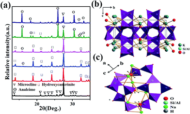 |
| | Fig. 2 (a) XRD patterns of K-feldspar and the solid products formed under various concentrations of Na2SiO3 (RC15, RC-N-1–RC-N-5, from bottom to top); the crystal structure of microcline (b) and analcime (c). | |
The chemical compositions of the solid products at 250 °C under various concentrations are listed in Table 2. When the concentration of Na2SiO3 in solution was in the range of 3.90 to 3.19 mol kg−1, the mass fraction of SiO2 in the solid products was between 45.16 and 55.25% corresponding to the mass fraction of SiO2 in the chemical formula of Na8Al6Si6O24(OH)2(H2O)2 (37.19%) lower than that in NaAlSi2O6·H2O (54.54%). Moreover, the variation of Al2O3 was between 26.44 and 22.35% and Na2O was between 16.60 and 11.00% which is consistent with the mass fraction of Al2O3 and Na2O in the chemical formula of Na8Al6Si6O24(OH)2(H2O)2 (31.6% and 25.6%) higher than that in NaAlSi2O6·H2O (23.18% and 14.09%). Compared to the RC15, the mass fraction of K2O in RC-N-1–RC-N-5 (2.93–4.68%) is far lower than that in RC15 (15.31%), demonstrating that most K2O has been transferred from the solid to liquid.
Table 2 Chemical compositions of the solid products prepared at 250 °C under various concentrations
| Samples |
Concentrations (Na2SiO3)/mol kg−1 |
SiO2/wt% |
Al2O3/wt% |
Na2O/wt% |
K2O/wt% |
| RC15 |
— |
64.8 |
17.35 |
0.66 |
15.31 |
| RC-N-1 |
3.90 |
45.16 |
26.44 |
16.60 |
2.93 |
| RC-N-2 |
3.19 |
48.02 |
23.06 |
14.44 |
4.33 |
| RC-N-3 |
2.85 |
46.27 |
22.40 |
12.66 |
4.68 |
| RC-N-4 |
2.57 |
54.22 |
23.43 |
11.00 |
4.48 |
| RC-N-5 |
2.34 |
55.25 |
22.35 |
11.00 |
4.32 |
Fig. 2b and c present the crystal structures of microcline and analcime. The crystal structure of microcline is made up of a three dimensional framework built by [AlO4] and [SiO4] tetrahedrons, which share their oxygen atoms to form 4 membered-rings along the a axis. There exist larger cavities occupied by cation (K+) with a larger radius among the rings. As shown in Fig. 2c, the crystal structure of analcime consists of 4, 6 and 8 rings built by [SiO4] and [AlO4]. Na+ surrounded by a distorted octahedron is connected with four oxygen atoms and two water molecules located in the continuous channels consisting of [AlO4] and [SiO4] tetrahedrons along the triply screw axis. The structure of hydroxycancrinite has been reported few times so far.
In order to understand the crystal structure of hydroxycancrinite, Rietveld structure refinements of hydroxycancrinite were performed based on the GSAS program using the powder XRD data. Therefore, hydroxycancrinite with a better crystallinity was synthesized by increasing the concentration of Na2SiO3·9H2O, the XRD Rietveld fit pattern of which is displayed in Fig. 3a and the final refined unit cell parameters are summarized in Table 3. It is obvious that all the Bragg diffraction lines of hydroxycancrinite are assigned to the hexagonal system with a space group P63/m. In order to understand the microstructure of this hydroxycancrinite, the TEM and crystal structure are depicted in Fig. 3b and c. The TEM image displayed in Fig. 3b confirms the rod structure of hydroxycancrinite. The crystal structure of hydroxycancrinite is composed of a three dimensional framework consisting of [AlO4] and [SiO4] connected alternately and sharing their corner-oxygen atoms to form 4, 6 and 12 membered rings. The 3-fold axis runs through the center of these rings. The cages formed by the six-membered rings are in parallel with the c axis. Water molecule (O8) is disordered about the 3-fold axis in each cage because of the hydrogen bonding. One interstitial cation site, Na1 atoms with the form of a [Na·H2O]+ cluster are located in the cage on the 3-fold axis and are slightly displaced from the plane of the six-membered rings.29 The other interstitial cation sites, Na2, are situated in the larger channel containing large anionic groups, such as OH−(O7), CO32−, SO42− and H2O.
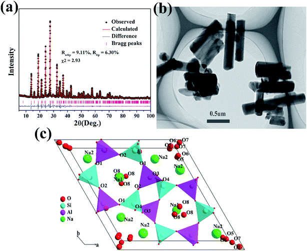 |
| | Fig. 3 (a) Experimental, calculated XRD patterns and their difference for the Rietveld fits of hydroxycancrinite using the GSAS program; (b) TEM of hydroxycancrinite (c) crystal structure of the hydroxycancrinite based on the refinement result. | |
Table 3 The refined positions of all the atoms and the lattice parameters of hydroxycancrinite
| Sample: hydroxycancrinite |
| Lattice constants are: a = b = 12.7705(8) Å, c = 5.1903(0) Å; α = β = 90°; γ = 120°; V = 733.06(9) Å3 |
| Atom |
x |
y |
z |
Occupancy |
| Na1 |
2/3 |
1/3 |
0.1246(5) |
1.00 |
| Na2 |
0.1378(3) |
0.2670(1) |
0.2952(6) |
1.00 |
| Al1 |
0.0762(5) |
0.4196(2) |
0.7503(6) |
1.00 |
| Si1 |
0.3298(1) |
0.4125(1) |
1/4 |
1.00 |
| O1 |
0.2064(3) |
0.4051(4) |
0.6801(2) |
1.00 |
| O2 |
0.1149(6) |
0.5690(5) |
0.7193(2) |
1.00 |
| O3 |
0.0241(6) |
0.3562(4) |
0.0590(7) |
1.00 |
| O4 |
0.3190(4) |
0.3611(5) |
0.0421(9) |
1.00 |
| O5 |
0.0558(7) |
0.1014(5) |
0.6716(6) |
0.17 |
| O6 |
0.0562(4) |
0.1089(7) |
0.9456(1) |
0.17 |
| O7 |
0 |
0 |
0.6902(6) |
0.33 |
| O8 |
0.6105(1) |
0.3061(5) |
0.6863(3) |
0.33 |
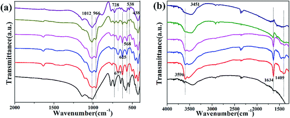
The FTIR spectra of the solid products at 250 °C under various concentrations are given in Fig. 4a and b. Analcime has FTIR wavenumbers of 449, 740, 1026, 1639, and 3620 cm−1 according to the report of Markovic et al.30 As shown in Fig. 4a, there are absorption bands of water molecules at 3569 and 3451 cm−1 broadening and overlapping with each other as the concentration of Na2SiO3 decreases, the reason of which is that there exists –OH in the structure of hydroxycancrinite (Na8Al6Si6O24(OH)2·H2O) but not in analcime (NaAlSi2O6·2H2O). The gradual disappearance of the sharp bands at 1634 cm−1 is also attributed to the impact of –OH. The band appearing at 1012 cm−1 is associated with the T–O (T = Al, Si) asymmetric stretching vibrations and is divided into two bands (1012 and 966 cm−1) in the structure of hydroxycancrinite. The bands at 728 and 438 cm−1 are owed to the existence of T–O (T = Al, Si) symmetric stretching vibrations and the bending vibrational coupling of T–O–T (T = Al, Si) as well as Na–O, respectively. Three stronger bands at 677, 625 and 568 cm−1 of hydroxycancrinite generated by the bending vibration of O–Si(Al)–O grow weaker and weaker as the concentration of Na2SiO3 decreases. In addition, the bands at 538 cm−1 are matched with the structure of microcline, indicating there still exists a little amount of microcline in accordance with the description in Fig. 2a.31
 |
| | Fig. 4 FTIR spectra of K-feldspar and the solid products formed under various concentrations of Na2SiO3 (RC15, RC-N-1–RC-N-5, from bottom to top) (a) 400–2000 cm−1, (b) 1300–4000 cm−1. | |
3.2. Reaction mechanism
The results indicated that the crystal structure of K-feldspar was destroyed under the hydrothermal reaction in our experiment, analcime and hydroxycancrinite were formed owing to the various concentrations of Na2SiO3. The dissolution of feldspar under hydrothermal conditions has been widely studied in recent years.32 Fu et al.33 reported that the dissolution of silicate minerals and the precipitation of secondary minerals are integrated processes in the hydrothermal alteration of rocks. As samples obtained in this paper, the dissolution of K-feldspar as well as the synthesis of analcime and hydroxycancrinite is an integrated process and could not be separated from each other. Gautier et al.34 argued that the Al–O bond was more easily broken than the Si–O bond during the dissolution of K-feldspar. It is agreed that the formation of secondary phases is an interface coupled dissolution–reprecipitation process mainly in the presence of a fluid phase and followed by repolymerization of the relict silica framework.35,36
Reaction of K-feldspar and Na2SiO3 can be described using the following chemical equations:
| | |
Na2SiO3·9H2O → 2Na+ + SiO32− + 9H2O
| (1) |
| | |
4Na+ + 2SiO32− + H2O → Na2Si2O5↓ + 2Na+ + 2OH−
| (2) |
| | |
KAlSi3O8 + 6OH− + 2H2O → K+ + Al(OH)4− + 3H2SiO42−
| (3) |
| | |
8Na+ + 6Al(OH)4− + 18H2SiO42− → Na8Al6Si6O24(OH)2·2(H2O)↓ + 12SiO32− + 10H2O
| (4) |
| | |
Na+ + Al(OH)4− + 2H2SiO42− → NaAlSi2O6·H2O↓ + 4OH− + 2H2O
| (5) |
By analyzing eqn (2), there exists another solid precipitation Na2Si2O5 not shown in Fig. 2a except for hydroxycancrinite and analcime, the reason for which may be that Na2Si2O5 decomposes into Na2SiO3 and SiO2. Every ionic concentration in the filtrate of samples RC-N-1–RC-N-5 is listed in Table 4. The concentration of Al3+ decreased from 10.1157 to 7.0804 mol L−1 while the concentration of Na2SiO3 increased, suggesting that the building of analcime needed more Al than that of hydroxycancrinite. The morphology of the K-feldspar grains and the solid samples generated in varying concentrations of Na2SiO3 is investigated using SEM (Fig. 5a–e). As shown in Fig. 5a, the morphology of the K-feldspar is tabular with no fixed shapes (Fig. 5a). The solid products (RC-N-1–RC-N-3) obtained in the Na2SiO3 concentrations of 3.9–3.19 mol kg−1 show rods with a length of around 2 μm and those obtained in the concentrations of 2.85–2.34 mol kg−1 present tetragonal trisoctahedrons. K-feldspar is dissolved in alkali solution and the ions of Na+, [Al(OH)4]−, SiO32− as well as H2SiO42− are precipitated to hydroxycancrinite (Fig. 5b–d) with a uniform rod morphology. When the concentration of Na2SiO3 is decreased to 2.57 mol kg−1, the concentration of ions is too small to build the structure of hydroxycancrinite, so the tetragonal trisoctahedron analcime (e and f) with a uniform size is formed. Fig. 5g displays the schematic illustration of the reaction process. According to the analysis above, we can conclude that the dissolution of K-feldspar as well as the formation of analcime and hydroxycancrinite is an interface coupled dissolution–reprecipitation process mainly in the presence of Na+, [Al(OH)4]−, [H2SiO4]2− and SiO32 and followed by a repolymerization process of the silicate framework.
Table 4 Ionic concentration in the filtrate of samples RC-N-1–RC-N-5
| Samples |
Concentration (Na2SiO3)/mol kg−1 |
SiO2 (mol L−1) |
Al3+ (mmol L−1) |
Na+ (mol L−1) |
K+ (mol L−1) |
ηK (%) |
| RC-N-1 |
3.90 |
0.4000 |
10.1157 |
0.4839 |
0.0560 |
85.89 |
| RC-N-2 |
3.19 |
0.3833 |
9.3118 |
0.4839 |
0.0494 |
75.77 |
| RC-N-3 |
2.85 |
0.3767 |
7.5863 |
0.5097 |
0.0470 |
72.18 |
| RC-N-4 |
2.57 |
0.3633 |
7.2843 |
0.4839 |
0.0470 |
72.18 |
| RC-N-5 |
2.34 |
0.3550 |
7.0804 |
0.4839 |
0.0470 |
72.18 |
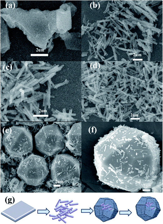 |
| | Fig. 5 SEM images of the K-feldspar and solid products formed under various concentrations of Na2SiO3 (RC15, RC-N-1–RC-N-5) (a–f); (g) schematic illustration of the product changes in the reaction. | |
4. Conclusion
In summary, the effect and mechanism of the concentration of Na2SiO3 on the formation of analcime and hydroxycancrinite were studied in this paper. By the XRD results, it is shown that analcime and hydroxycancrinite were synthesized successively when the concentration of Na2SiO3 decreased. In the structure of hydroxycancrinite, [Na·H2O]+ clusters are formed by Na1 atoms and water molecules (O8) situated in the center of the six-membered rings, as well as Na2 atoms and larger anionic groups OH−(O7) are seated in the larger channel according to the results of the Rietveld analysis. In terms of the analytic results of FTIR, gradual disappearing of sharp absorption bands in 1634 cm−1 is attributed to the vanishment of OH from Na8Al6Si6O24(OH)2·2H2O to NaAlSi2O6·H2O. According to the analysis of the dissolution mechanism, the formation of analcime and hydroxycancrinite is an interface coupled dissolution–reprecipitation process and a repolymerization process of the silicate framework in the presence of Na+, [Al(OH)4]−, [H2SiO4]2− and SiO32−.
Acknowledgements
The present work was supported by the Fundamental Research Funds for the Central Universities (2652014017, 2652015371 & 2652015422).
References
- C. J. Jacobsen, C. Madsen, J. Houzvicka, I. Schmidt and A. Carlsson, Mesoporous zeolite single crystals, J. Am. Chem. Soc., 2000, 29, 7116–7117 CrossRef.
- J. Ju, C. Zeng, L. Zhang and N. Xu, Continuous synthesis of zeolite NaA in a microchannel reactor, Chem. Eng. J., 2006, 2, 115–121 CrossRef.
- Y. Yokomori and S. Idaka, The crystal structure of analcime, Microporous Mesoporous Mater., 1998, 4, 365–370 CrossRef.
- T. Wajima and Y. Ikegami, Synthesis of crystalline zeolite-13X from waste porcelain using alkali fusion, Ceram. Int., 2009, 7, 2983–2986 CrossRef.
- A. Y. Atta, B. Y. Jibril, B. O. Aderemi and S. S. Adefila, Preparation of analcime from local kaolin and rice husk ash, Appl. Clay Sci., 2012, 61, 8–13 CrossRef CAS.
- S. N. Azizi and M. Yousefpour, Synthesis of zeolites NaA and analcime using rice husk ash as silica source without using organic template, J. Mater. Sci., 2010, 20, 5692–5697 CrossRef.
- D. I. Petkowicz, R. T. Rigo, C. Radtke, S. B. Pergher and J. H. dos Santos, Zeolite NaA from Brazilian chrysotile and rice husk, Microporous Mesoporous Mater., 2008, 1, 548–554 CrossRef.
- E. Z. Hegazy, I. H. A. El Maksod and R. A. El Enin, Preparation and characterization of Ti and V modified analcime from local kaolin, Appl. Clay Sci., 2010, 3, 149–155 CrossRef.
- X. Querol, N. Moreno, J. C. Umana, A. Alastuey, E. Hernández, A. Lopez-Soler and F. Plana, Synthesis of zeolites from coal fly ash: an overview, Int. J. Coal Geol., 2002, 1, 413–423 CrossRef.
- S. Tangkawanit, K. Rangsriwatananon and A. Dyer, Ion exchange of Cu2+, Ni2+, Pb2+ and Zn2+ in analcime (ANA) synthesized from Thai perlite, Microporous Mesoporous Mater., 2005, 1, 171–175 CrossRef.
- A. V. Borhade and S. G. Wakchaure, One-pot synthesis, X-ray diffraction and MAS NMR spectroscopic study of gallosilicate nitrate cancrinite Na8[GaSiO4]6(NO3)4(H2O)6, E-J. Chem., 2010, 2, 369–376 CrossRef.
- M. Fechtelkord, B. Posnatzki and B. U. H. L. Josef-Christian, Characterization of basic cancrinite synthesized in a butanediol–water system, Eur. J. Mineral., 2003, 15, 589–598 CrossRef CAS.
- R. M. Barrer, J. F. Cole and H. Villiger, Chemistry of soil minerals. Part VII. Synthesis, properties, and crystal structures of salt-filled cancrinites, J. Chem. Soc. A, 1970, 1523–1531 RSC.
- J. C. Buhl, Synthesis and characterization of the basic and non-basic members of the cancrinite-natrodavyne family, Thermochim. Acta, 1991, 178, 19–31 CrossRef CAS.
- H. W. Ma, Potassic rocks in China: resource and clean utilization techniques, Chemical Industry Press, 2010, pp. 3–13 Search PubMed.
- H. W. Ma, J. Yang, S. Q. Su, M. T. Liu, H. Zheng, Y. B. Wang, H. B. Qi, P. Zhang and W. G. Yao, 20 years advances in preparation of potassium salts from potassic rocks: a review, Acta Geol. Sin. (Engl. Ed.), 2015, 6, 2058–2071 Search PubMed.
- M. Y. Bakr, A. A. Zatout and M. A. Mouhamed, Orthoclase, gypsum and limestone for production of aluminum salt and potassium salt, Interceram, 1979, 1, 34–35 Search PubMed.
- L. Shi and D. S. Chen, Thermodynamic analysis of microcline–CaSO4–CaCO3 system, J. South China Univ. Technol., Nat. Sci., 2007, 5, 94–99 Search PubMed.
- T. X. Hu and J. G. Yu, Experimental study on dissolution of K-feldspar with CaCl2 and NaCl for extraction of potassium, Chin. J. Process Eng., 2010, 4, 701–705 Search PubMed.
- H. W. Ma, S. Q. Su, J. Yang, B. Y. Cai, M. T. Liu, W. G. Yao and H. Peng, Preparation of potassium sulfate from K-feldspar by hydrothermal alkaline method: reaction principle and process evaluation, CIESC J., 2014, 6, 2363–2371 Search PubMed.
- S. Q. Su, H. W. Ma, J. Yang, P. Zhang and Z. Luo, Synthesis of kalsilite from microcline powder by alkali-hydrothermal process, Int. J. Miner., Metall. Mater., 2014, 28, 826–831 Search PubMed.
- Y. Q. Liu, H. T. Xia and H. W. Ma, Kinetics of hydrothermal dissolution of potassium feldspar with calcium hydroxide, Adv. Mater. Res., 2012, 549, 65–69 CrossRef CAS.
- H. W. Ma, J. Yang, S. Q. Su, M. T. Liu, H. Zheng, Y. B. Wang, H. B. Qi and P. Zhang, 20 years advances in preparation of potassium salts from potassium rocks: a review and prospect, Earth Sci. Front., 2014, 5, 236–254 Search PubMed.
- Y. M. Nie, H. W. Ma, H. Liu, P. Zhang, M. Y. Qiu and L. Wang, Reactive mechanism of potassium feldspar dissolution under hydrothermal condition, J. Chin. Ceram. Soc., 2006, 7, 846–850 Search PubMed.
- A. C. Larsen and R. B. Von Dreele, General Structure Analysis System, LANSCE, MSH805, Los Alamos National Laboratory, Los Alamos, NM, 1994 Search PubMed.
- National Standards of the People’s Republic of China for chemical analysis for silicate rocks (GB T/14506) (2011).
- X. Ma, J. Yang, H. W. Ma, C. J. Liu and P. Zhang, Synthesis and characterization of analcime using quartz syenite powder by alkali-hydrothermal treatment, Microporous Mesoporous Mater., 2015, 201, 134–140 CrossRef CAS.
- J. Y. Yuan, J. Yang, H. W. Ma and C. J. Liu, Crystal structural transformation and kinetics of NH4+/Na+ ion-exchange in analcime, Microporous Mesoporous Mater., 2016, 222, 202–208 CrossRef CAS.
- H. D. Grundy and I. Hassan, The crystal structure of a carbonate-rich cancrinite, Can. Mineral., 1982, 2, 239–251 Search PubMed.
- S. Markovic, V. Dondur and R. Dimitrijevic, FTIR spectroscopy of framework aluminosilicate structures: carnegieite and pure sodium nepheline, J. Mol. Struct., 2003, 1, 223–234 CrossRef.
- D. I. Godfrey-Smith and M. Cada, IR stimulation spectroscopy of plagioclase and potassium feldspars, and quartz, Radiat. Prot. Dosim., 1996, 379–385 CrossRef CAS.
- S. Q. Su, H. W. Ma and X. Chuan, Hydrothermal dissolution of K-feldspar in KOH–NaOH–H2O medium, Hydrometallurgy, 2015, 156, 47–52 CrossRef CAS.
- Q. Fu, P. Lu, H. Konishi, R. Dilmore, H. Xu, W. E. Seyfried and C. Zhu, Coupled alkali-feldspar dissolution and secondary mineral precipitation in batch systems: 1. New experiments at 200 °C and 300 bars, Chem. Geol., 2009, 3, 125–135 CrossRef.
- J. M. Gautier, E. H. Oelkers and J. Schott, Experimental study of K-feldspar dissolution rates as a function of chemical affinity at 150 °C and pH 9, Geochim. Cosmochim. Acta, 1994, 21, 4549–4560 CrossRef.
- R. Hellmann, R. Wirth, D. Daval, J. P. Barnes, J. M. Penisson, D. Tisserand and R. L. Hervig, Unifying natural and laboratory chemical weathering with interfacial dissolution–reprecipitation: a study based on the nanometer-scale chemistry of fluid–silicate interfaces, Chem. Geol., 2012, 294, 203–216 CrossRef.
- H. W. Nesbitt and W. M. Skinner, Early development of Al, Ca, and Na compositional gradients in labradorite leached in pH 2 HCl solutions, Geochim. Cosmochim. Acta, 2001, 5, 715–727 CrossRef.
|
| This journal is © The Royal Society of Chemistry 2016 |
Click here to see how this site uses Cookies. View our privacy policy here. 


