Maxime Fréneauab,
Norbert Hoffmannb,
Jean-Pierre Vorsc,
Pierre Genixc,
Claire Richardad and
Pascal de Sainte Claire*ae
aEquipe Photochimie CNRS, UMR 6296, ICCF, F-63178 Aubière, France
bCNRS, Université de Reims Champagne-Ardenne, ICMR, Equipe de Photochimie, UFR Sciences, B.P. 1039, 51687 Reims, France
cBayer S.A.S, 14 Impasse Pierre Baizet, 69263 Lyon, France
dUniversité Clermont Auvergne, Université Blaise Pascal, Institut de Chimie de Clermont-Ferrand, BP 10448, F-63000 Clermont-Ferrand, France
eUniversité Clermont Auvergne, SIGMA Clermont, Institut de Chimie de Clermont-Ferrand, BP 10448, F-63000 Clermont-Ferrand, France. E-mail: pascal.de_sainte-claire@sigma-clermont.fr; Tel: +33 4 73 40 76 27
First published on 28th June 2016
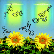
The competitive photoisomerization and photodegradation reactions of a tetrazoline oxime ether showing important fungicidal activity are investigated by Time Dependent-Density Functional Theory (TD-DFT) and Complete Active Space Self-Consistent Field (CASSCF) calculations of ground state and excited state (singlet, S and triplet, T) potential energy surfaces. Two key experimental results reported previously are explained in this study: (1) why only E isomers undergo N–O bond scission and photodegradation and (2) what is the mechanism for deactivation of the Z isomers. In addition, the reactive pathway that involves intersystem crossing is shown to be competitive with reactions in the singlet state. Our results demonstrate that the photoreactivity (photodegradation or photoisomerization) of the eight conformers studied here is clearly related to the differences between the respective electronic configurations of the lowest singlet excited state. It is shown that the addition of a bulky substituent on the pyridyl group prevents E isomers from being photodegraded.
Our recent results have shown that (1) the lower absorptivity of E compared to Z favors its accumulation and (2) the photodegradation proceeds exclusively from E (Z undergoes only photoisomerization) and starts by the cleavage of the N–O bond into iminyl and alkoxyl radicals (see Fig. 2). These results explained the experimental loss of biological activity of this species. In this study, the photoisomerization and photodegradation mechanisms are discussed on the basis of theoretical calculations in the ground state and low-lying excited states.
![]() | ||
Fig. 2 The photoreaction scheme for Z (Ph = phenyl, Tz = tetrazole, Py = pyridyl, R = NHCOOC4H5). Experimental1 rate constants are kZ→E = 0.13 min−1, kE→Z = 0.07 min−1, kE→Photoproducts = 0.011 min−1. |
The simpler model structure Z′ (see Fig. 1) was also studied in order to investigate the role of the pyridyl group (Py) in Z.
The photoisomerization17 of conjugated isomers generally proceeds through intersystem crossing (isc) via T1 or adiabatic singlet deactivation (see Fig. 3). In the latter case, S1 reaches the conical intersection (CI) between S0 and S1. Both isomers are obtained in this manner. The S1 state may have n/π*18 or π/π* character.19–23 In case of isc, the triplet state T1 is reached from S1. The geometry of the minimum energy structure in T1 is one where δ ∼ 90 deg. (see Fig. 1 for definition of δ). Z and E isomers are obtained through further T1 → S0 isc deactivation.24
![]() | ||
Fig. 3 The photoisomerization mechanism. The conical intersection (S0/S1) is labeled CI. Dihedral values are given as indicative purposes. Arrows describe the singlet state mechanism. See Fig. 1 for definition of δ. |
The photodegradation of oxime species may also compete with the photoisomerization mechanism. Experimental analyses1,3,10–15 and theoretical studies18 of photodegradation products gave evidence of N–O bond dissociation as one of the main degradation pathway. The photodissociation mechanism of the N–O bond can proceed through conical intersections that arise from the diabatic crossing of excited singlet states that have significantly different electronic character (π/π* and σNO/).18 If a local minimum energy structure is found in the S1 state, an activation energy barrier is expected for N–O bond dissociation. Photodissociation may also be obtained through intersystem crossing. Finally, photocyclization products were also reported in photoexcitation experiments of conjugated photoisomers.23,25
S0a | S1(FC)b | λ(S1)c | f(S1)d | λ(S2)c | f(S2)d | PhRe | S0a | S1(FC)b | ||
---|---|---|---|---|---|---|---|---|---|---|
a Energies in kcal mol−1. Reference is the most stable species (Z-b, E-a or Zm-b). The relative weight of each conformer is given in parentheses. See text for calculation method.b Energy in kcal mol−1 of the S1 state for the Franck–Condon structure.c Energy level in nm of the S1 and S2 states for the Franck-Condon structure.d Oscillator strengths of S1 and S2.e Photoreactivity: I for photoisomerization and P for photodegradation. | ||||||||||
Z-a | 0.61 (13.7%) | 87.80 | 328 | 0.0005 | 287 | 0.2658 | I | Zm-a | 1.93 (2.0%) | 79.47 |
Z-b | 0.0 (38.3%) | 100.22 | 285 | 0.3813 | 277 | 0.0112 | I | Zm-b | 0.0 (52.1%) | 94.18 |
Z-c | 0.62 (13.4%) | 96.76 | 297 | 0.0126 | 285 | 0.4390 | P | Zm-c | 1.15 (7.5%) | 87.35 |
Z-d | 0.06 (34.6%) | 98.87 | 289 | 0.4713 | 276 | 0.0019 | I | Zm-d | 0.18 (38.4%) | 93.99 |
E-a | 0.0 (68.4%) | 97.61 | 293 | 0.0657 | 284 | 0.1819 | P | |||
E-b | 0.69 (21.4%) | 97.16 | 296 | 0.1390 | 285 | 0.1357 | P | |||
E-c | 1.51 (5.4%) | 97.06 | 289 | 0.2087 | 283 | 0.0076 | I | |||
E-d | 1.58 (4.8%) | 101.55 | 286 | 0.1906 | 281 | 0.0040 | I |
![]() | ||
Fig. 4 The low energy structures of the Z isomers in the ground state at the B3LYP/6-31+G(d,p) level. (b) is the most stable Z isomer (see Table 1). The N*–H* bond distance is 2.270 Å in structure (a) (see also Fig. 1). The CH3–NPy bond distances are 2.825 Å and 3.015 Å in (b) and (d), respectively. |
![]() | ||
Fig. 5 The low energy structures of E isomers in the ground state at the B3LYP/6-31+G(d,p) level. (a) is the most stable isomer (see Table 1). The NPy–HPh bond distance in structure (c) is 3.538 Å. |
![]() | ||
Fig. 6 Experimental and theoretical UV spectra of Z (top) and E (bottom) isomers, and position of electronic excited states of the most stable isomers, i.e. Z-b (top) and E-a (bottom). Oscillator strengths are represented by vertical lines. The positions of S1 and S2 are also indicated. Theoretical spectra are averaged over the isomers according to their respective weight (see Table 1). |
Finally, it was found that the electronic configuration of S1 was similar for Z-a and Z-c on the one hand, and Z-b and Z-d on the other. The phototransformation of all these conformers will be studied in this work.
Thus, the eight isomers investigated in this work (Fig. 4 and 5) can be separated into two sets: one where the first excited singlet state arose from promotion of a Py electron to the Ph/Ox/Tz part of the molecule, thus an electronic transfer through the oxime linkage (Z-a, Z-c, E-a, E-b), and the second group (Z-b, Z-d, E-c, E-d) where S1 was characterized by an electronic transition that remained on the Ph/Ox/Tz part of the species. This is illustrated by the oscillator strengths given in Table 1. Large f(S1) and small f(S2) characterize species that belong to the second group (Z-b, Z-d, E-c, E-d), whereas small f(S1) and large f(S2) characterize species that belong to the first set (Z-a, Z-c, E-a, E-b; note that in E-b both S1 and S2 involve excitation from orbitals that have Py and Ph/Ox/Tz character, thus large f(S1) and large f(S2)). Thus, different phototransformation mechanisms are expected for the two sets of species. Last, although S1 was similar for Z-a and Z-c/E-a/E-b, it will be shown below that Z-a reacted singularly because of the hydrogen bonding between the Py and Tz parts of the molecule.
Finally, the model molecule Z′ was also studied to investigate the role of the Py substituent on the phototransformation mechanisms.
Assuming photodegradation is governed by the reactivity of the first excited state, the difference of photoreactivity observed between Z and E isomers is strongly dependent on S1 electronic configuration, as discussed in the next section.
Optimization of Z-c in S1 yielded a minimum-energy structure with significant increase of the N–O bond (RN–O(S0) = 1.390 Å and RN–O(S1) = 1.477 Å). The Minimum Energy Path (MEP) was followed for fixed N–O distances and interpolated in order to estimate the energy barrier for bond dissociation and thus photodegradation. The electronic energy barrier was 6.8 kcal mol−1 at the TD-B3LYP/6-31+G(d,p) level (see ESI†). Thus, rapid photodegradation through N–O bond scission is expected for Z-c. This conformer amounts to 13.4% of the Z isomers (see Table 1).
Z-a reacted singularly in S1 because of significant hydrogen bonding between the Py and Tz parts of the molecule (see Fig. 4a). Considering that the electronic configuration of S1 was similar for Z-a and Z-c, Z-a was expected to give photodegradation products through N–O bond scission. However, optimization of Z-a in the S1 state yielded a structure where the hydrogen atom H* in Fig. 1 was shifted to N* (Tz). S0 and S1 are close-lying in this region of the PES, and the DFT method is not suited to identify the respective stationary point. State averaged CASSCF calculations were performed on Zm (active space molecular orbitals are given in ESI section†) to explore carefully this region of the PES. CAS(8,7)/6-31G optimization of Zm-a yielded a biradical minimum energy structure where the N–O and CN bonds slightly increased by resonance effects (see Fig. 7). Optimization of this structure in S0 yielded the Z isomer Zm-a (see geometries in ESI†). Thus, the photoexcitation of Z-a is formally unreactive.
![]() | ||
Fig. 7 The photoreaction path of Zm-a. Zm-a(S1) is the biradical minimum structure obtained at the CAS(8,7)/6-31G level in the S1 state. Cartesian coordinates of Zm-a(S1) are given in ESI† section. Hydrogen bond length between N* and H* (see Z in Fig. 1) is also indicated. The N–O and C![]() |
Last, optimization of the Z isomers in T1 yielded the expected minimum energy structure (δ ∼ 90 deg.; see Fig. 3 and ESI† for geometries). Photoisomerization is thus expected in case of significant intersystem crossing.
In conclusion, only a minor part of the Z isomers (Z-c) undergoes photodegradation, as found in our recent experimental study.1
Optimization of E-b in S1 gave a minimum energy structure with a geometry similar to E-a. The structure of E-b is roughly obtained by 180 deg. rotation of the NOxOOx–CCPy dihedral angle in E-a. The NO bond length is large as well (RN–O(E-b) = 1.491 Å). In addition, electronic configurations of the respective S1 states were similar for E-a and E-b (i.e. Py → Ph/Ox/Tz electronic transition). Thus similar photoreactivity is expected for E-a and E-b.
Contrarily, excited state optimization of E-c and E-d yielded the CI geometry in Fig. 3. Thus, photoisomerization is the preferred pathway for E-c and E-d, as expected from examination of electronic configurations (Ph/Ox/Tz localized).
All these results provide a coherent picture for the photodegradation of the oxime species investigated in this work and may be summarized as follows: isomers react according to the specificity of their first excited electronic state. If S1 is characteristic of an electronic transition that spans regions of the molecule that are separated by the oxime bond (here Py and Ph/Ox/Tz), photodegradation is observed. Contrarily, if it is localized on the Ph/Ox/Tz substituents, photoisomerization is the preferred pathway. According to our results, Z-a, Z-b and Z-d undergo photoisomerization, i.e. 86.6% of the Z isomers (see Table 1). Contrarily, E-a and E-b are photodegraded through N–O bond scission, thus 89.8% of the E isomers (see Table 1). This is in excellent agreement with our previously reported experimental results.1
Full optimization and relaxed N–O scan calculations were also performed to investigate the reactivity in the T1 state of the most abundant E isomer E-a. The initial geometry was that of the minimum energy structure found in S1. T1 state optimization yielded the CI structure (Fig. 3), thus the photoisomerization pathway. Note that T2 and S1 are nearly degenerate in the region of the minimum energy structure in S1, and an intersystem crossing mechanism may involve T2, i.e. S1/T2 isc and T2/T1 relaxation. Nevertheless, the relaxed N–O scan in the T1 state in the direction of increased bond length showed that an excess energy of 3.4 kcal mol−1 (from isc) was sufficient to overcome the N–O dissociation barrier. Thus, photodegradation from the triplet state may not be ruled out. The experimental ratio of the photoisomerization and photodegradation rate constants for the E isomer was kE→Z/kE→Photoproducts = 6.4 in continuous irradiation conditions.1 Our theoretical results show that the preferred pathway in S1 is photodegradation, while photoisomerization (through isc) is favored from T1. The experimental rate constants indicate slow photodegradation in comparison to isc. However, although photodegradation is a slow process, reactants will inevitably undergo complete photodegradation in continuous irradiation conditions, in agreement with our calculations.
(1) the S1 electronic configuration at the ground state minimum energy structure (mesh area in Fig. 8) is consistent with the above theoretical results for all the conformers: E-a, E-b and Z-c underwent photodegradation (i.e. N–O bond scission; red line is highest in Fig. 8 for the respective geometries), while E-c, E-d, Z-b and Z-d followed the isomerization pathway (blue line is highest).
(2) The πOx → diabatic state (blue line) exhibits small changes in comparison to the other state. Thus, it is interesting to understand the origin of the variations of the πPy →
state. First, in all cases, for δ3 ∼ ±180 deg. (see Newman representations in Fig. 8, far-right and far-left), S1 and S2 belong to πPy − πOx →
and πPy + πOx →
transitions, respectively (πPy − πOx and πPy + πOx are antibonding and bonding, respectively, between Py and the CN bond of the oxime group). In this configuration, the Py plane bisects the N–O bond, thus allowing mixing of the πPy and πOx orbitals. Destabilization of πPy − πOx (S1), is obtained in this case. A similar effect, although smaller, is also observed for δ3 ∼ 0 deg. (NPy pointing toward the N–O bond): the MO is more diffuse on C** (see Fig. 1) than on NPy (see Fig. SI-8†), and the variation of the respective diabatic state in this region is not as important.
(3) strong destabilization of the other diabatic state is seen for δ3 ∼ ±60 deg. (red line in Fig. 8). In this geometric configuration, the CPy–NPy and N–O bonds parallel each other, with NPy pointing toward the oxime bond for δ3 ∼ +60 deg. (see Newman representations in Fig. 8 for Z-d). The πOx MO is destabilized by the antibonding contribution of the lone pair on NPy and the σ(CH2–CPy) bond on the one hand, and πOx on the other (this MO is shown in Fig. SI-9† for E-a).
In this study, the experimental photoreactivity of oxime species Z was explained on a theoretical basis. Structures where δ3 ∼ ±180 deg. are bound to undergo photodegradation. This effect can be prevented by addition of a bulky substituent on C** (Py cycle, see Fig. 1) because electronic repulsion between this substituent and the Ox/Ph/Tz part of the molecule favors conformations where −120 < δ3 < 120 deg. Moreover, conformations where CPy–NPy is parallel to the N–O bond (e.g. δ3 = −69 deg. in E-c, or δ3 = +66 deg. in E-d) significantly enhances the probability of photoisomerization. This effect is achieved by hydrogen bonding between (1) NPy and the substituent borne by Tz for the E isomers (methyl in our case) or (2) NPy and Ph for the Z isomers. In Z-c, the methyl substituent on Tz and the Py group are on opposite sides of the CN–O–C plane. Contrarily, in Z-b, these substituents are on the same side of the C
N–O–C plane. Hydrogen bonding is stabilizing and Z-b is more stable than Z-c. The addition of a hydrogen bonding substituent on Ph is necessary if significant photodegradation is detected from the Z isomers.
Last, S1 optimization of Z′ (Fig. 1) followed the photoisomerization pathway to the conical intersection structure CI in Fig. 3. This was expected because the Py group was not present in this species. Optimization of E′ in the S1 state leaded to the conical intersection (CI in Fig. 3) and photoisomerization.
Investigation of the photoreactivity in the triplet state showed that photodegradation of E isomers may not be ruled out if excess energy released from isc is available to cross the small activation barrier in T1.
Footnote |
† Electronic supplementary information (ESI) available. See DOI: 10.1039/c6ra07712a |
This journal is © The Royal Society of Chemistry 2016 |