DOI:
10.1039/C6RA07681E
(Paper)
RSC Adv., 2016,
6, 65756-65762
POSS–PU electrospinning nanofibers membrane with enhanced blood compatibility
Received
24th March 2016
, Accepted 20th June 2016
First published on 22nd June 2016
Abstract
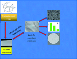
Open cage polyhedral oligomeric silsesquioxane (POSS) was firstly used to modify polyurethane (PU) to prepare a POSS–PU composite. Then a POSS–PU nanofiber membrane was prepared by electrospinning technology. The hydrophilic/hydrophobic properties, fiber morphology and biocompatibility of the POSS–PU nanofibers membrane were investigated. Contact angle increased by 24.3° for 2 wt% POSS–PU nanofibers membrane compared to PU. The ability of the nanofibers membrane surface to repel proteins and platelets was assessed by using platelet adhesion test and BSA static protein-adsorption experiment. Platelet adsorption amount obviously decreased compared with PU and very few platelet was adhered on the surfaces of nanofibers membranes when 1 wt% POSS was added. Protein adsorption was also decreased with addition of POSS. Hemolysis tests showed that hemolysis rate localized in the desired range of values (<5%) for all nanofibers membrane. Moreover, the antibacterial activity of nanofibers membranes was obviously improved after addition of POSS.
1 Introduction
Polyurethane (PU) is widely used in biomedical applications for artificial organs, medical devices and disposable clinical apparatus due to its relative ease of fabrication into devices, excellent mechanical properties and blood biocompatibility.1–4 However, plasma proteins and platelets are rapidly adsorbed on PU when it comes into contact with blood.5,6 Therefore, many efforts are made to improve the surface biocompatibility of PU by chemical modification or the introduction of surface active additives. Chen et al. directly coated carbon nanotube arrays (ACNT) with fluorinated poly(carbonate urethane) (FPCU), leading to a nanostructured biomaterial surface of low surface free energy. Compared to a flat FPCU surface, almost no platelet adhered on the nanostructured hydrophobic surface.7 Hsiao et al. prepared the pp-HC (the film consisting of hexamethyldisiloxane and fluorocarbons) to modify the surface of polyurethane (PU). The superhydrophobic pp-HC-coated PU exhibited extremely low platelet adhesion even after a long incubation time.8
It was demonstrated that silicon polymers have both anti-protein and anti-platelet functions.9 However, common silicon-containing vessels are rigid and have low elasticity with a tendency to form intimal hyperplasia more rapidly.10 Unlike other common silicon molecules, polyhedral oligomeric silsesquioxanes (POSS) with 1–3 nm in diameter can be used to prepare organic–inorganic hybrid materials with excellent viscoelastic properties.11–13 Therefore, biomaterials containing POSS may overcome disadvantage of common silicon-containing biomaterials. It is known that POSS have the bioactivity and other physical properties of porous Si as a nanoscale hollow cage structure of Si.14 It has been also demonstrated that POSS have excellent biocompatibility.15–17 The POSS consisting of Si–O–Si and Si–C groups has similar chemistry to silicone elastomers (poly(dimethylsiloxane), PDMS). In particular, owing to the biocompatibility and ability of POSS to incorporate additional polymers, POSS nanostructures have been shown to offer high potential in several biomedical applications such as drug delivery systems, dental composites, biosensors, biomedical devices, antibacterial agents and tissue engineering products.18 The incorporation of POSS into PU as building block or pendent-cage fillers can reduce platelet and protein adsorption to PU due to the decrease of surface tension.19–23 It is known that hydrophobic surface with low surface energy resists the adsorption of blood proteins and platelet adhesion.24–27 The trans-cyclohexanediol isobutyl-POSS as the “pendant-type” was attached to polyurethane and the composite was used as heart valve leaflet and artificial capillary bed.10,21,27
Previous researches of POSS/PU hybrid composite all used the enclosed-cage POSS.28 However, the effect of POSS with open cage-like on the blood compatibility of the PU electrospun nanofibers membrane is few researched. Polymer in electrospun form is a promising candidate for tissue engineering,29 wound dressings,30 high performance filters,31 vascular grafts and other applications32–35 due to its intrinsically high specific surface area, in-principle extreme length, high surface-area and tunable porosity, intrinsic three-dimensional (3D) topography, and functional properties.36
Aromatic polyurethane based on 4,4′-diphenylmethane diisocyanate (MDI), polytetramethylene glycol (PTMEG) and 1,4-butanediol (BDO) has good biological compatibility and mechanical properties.37 Moreover, these materials are cheap and easy to get. The object of this paper is to prepare PU nanofibers membrane with improved biocompatibility by incorporating very small amounts of reactive POSS molecules as an integral part of polyurethane chain segments. In this study, open cage-like trisilanolisobutyl POSS was firstly covalently attached with the PU backbone.38,39 Then POSS–PU nanofibers membrane was prepared by electrospinning technology. Furthermore, the platelet adsorption, protein adsorption and antibacterial property on the POSS–PU nanofibers membrane were evaluated.
2 Experimental
2.1 Materials
Polytetramethylene glycol (PTMEG, Mn = 2000), 4,4′-diphenylmethane diisocyanate (MDI, purity 98%) and 1,4-butanediol (BDO, purity 99%) were obtained from Aladdin. N,N-Dimethylformamide (DMF) and tetrahydrofuran (THF) were purchased from Tianjin Kermio Chemical Reagent corporation. Trisilanolisobutyl POSS was received from Hybrid Plastics. N,N-Dimethylformamide (DMF), tetrahydrofuran (THF) and 1,4-butanediol (BDO) were further purified by distillation.
2.2 Synthesis of POSS–PU composites with different POSS content
PTMEG (50 g) and trisilanolisobutyl POSS (0.7 g) were added into a 250 mL three-necked round bottomed flask equipped with a mechanical stirrer. The mixture was heated to 110 °C to dissolve POSS and dehydrate under vacuum for 2 h. MDI (19.04 g) was added after the mixture was cooled to 60 °C. The reaction solution was stirred at 80 °C under a nitrogen atmosphere for 2 h to obtain a prepolymer. The NCO group content was then determined and chain extension was carried out by the addition of desired amounts of BDO (4.85 g). The mixture was poured into the mould and cured at 100 °C for 12 h to prepare POSS–PU (1 wt% POSS) nanocomposites. POSS–PU nanocomposites with different content of POSS was prepared by adjusting the content of POSS. POSS–PU nanocomposite with 0 wt%, 1 wt% and 2 wt% POSS were denoted as PU, 1% POSS–PU and 2% POSS–PU, respectively.
2.3 POSS–PU nanofiber membrane preparation
POSS–PU homogeneous spinning solution with different POSS content were formed by POSS–PU dissolving in DMF/THF (w/w = 1
:
2). Then neat PU and POSS–PU nanofibers membrane were fabricated under the optimal electrospun conditions of 12% POSS–PU solution concentration, 0.8 mL h−1 extruding speed, 20 kV voltage and 20 cm tip-to-collector distance. POSS–PU nanofibers with 0 wt%, 1 wt% and 2 wt% POSS were denoted as PU0, PU1 and PU2, respectively.
2.4 Characterization
2.4.1 Determination of POSS–PU molecular weight. The molecular weights of the POSS–PU nanocomposites with different POSS content were determined by GPC. The columns were connected to a refraction index detector using THF as solvent, and the calibration was based on polystyrene standards.
2.4.2 Fourier infrared (FTIR) test of nanofiber membrane. A VECTOR 22 FTIR spectrometer was used at room temperature to characterize the molecular vibration of the hybrid films within the range 400–4000 cm−1. The nano fiber membrane was tested by the attenuated total reflection mode.
2.4.3 SEM characterization of nanofiber. Surface morphology and structure of nanofiber membrane was observed by FEI QUANTA 200 scanning electron microscope. Image-Pro Plus 6 software was employed to measure the fiber diameter. Average fiber diameter was calculated by measuring the diameter of 100 fibers at the different positions on SEM photos.
2.4.4 TEM characterization of nanofiber. The phase morphology of nanofiber membrane was characterized by JEM 2100 transmission electron microscope. The samples were mounted on Cu TEM grids.
2.4.5 Water contact angle measurement. The hydrophilic/hydrophobic property of the nanofiber membrane was evaluated by the sessile drop method using a video contact angle instrument (JY-82 contact angle measurement) at room temperature. The contact angle was calculated with the drop shape analysis. Each sample was measured at five different locations and the contact angle values were averaged.
2.4.6 Platelet adhesion test. Fresh blood of human was centrifuged at 2000 rpm for about 15 min to prepare platelet-rich plasma (PRP) for the platelet adhesion experiment. The nanofiber membrane were placed in 24-well tissue culture plate and immersed in PBS (pH 7.4) for 12 h. After removing PBS, the membranes were incubated at 37 °C for 60 min in new wells containing the PRP. After being rinsed with PBS three times to remove any non-adhered platelets, the disks were transferred into new wells and fixed for 30 min in 2.5% glutaraldehyde, then the platelets adhering to the samples were dehydrated in an ethanol grade series (50%, 60%, 70%, 80%, 90% and 100%) for 30 min. After each membrane was allowed to dry at room temperature, the disks were coated with gold for examination in FEI QUANTA 200 scanning electron microscope (SEM).
2.4.7 Hemolysis test. The hemolysis experiment was carried out in the following steps.40–42 3.8 wt% sodium citrate solution was added to 2 mL fresh human blood at 1
:
9 ratio, then anticoagulant human blood was diluted with 0.9% sodium chloride solution by 1
:
1.25 ratio. Each of the three fiber membrane types was dipped into 15 mL centrifuge tubes containing 10 mL 0.9 wt% sodium chloride solution previously incubated in a water bath at 37 °C for 30 min. Next, 0.2 mL of the aforementioned diluted blood was added to the tubes, and the mixture was incubated at 37 °C for 60 min. A solution of 0.9 wt% sodium chloride and 10 mL deionized H2O were used as negative and positive controls, respectively. After incubation, all the test tubes were centrifuged at 1000 rpm for 10 min. The supernatant liquid was taken into the cuvette, and the absorbance was measured at 545 nm wavelength by spectrophotometer. The percent of hemolysis was calculated as the following equation.
| Hemolytic rate (%) = (X1 − X3)/(X2 − X3) × 100% |
where X1, X2 and X3 are the absorbance values of the test sample, positive control (water), and negative control (sodium chloride solution), respectively.
2.4.8 BSA static protein-adsorption experiments. For static protein-adsorption tests,43–45 1 mg mL−1 BSA solution was prepared in PBS (pH 7.4). Fiber membranes were cut into small pieces (1 cm × 1 cm) and immersed in 10 mL 1 mg mL−1 BSA solution in a test tube. BSA adsorption was conducted under vibration at 37 °C for 3 h to allow for adsorption equilibrium. The experiments were performed with three measurements for each sample. BSA content was measured using a spectrophotometer at a wavelength of 280 nm and then the amount of adsorbed BSA on membranes was calculated. For each membrane, the reported data is the average of three measurements.
2.4.9 Antibacterial assay. The antibacterial property of the fibers membrane was tested by using E. coli. The bacteria were cultivated in liquid lysogeny broth (LB) medium (containing 10 g L−1 peptone, 5 g L−1 yeast extract and 10 g L−1 sodium chloride, pH = 7.0) and then placed in an incubator-shaker overnight at 37 °C. Following that, the E. coli (1 mL) was pipetted from the overnight phase into another flask containing 50 mL of freshly prepared LB. The mixture was then cultured at 37 °C in an incubator shaker for another 5 h to obtain bacterial suspension at exponential growth phase. This was done because the bacterial suspension at exponential growth phase was believed to have higher activity and be more viable than other growth phases. Then, the bacterial suspension was diluted with the LB liquid culture medium to 1 × 10⁶ colony forming units (CFU) per mL. 30 μL of the diluted bacterial suspension was pipetted out from the flask and spread onto the surfaces of electrospun fibers membrane. After placing the membranes on the nutritive agar plate, the plates were sealed and incubated at 37 °C for 12 h. Finally, the numbers of the CFU on membrane surface were counted.
3 Results and discussion
3.1 The synthesis and characterization of POSS–PU
POSS–PU prepolymer was obtained by polycondensation reaction between PTMEG, trisilanolisobutyl POSS and MDI. Then 1,4-butanediol was added into prepolymer and POSS–PU nanocomposite was prepared by chain extension (Fig. 1). POSS–PU nanocomposite can dissolve in DMF or THF. Only part hydroxyl groups of trisilanolisobutyl POSS take part in the polymerization due to steric hindrance of POSS. The weight-average (Mw) and number-average (Mn) molecular weights of the POSS–PU hybrids are listed in Table 1. When the POSS concentration is 1 wt%, the molecular weight of POSS–PU increase compared to PU homopolymer, indicating that the introduction of POSS can improve the molecular weight of polyurethane due to extender chain effect of POSS.14 Notably, the molecular weight of POSS–PU is lower than 1% POSS–PU when the POSS concentration increases to 2 wt%. The reason that molecular weight of POSS–PU with 2 wt% POSS is lower than that of POSS–PU with 1 wt% POSS may be attributed to the stronger steric hindrance and aggregation of POSS in higher concentration during the polymerization process,38 which hindered the full extension of POSS molecules.
 |
| | Fig. 1 The synthetic route of POSS–PU. | |
Table 1 The molecular weight of POSS–PU with different POSS concentrations
| Sample |
Mn (g mol−1) |
Mw (g mol−1) |
Mw/Mn |
| PU |
6062 |
29 105 |
4.829 |
| 1% POSS–PU |
14 303 |
53 080 |
3.711 |
| 2% POSS–PU |
8944 |
20 772 |
2.322 |
3.2 Fourier transform infrared spectroscopy of nanofibers membrane
The FTIR spectra of the PU0, PU1 and PU2 are shown in Fig. 2. The characteristic peak of isocyanate group (–NCO stretching, 2270 cm−1) disappears, demonstrating the complete reaction of isocyanate groups. The characteristic absorption bands of all the nanofibers membrane are at 3400 to 3230 cm−1 (N–H stretching) and 1702 cm−1 (C–O stretching). The absorption band of C–O–C is at 1102 cm−1 for the PU0, while the absorption peak slightly shift to the left due to the overlap of C–O–C and Si–O–Si peak.39 The absorption bands of N–H in the PU0 is at 1532 cm−1, while the blue shift of absorption bands occurs after the addition of POSS, indicating the decrease of urea bond.46 The band at 991 cm−1 is attributed to the Si–OH,47,48 confirming that not all hydroxyl groups of trisilanolisobutyl POSS take part in the polymerization, which consists with the result that silsesquioxane skeletal deformation band is present at 560 cm−1.
 |
| | Fig. 2 FTIR spectra of (a) PU0, (b) PU1 and (c) PU2. | |
3.3 Morphology analysis of nanofibers membrane and phase morphology analysis of nanofibers with different POSS
Electrospun nanofibers membranes with different POSS concentrations were prepared after optimizing the spinning condition. The SEM images and corresponding fiber diameter distributions of nanofibers membrane are shown in Fig. 3. It can be found that uniform and smooth nanofibers without beads are obtained and they randomly distribute to form the fibrous web, indicating that the presence of POSS does not affect the spinnability of the solutions. The average fiber diameters of PU0, PU1 and PU2 are 890 nm, 670 nm and 840 nm, respectively. The reason that POSS can decrease the fiber diameter is related to both the parameters of the electrospinning process and the nature of the PU solution including rheological behavior, surface charge density of polymer solution and solvent characteristics.49 Besides, POSS diminishes the surface tension of liquids that is responsible to the solution splits into fine fibers.50,51 Moreover, POSS containing Si atoms can increase the conductivity of the solutions to promote the elongation force of the ejected jets and favor the formation of thinner nanofibers.52 Therefore, the electric conductivity is also an important factor on fiber diameter. It can be also seen that aggregation of POSS appears on surface of the PU1 and PU2. Moreover, the aggregation of POSS is more obvious with the increasing of POSS content, which is consistent with the previous results.53–55
 |
| | Fig. 3 SEM images and corresponding fiber diameter distributions of nanofibers with different POSS content (a and d) is PU0, (b and e) is PU1, (c and f) is PU2. | |
In order to characterize the structural features of electrospun nanofibers and the dispersity of POSS in PU nanofibers, TEM was performed. TEM micrographs of PU0, PU1 and PU2 are shown in Fig. 4. It can be seen that the surface of nanofibers is uniform and smooth. More aggregated POSS particles can be observed in PU2 compared with PU1, which is consistent with the results of SEM.
 |
| | Fig. 4 TEM micrographs of PU0, PU1 and PU2. | |
3.4 Effect of POSS on the hydrophilic/hydrophobic properties of nanofibers membrane
Hydrophobic and hydrophilic characteristics of polymer surface have an important influence on haemocompatibility that directly affects the interaction between the material surface and the biological system. Contact angle is considered to determine the wettability of material. Contact angles of PU0, PU1 and PU2 are shown in Fig. 5. The results indicate that the content of POSS had a major impact on the surface of electrospun fibers membrane. Water contact angle of PU2 is 132.9° that is much larger than 108.6° of PU0, which originates from the presence of the weak polarity and distinctly hydrophobic POSS molecules in the polymer.10,56
 |
| | Fig. 5 Water contact angles of PU0, PU1 and PU2 electrospun nanofibers membrane. | |
3.5 Hemolysis properties of nanofibers membrane
Hemolysis of blood is an important parameter of blood compatibility for implant material. Good blood compatibility not only has low platelet adsorption but also no hemolytic phenomenon. Hemolysis induces the release of hemoglobin into plasma due to the damage of erythrocytes membranes, which directly affects the blood compatibility of material. The disruption of erythrocytes caused by materials is also associated with the plasma protein adsorption on the material surfaces. The results of hemolysis rate are shown in Fig. 6. Hemolysis rate indicates the extent of erythrocytes lysed after the sample contacts with whole blood. The smaller the hemolytic rate is, the better the blood compatibility of the biomaterials is. Hemolysis rate of the ideal biomaterials should be less than 5%.57 It can be observed that the hemolysis rate reduces when incorporating of the POSS to polyurethane.
 |
| | Fig. 6 Hemolysis rate of the nanofibers membrane in contact with blood in vitro. | |
3.6 Protein adsorption and platelet adhesion on the nanofibers membrane
Plasma protein adsorption is a key parameter to determine the thrombogenicity of materials. The human plasma contains a variety of proteins including serum albumin (HSA 45 mg mL−1), total immunoglobulin G (IgG 10 mg mL−1), fibrinogen (Fib 3 mg mL−1), transferrin (Tr 3 mg mL−1) and total immunoglobulin A (IgA 1 mg mL−1).58 When blood contacts with the material, protein adsorption firstly occurs, which can provoke the adhesion of platelets, white blood cells and some red blood cells onto the plasma protein layer. Some materials such as ADP and ATP caused by the aggregated platelets would lead to more platelets aggregation on the surface and result in more plugs. Finally, the system will form the thrombin, non-soluble fibrin network or thrombus.59 Thus, the protein adsorption should be characterized to evaluate blood compatibility of artificial surfaces.
Here, BSA was chosen as the model protein due to that HSA concentration in plasma is the highest and BSA is similar to its structure. Fig. 7 shows the amounts of adsorbed protein on the PU0, PU1 and PU2. The adsorption of BSA proteins obviously decreased as the amounts of POSS in the polyurethane matrices increased. The adsorption amount of PU0, PU1 and PU2 is 336.36 μg cm−2, 159.50 μg cm−2 and 115.70 μg cm−2, respectively.
 |
| | Fig. 7 The BSA protein adsorption of the nanofibers membrane. | |
Platelet adhesion is also one of the most important factors for blood coagulation on biomaterial surface.60 Platelet adhesion was studied to determine the potential blood compatibility of the materials. The SEM photographs of the platelet adhesion for the PU0, PU1 and PU2 are shown in Fig. 8. It can be observed that many adhered platelets are on the surface of PU0. In contrast, little platelet is adhered on the surface of nanofibers membranes with 1 wt% POSS. Therefore, the surface platelet compatibility of nanofibers membranes was obviously improved when POSS was added.
 |
| | Fig. 8 SEM of platelet adsorption on nanofibers membrane (a) PU0 and (b) PU1. | |
On one hand, the decreasing protein and platelet adsorption can be attributed to the decrease of urethane and urea concentrations in PU when POSS was added. Groth et al. confirmed that platelet and protein adsorption increase with urethane and urea concentrations.61 In our study, POSS–PU was synthesized by modification of the POSS molecule integrated with PU within the hard segments. Silicon-rich POSS nanocages occupying the hard segment of the nanocomposite reduced urethane and urea concentration, which can contribute to decrease the protein and platelet adsorption.20 On the other hand, the incorporation of POSS into nanocomposite could lower its surface tension.22,23 The variable surface tensions benefit to resist protein and platelet adsorption, and decreases the binding strength of platelets to the polymer.62 Previous research indicated that hydrophobic phospholipid moieties on biomaterial surfaces suppresses the adsorption of BSA and bovine plasma fibrinogen and the adhesion and aggregation of platelets.63 The improvement of hydrophobicity for nanofibers membrane has been verified by the increasing water contact.
3.7 Antibacterial properties of nanofibers membrane
When material is used in vivo, the microorganism infection may occur. Therefore, an antibacterial experiment was used to evaluate the antibacterial property of nanofibers. The results are presented in Fig. 9. It can be seen that after incubation for 12 h, the bacterial growth is obvious on PU0 surface. The number of E. coli on PU1 is obviously less than that of PU0. Notably, there was nearly no bacterial colony on the PU2, indicating that nanofibers membrane modified by POSS can suppress bacteria growth.
 |
| | Fig. 9 Photographs of E. coli growth on the surfaces of nanofibers membrane (a) PU0 (b) PU1 and (c) PU2. | |
The lower surface tension caused by incorporating POSS could also decrease bacterial adhesion, thus leading to an antibacterial surface action.64 In addition, hydrophobic groups have a certain antibacterial effect due to that hydrophobic substrates can form self-cleaning surfaces by an effect called the “lotus leaf” which can resist contamination of bacteria.65–67 Moreover, bacteria tends to adhere to a high energy substrate.56 Therefore, hydrophobic POSS that can decrease free energy of the surface may also contribute to inhibit the adhesion of bacteria.
4 Conclusions
A novel POSS–PU electrospun nanofibers membrane was prepared by electrospinning technology. Contact angle increased with POSS content increasing. Platelet adsorption amount and protein adsorption of electrospun nanofibers membrane decreased significantly after POSS addition. The growth of bacteria was inhibited after POSS addition. POSS–PU electrospun nanofibers membrane show good biocompatibility. POSS–PU electrospun nanofibers membrane is a promising candidate to suppress thrombus formation and microbial invasion.
Acknowledgements
We gratefully acknowledge the financial support of National Natural Science Foundation (31371014), the Tianjin Research Program of Application Foundation and Advanced Technology (13JCZDJC32500) and National Key Technology R&D Program (2011BAE10B01).
Notes and references
- C. Lluch, G. Lligadas, J. C. Ronda, M. Galià and V. Cádiz, Macromol. Biosci., 2013, 13, 614–622 CrossRef CAS PubMed.
- B. Das, P. Chattopadhyay, M. Mandal, B. Voit and N. Karak, Macromol. Biosci., 2013, 13, 126–139 CrossRef CAS PubMed.
- V. Chiono, S. Sartori, A. Rechichi, C. Tonda-Turo, G. Vozzi, F. Vozzi, M. D'Acunto, C. Salvadori, F. Dini, G. Barsotti, F. Carlucci, S. Burchielli, S. Nicolino, C. Audisio, I. Perroteau, P. Giusti and G. Ciardelli, Macromol. Biosci., 2011, 11, 245–256 CrossRef CAS PubMed.
- J. Kucinska-Lipka, I. Gubanska, H. Janik and M. Sienkiewicz, Mater. Sci. Eng., C, 2015, 46, 166–176 CrossRef CAS PubMed.
- J. M. Grunkemeier, W. B. Tsai, C. D. McFarland and T. A. Horbett, Biomaterials, 2000, 21, 2243–2252 CrossRef CAS PubMed.
- Z. Ren, G. Chen, Z. Wei, L. Sang and M. Qi, J. Appl. Polym. Sci., 2013, 127, 308–315 CrossRef CAS.
- L. Chen, D. Han and L. Jiang, Colloids Surf., B, 2011, 85, 2–7 CrossRef CAS PubMed.
- C. R. Hsiao, C. W. Lin, C. M. Chou, C. J. Chung and J. L. He, Appl. Surf. Sci., 2015, 346, 50–56 CrossRef CAS.
- T. Okada, M. K. Chaudhury and S. L. Cooper, J. Biomed. Mater. Res., 1995, 29, 535–548 CrossRef PubMed.
- A. G. Kidane, G. Burriesci, M. Edirisinghe, H. Ghanbari, P. Bonhoeffer and A. M. Seifalian, Acta Biomater., 2009, 5, 2409–2417 CrossRef CAS PubMed.
- Q. H. Zhang, X. Huang, X. L. Wang, X. D. Jia and K. Xi, Polymer, 2014, 55, 1282–1291 CrossRef CAS.
- S. Alexandris, A. Franczyk, G. Papamokos, B. Marciniec, K. Matyjaszewski, K. Koynov, M. Mezger and G. Floudas, Macromolecules, 2015, 48, 3376–3385 CrossRef CAS.
- B. X. Fu, M. Y. Gelfer, B. S. Hsiao, S. Phillips, B. Viers, R. Blanski and P. Ruth, Polymer, 2003, 44, 1499–1506 CrossRef CAS.
- W. Wang, Y. Guo and J. U. Otaigbe, Polymer, 2009, 50, 5749–5757 CrossRef CAS.
- X. Le, G. E. J. Poinern, N. Ali, C. M. Berry and D. Fawcett, Int. J. Biomater., 2013, 2013, 16 Search PubMed.
- J. Raghunath, H. Zhang, M. J. Edirisinghe, A. Darbyshire, P. E. Butler and A. M. Seifalian, Biotechnol. Appl. Biochem., 2009, 52, 1–8 CrossRef CAS PubMed.
- J. Li, Z. Zhou, L. Ma, G. Chen and Q. Li, Macromolecules, 2014, 47, 5739–5748 CrossRef CAS.
- H. Ghanbari, B. G. Cousins and A. M. Seifalian, Macromol. Rapid Commun., 2011, 32, 1032–1046 CrossRef CAS PubMed.
- A. G. Kidane, G. Burriesci, M. Edirisinghe, H. Ghanbari, P. Bonhoeffer and A. M. Seifalian, Acta Biomater., 2009, 5, 2409–2417 CrossRef CAS PubMed.
- R. Y. Kannan, H. J. Salacinski, J. D. Groot, I. Clatworthy, L. Bozec, M. Horton, P. E. Butler and A. M. Seifalian, Biomacromolecules, 2006, 7, 215–223 CrossRef CAS PubMed.
- R. Y. Kannan, H. J. Salacinski, M. J. Edirisinghe, G. Hamilton and A. M. Seifalian, Biomaterials, 2006, 27, 4618–4626 CrossRef CAS PubMed.
- A. K. Nanda, D. A. Wicks, S. A. Madbouly and J. U. Otaigbe, Macromolecules, 2006, 39, 7037–7043 CrossRef CAS.
- S. A. Madbouly, J. U. Otaigbe, A. K. Nanda and D. A. Wicks, Macromolecules, 2007, 40, 4982–4991 CrossRef CAS.
- T. Hasebe, S. Nagashima, A. Kamijo, M. W. Moon, Y. Kashiwagi, A. Hotta, K. R. Lee, K. Takahashi, T. Yamagami and T. Suzuki, Diamond Relat. Mater., 2013, 38, 14–18 CrossRef CAS.
- V. Kumar, J. Pulpytel, G. Giudetti, H. Rausche, F. Rossi and F. A. Khonsari, Plasma Processes Polym., 2011, 8, 373–385 CrossRef CAS.
- M. V. Pergal, J. Nestorov, G. Tovilović, S. Ostojić, D. Gođevac, D. Vasiljević-Radović and J. Djonlagić, J. Biomed. Mater. Res., Part A, 2014, 102, 3951–3964 CrossRef PubMed.
- H. Ghanbari, A. G. Kidane, G. Burriesci, B. Ramesh, A. Darbyshire and A. M. Seifalian, Acta Biomater., 2010, 6, 4249–4260 CrossRef CAS PubMed.
- S. W. Kuo and F. C. Chang, Prog. Polym. Sci., 2011, 36, 1649–1696 CrossRef CAS.
- H. Q. Li, Y. S. Wong, F. Wen, K. W. Ng, G. K. L. Ng, S. S. Venkatraman, F. Y. C. Boey and L. P. Tan, Macromol. Biosci., 2013, 13, 299–310 CrossRef CAS PubMed.
- M. Abrigo, S. L. McArthur and P. Kingshot, Macromol. Biosci., 2014, 14, 772–792 CrossRef CAS PubMed.
- W. Sambaer, M. Zatloukal and D. Kimmer, Chem. Eng. Sci., 2011, 66, 613–623 CrossRef CAS.
- H. Savoji, A. Hadjizadeh, M. Maire, A. Ajji, M. R. Wertheimer and S. Lerouge, Macromol. Biosci., 2014, 14, 1084–1095 CrossRef CAS PubMed.
- M. Gorji, A. Jeddi and A. A. Gharehaghaji, J. Appl. Polym. Sci., 2012, 125, 4135–4141 CrossRef CAS.
- M. Ignatova, N. Manolova and I. Rashkov, Macromol. Biosci., 2013, 13, 860–872 CrossRef CAS PubMed.
- G. Yang, J. Wang, L. Li, S. Ding and S. B. Zhou, Macromol. Biosci., 2014, 14, 965–976 CrossRef CAS PubMed.
- L. Persano, A. Camposeo, C. Tekmen and D. Pisignano, Macromol. Mater. Eng., 2013, 298, 504–520 CrossRef CAS.
- I. R. Clemitson, Castable polyurethane elastomers, CRC Press, 2008 Search PubMed.
- L. Nayyer, M. Birchall, A. M. Seifalian and G. Jell, Nanomedicine, 2014, 10, 235–246 CAS.
- R. Pan, R. Shanks and L. Wang, Polym. Bull., 2014, 71, 2453–2464 CrossRef CAS.
- C. He, M. Wang and X. Cai, Appl. Surf. Sci., 2011, 258, 755–760 CrossRef CAS.
- J. Yuan, S. Lin and J. Shen, Colloids Surf., B, 2008, 66, 90–95 CrossRef CAS PubMed.
- Q. Zhang, Y. Liu and K. C. Chen, Colloids Surf., 2013, 102, 354–360 CrossRef CAS PubMed.
- H. Meng, Q. Cheng and C. Li, Appl. Surf. Sci., 2014, 303, 399–405 CrossRef CAS.
- L. P. Zhu, X. X. Zhang and L. Xu, Colloids Surf., B, 2007, 57, 189–197 CrossRef CAS PubMed.
- S. M. G. Demneh, B. Nasernejad and H. Modarres, Colloids Surf., B, 2011, 88, 108–114 CrossRef CAS PubMed.
- Q. Zhang, H. He, K. Xi, X. Yu and X. Jia, Macromolecules, 2011, 44, 550–557 CrossRef CAS.
- D. Jiang, L. Xing and L. Liu, Compos. Sci. Technol., 2015, 117, 168–175 CrossRef CAS.
- B. P. Nair and C. Pavithran, Langmuir, 2009, 26, 730–735 CrossRef PubMed.
- Z. M. Huang, Y. Z. Zhang, M. Kotak and S. Ramakrishna, Compos. Sci. Technol., 2003, 63, 2223–2253 CrossRef CAS.
- E. S. Cozza, O. Monticelli, O. Cavalleri and E. Marsano, Polym. Adv. Technol., 2012, 23, 1252–1257 CrossRef CAS.
- E. S. Cozza, Q. Ma, O. Monticelli and P. Cebe, Eur. Polym. J., 2013, 49, 33–40 CrossRef CAS.
- E. S. Cozza, O. Monticelli and E. Marsano, Macromol. Mater. Eng., 2010, 295, 791–795 CrossRef CAS.
- R. M. D. Soares, V. L. Patzer, R. Dersch, J. Wendorff, N. P. Silveira and P. Pranke, Int. J. Biol. Macromol., 2011, 49, 480–486 CrossRef CAS PubMed.
- Y. M. Ha, T. Amna, M. H. Kim, H. C. Kim, M. S. Hassan and M. S. Khil, Colloids Surf., B, 2013, 102, 795–802 CrossRef CAS PubMed.
- A. Fina, D. Tabuani, T. Peijs and G. Camino, Polymer, 2009, 50, 218–226 CrossRef CAS.
- C. P. Teng, K. Y. Mya, K. Y. Win, C. C. Yeo, M. Low, C. He and M. Y. Han, NPG Asia Mater., 2014, 6, e142 CrossRef.
- C. He, M. Wang, X. Cai, X. B. Huang, L. Li, H. M. Zhu, J. Shen and J. Yuan, Appl. Surf. Sci., 2011, 258, 755–760 CrossRef CAS.
- E. A. Vogler, Biomaterials, 2012, 33, 1201–1237 CrossRef CAS PubMed.
- K. Ishihara, K. Fukumoto, Y. Iwasaki and N. Nakabayashi, Biomaterials, 1999, 20, 1553–1559 CrossRef CAS PubMed.
- M. B. Gorbe and M. V. Sefton, Biomaterials, 2004, 25, 5681–5703 CrossRef PubMed.
- T. Groth, E. J. Campbell, K. Herrmann and B. Seifert, Biomaterials, 1995, 16, 1009–1015 CrossRef CAS PubMed.
- J. H. Silver, J. C. Lin, F. Lim, V. A. Tegoulia, M. K. Chaudhury and S. L. Cooper, Biomaterials, 1999, 20, 1533–1543 CrossRef CAS PubMed.
- E. J. P. Jansen, R. E. J. Sladek, H. Bahar, A. Yaffe, M. J. Gijbels, R. Kuijer, S. K. Bulstra, N. A. Guldemond, I. Binderman and L. H. Koole, Biomaterials, 2005, 26, 4423–4431 CrossRef CAS PubMed.
- S. D. Knorr, E. C. Combe, L. F. Wolff and J. S. Hodges, Dent. Mater., 2005, 21, 272–277 CrossRef CAS PubMed.
- J. Meng, X. Zhang, L. Ni, Z. Tang, Y. F. Zhang, Y. J. Zhang and W. Zhang, Desalination, 2015, 359, 156–166 CrossRef CAS.
- K. Page, M. Wilson and I. P. Parkin, J. Mater. Chem., 2009, 19, 3819–3831 RSC.
- I. A. Larmour, S. E. J. Bell and G. C. Saunders, Angew. Chem., 2007, 119, 1740–1742 CrossRef.
|
| This journal is © The Royal Society of Chemistry 2016 |
Click here to see how this site uses Cookies. View our privacy policy here.