DOI:
10.1039/C6RA07577K
(Paper)
RSC Adv., 2016,
6, 55406-55414
A graphene oxide incorporated TiO2 photoanode for high efficiency quasi solid state dye sensitized solar cells based on a poly-vinyl alcohol gel electrolyte
Received
23rd March 2016
, Accepted 27th May 2016
First published on 1st June 2016
Abstract
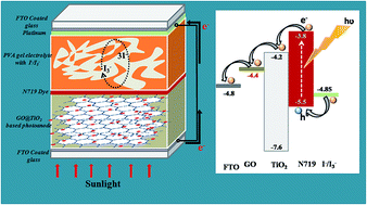
An effective self assembly method was used to synthesize hybrid nanocomposites of TiO2 nanoparticles on graphene oxide (GO) sheets for application in Dye Sensitized Solar Cells (DSSCs). The successful incorporation of TiO2 on the GO sheets was confirmed by X-ray diffraction (XRD), Energy Dispersive X-ray (EDX), Raman and UV-visible spectroscopy. The morphology and size of the TiO2 nanoparticles on the GO sheets were analyzed by scanning electron microscope (SEM) and transmission electron microscope (TEM) analysis. This study is concerned with the effects of different GO contents of the photoanode on the energy conversion efficiency of the PVA gel electrolyte based DSSCs. DSSCs based on the GO@TiO2 nanocomposite photoanode with an optimum GO content of 2.5 wt% showed a short circuit current density (Jsc) of 7.67 mA cm−2, an open circuit voltage (Voc) of 0.76 V and photoconversion efficiency of 3.97% which is much higher than that of the pure TiO2 nanoparticles.
1. Introduction
The dye sensitized solar cell is the most studied device of the last two decades as the most promising alternative to silicon solar cells because of its low cost, facile fabrication process and high efficiency.1,2 In 1991, Grätzel and O'regan assembled and demonstrated the first dye sensitized solar cell consisting of a photoanode of transparent fluorine doped tin oxide glass (FTO), a wide band gap nanocrystalline n-type semiconductor, a visible-light absorber, a redox couple electrolyte and a counter electrode of platinum. The performance of photoelectric conversion of DSSCs depends on four key factors: abundant photoelectron generation by dyes, rapid electron transfer at the photoanode, fast ion diffusion in the electrolyte and efficient reduction of redox media at the counter electrode. Amongst all components in DSSCs, the photoanode material plays a very important role in the dye loading, electron injection, transportation and collection, and in turn significantly influences the photoconversion efficiency.3–6
The photoanode of DSSCs is made of nanoparticles, which was sintered into a mesoporous network on FTO. Electron transports in the disordered TiO2 nanoparticles with a random motion resulting in the possibility of charge recombination and thus reduces the photocurrent and the performance of the device.7,8 Therefore, in order to improve the performance of DSSCs designing of photoanode in a manner is very important so that photogenerated electrons can be transported across the TiO2 nanoparticle network, competing with the charge recombination.9–11 The choice of photoanode material is based on two main factors: energy gap of the semiconductor must match with that of the dye so that photo-generated electrons can easily transfer from dyes to semiconductor. Secondly, to harvest solar energy as much as possible, semiconductor must have a high surface area so that more dye molecules get adsorbed on it. To enhance the electron transport and to reduce the recombination, there are several kinds of photoanode materials that have been developed including doped TiO2,12,13 metal/metal oxide–TiO2 hybrid, composite and core–shell structures,14–16 porous TiO2 structures,17–21 and TiO2–carbon material.22–24
Now a days, the 2D carbon nanomaterial graphene has drawn much attention as it is a zero band gap material resulting in an excellent electrical conduction in two dimensions. Herein, we incorporate graphene oxide (GO) into TiO2 nanoparticles (GO@TiO2) so that GO can form some bridges and enhance the charge transport rate to prevent the charge recombination improving photoelectrical conversion efficiency. TiO2 nanoparticles are anchored compactly on the GO flakes through physisorption, electrostatic binding or charge transfer interaction. As GO is an excellent electrical conductor it behaves as an electron transfer medium to enhance the electron transport from conduction band of TiO2 and resulting in the reduced recombination.25 In DSSCs, triodide/iodide (I3−/I−) is used as redox couple electrolyte. The volatile nature of different organic solvents used in liquid electrolytes triggers leakage and evaporation thus restricting long term performance of DSSCs. To overcome these drawbacks people focused on alternatives to the liquid electrolyte. In recent times, polymer gel electrolyte offers a great area of research as it possess the properties of solid as well as liquid and the unique hybrid network structure results in lower leakage and evaporation with long term durability of the cell. The effect of such electrolyte in GO@TiO2 photoanode based DSSCs are yet to be studied. Inspired from the forgoing discussions, a series of quasi solid state DSSCs have been fabricated using different weight% of GO@TiO2 photoanode and an attempt has been made to replace the conventional liquid electrolyte with a PVA gel electrolyte. Among the various polymers poly (vinyl alcohol) (PVA) has been chosen because of its good film forming properties and physical properties, biocompatibility, and have good chemical resistance.
2. Experimental
2.1. Materials and methods
2.1.1. Materials. Poly(vinyl alcohol), glutaraldehyde, nitric acid (HNO3), lithium iodide (LiI), and iodine (I2) were purchased from Merck India. The chemicals tertiary butylpyridine (TBP), 1-methyl-3propyl imidazolium iodide (MPI), N-methyl 2-pyrrolidone (NMP), cis-bis(isothiocyanato)bis(2,2′-bipyridyl-4,4′-dicarboxylato) ruthenium(II)bis-tetrabutylammonium (N719) dye, acetonitrile, fluorine doped tin oxide (FTO) coated glasses (Sheet resistance: 15 Ω sq.−1) were purchased from Sigma Aldrich India.Sodium nitrate (NaNO3), potassium permanganate (KMnO4), sodium dodecyl sulfate (CH3(CH2)11OSO3Na), hydrogen peoxide (H2O2) were purchased from Merck India. Graphite powder (<20 μm, Sigma-Aldrich; 99% pure) were used without further purification.
2.1.2. Preparation of TiO2@GO nanocomposite.
2.1.2.1. Synthesis of graphene oxide. Graphene oxide was synthesized from natural graphite powder by using Hummers method.26 Graphite (5 g), NaNO3 (2.5 g) and H2SO4 (120 mL) were mixed in a 500 mL beaker and the mixture was stirred vigorously in an ice bath for 30 min. Under constant stirring 15 g of KMnO4 was added to the above suspension in a very controlled way so that the reaction temperature remains below 20 °C. The mixture was kept under stirring for 12 h at room temperature, followed by the addition of 150 mL of distilled water and kept for 24 h under constant stirring. Then 50 mL of 30% H2O2 was added to the mixture and stirred for another 6 h. Finally, the obtained product was washed with 5% HCl and followed by distilled water so that the pH of the filtrate becomes 7 and dried in vacuum oven. The obtained graphene oxide was then dispersed and exfoliated in deionized water by using an ultrasonic bath for 40 min. The obtained graphene oxide suspension was then used for the synthesis of GO@TiO2 nanocomposites.
2.1.2.2. Synthesis of GO@TiO2 nanocomposite. GO@TiO2 nanocomposites were synthesized by a previously reported self-assembly method.27 The GO suspension (1.3 g L−1) was made with the addition of sodium dodecyl sulfate. To this mixture an aqueous solution of 50 mL of 0.12 mol L−1 TiCl3 was added and kept for 1 h under constant stirring. To the mixture, 10 mL of 0.6 M Na2SO4 solution and 5 mL of 1 wt% H2O2 solution were added and stirring was continued for another 1 h at 90 °C. The final precipitates of the reactions were separated and washed with water and ethanol and then dried at 70 °C. Finally, the dried product was calcinated at 400 °C for 2 h. Five sets of GO@TiO2 nanocomposites viz. GO@TiO2-A, GO@TiO2-B, GO@TiO2-C, GO@TiO2-D & GO@TiO2-E having GO wt% of 0.25, 0.5, 1, 2.5 & 5 respectively were synthesized by taking different amounts of GO. The compositions of the prepared GO@TiO2 nanocomposites are given in Table 1.
Table 1 Composition for the synthesis of GO@TiO2 nanocomposites
TiO2@GO |
GO@TiO2-A (0.25 wt%) |
GO@TiO2-B (0.5 wt%) |
GO@TiO2-C (1 wt%) |
GO@TiO2-D (2.5 wt%) |
GO@TiO2-E (5 wt%) |
GO (ml) |
1.85 |
3.70 |
7.40 |
18.50 |
37 |
SDS (ml) |
0.11 |
0.22 |
0.44 |
1.10 |
2.20 |
2.1.3. Synthesis of PVA hydrogel. PVA hydrogel was prepared by dissolving 1 g of PVA polymer powder in 10 mL of deionized water and kept under constant stirring at a temperature of 80 °C until the PVA is completely soluble. The PVA solution was allowed to cool down to room temperature, then chemically cross linked by adding 1.5 mL of glutaraldehyde and allowed to form gel at 60–70 °C.28 The obtained PVA hydrogel was then washed thoroughly and dried in a vacuum oven at 60 °C. The product was collected for further analysis and applications.
2.1.4. Preparation of PVA gel electrolyte. The gel electrolyte was prepared by dipping 0.5 g of dried PVA hydrogel in liquid electrolyte for about 48 h so that the gels reach equilibrium absorption. The liquid electrolyte consists of 0.1 M lithium iodide (LiI), 0.6 M I2, 0.5 M tertiarybutylpyridine (TBP), 0.05 M 1-methyl-3propyl imidazolium iodide (MPI) in a mixed solvent of N-methyl 2-pyrrolidone (NMP) and acetonitrile 2
:
8 ratio.
2.1.5. Assembling of dye sensitized solar cell. FTO glass sheets (Sigma-Aldrich, sheet resistance: 15 Ω sq.−1) were first cleaned by ultrasonicating in a detergent and deionized water followed by treating with boiling acetone and isopropanol and finally dried under a nitrogen atmosphere. Then GO@TiO2 nanocomposites photoanode electrodes were prepared by depositing a paste of GO@TiO2 nanocomposites on FTO by doctor blade technique, which was annealed at 450 °C for 30 minutes to retain the texture. Finally the annealed photoanodes were sensitized by immersing into a 0.3 mM N719 dye solution prepared in a mixed solvent of acetonitrile and ethanol with a volume ratio of 1
:
1 for 24 h. After that the electrodes were washed properly with ethanol and dried in air before the final assembling of the devices. The platinum deposited FTO coated glass slide was used as the counter electrode. DSSCs was fabricated by placing the PVA gel electrolyte evenly in between the dye adsorbed photoanodes and Pt counter electrode. Before taking the photovoltaic measurements, all the devices were kept at 70 °C for 10 min so that the electrolyte can penetrate into dye adsorbed GO@TiO2 nanocomposites layer. A schematic presentation of the structure of quasi solid state DSSC is shown in Fig. 1.
 |
| Fig. 1 Schematic presentation of the GO@TiO2 nanocomposite based photoanode for quasi solid state DSSC. | |
2.1.6. Measurements.
2.1.6.1. X-ray diffractometer (XRD). The XRD measurements were carried out in a Rigaku X-ray diffractometer (Miniflex, UK) using CuKα (λ = 0.154 nm) radiation at a scanning rate of 2° min−1 with an angle (2θ) ranging from 10° to 80° to study the structural characteristics of the synthesized nanocomposites.
2.1.6.2. Ultraviolet-visible (UV-vis) spectroscopy. The optical property of MWCNT@TiO2 nanocomposites were measured by using UV-visible diffuse reflectance spectra in the range 200–800 nm using Shimadzu UV-2550 UV-visible spectrophotometer.
2.1.6.3. Scanning electron microscopy (SEM). The surface morphologies of the prepared samples were studied by using a Jeol-JSM-6390L V scanning electron microscope at an accelerating voltage of 5–15 kV. The surface of the samples was Pt-coated before the scanning.
2.1.6.4. Transmission electron microscopy (TEM). To study the distribution of the of particles within the nanocomposites, TEM analysis was done in a JEOL, JEM 2100 transmission electron microscope at an accelerating voltage of 200 kV.
2.1.6.5. Impedance spectroscopy measurement of devices. The impedance spectroscopy was performed on a SP-150 Potentiostat Galvanostat electrochemical impedance workstation at a constant temperature of 25 °C. The amplitude of the applied ac signal was of 90 to 264 V in the frequency range from 100 mHz to 100 kHz.
2.1.6.6. Photovoltaic test. The photocurrent voltage characteristic curves of the fabricated gel electrolyte based DSSCs was measured using a 100 mW cm−2 xenon arc lamp in ambient atmosphere. The performance parameters of the fabricated devices i.e. the cell fill factor (FF) and cell power conversion efficiency (η) were calculated by following eqn (1) and (2) respectively. |
 | (1) |
|
 | (2) |
where, Jmax and Vmax are the maximum current density and voltage respectively, at the point of maximum power of photocurrent density versus voltage plot. Jsc is the short circuit current density (mA cm−2), Voc is the open circuit voltage (V) and Pin is the intensity of the incident white light.
3. Results and discussion
3.1. Ultraviolet-visible (UV-vis) spectroscopy
UV-visible analysis is recorded in the range of 200–800 nm and the spectra of GO@TiO2 nanocomposites are shown in Fig. 2a. The presence of different amounts of GO influences the optical absorption for GO@TiO2 nanocomposites significantly. All the nanocomposites show the same absorption profile. However with increasing GO content in the nanocomposite absorption intensity in the UV region raises.29 A red shift to higher wavelength in the absorption edge of the GO@TiO2 nanocomposites has also been observed at about 400 nm, indicating a narrowing of the band gap of TiO2. Due to the incorporation of GO into the matrix of TiO2, the background absorption in the area 400–800 nm increases resulting in the difficulty to measure the red shift.
 |
| Fig. 2 (a) UV-vis absorption spectra for TiO2 and GO@TiO2 nanocomposites (b) Tauc's plot for optical band-gap calculation for TiO2 and GO@TiO2 nanocomposites. | |
Optical band gap calculations are carried out using the Tauc's relationship. The Tauc's equation for band gap calculations is described as,
where,
α is the absorption coefficient of the solid at a certain value of wavelength
λ,
h is Planck's constant,
C is the proportionality constant, ν is the frequency of light,
Eg is the band gap energy.
Fig. 2b shows the relationship of (αhν)1/2 vs. photon energy (hν = 1239/λ) for TiO2 and also the GO@TiO2 nanocomposites. The band gap for bare TiO2 is 3.20 eV, whereas the band gap of GO@TiO2 nanocomposites are 3.06, 3.0, 2.89, 2.84 & 2.80 eV corresponding to GO@TiO2-A, GO@TiO2-B, GO@TiO2-C, GO@TiO2-D & GO@TiO2-E respectively i.e. with increase GO content band gap decreases. This result supports the qualitative observation of a red shift in the absorption edge of GO@TiO2 nanocomposites as compared to the bare TiO2. GO contains many oxygen containing groups such as carboxyl, hydroxyl and epoxide which are covalently bonded to its layers surfaces. There is a possibility that some of the unpaired π electrons of GO can bonded with the free electrons present on the surface of TiO2 nanoparticles to form Ti–O–C structure, resulting in the shifting of valence band and reduction in the band gap.30
3.2. X-ray diffractometry (XRD)
The X-ray diffraction patterns for the GO and the GO@TiO2 nanocomposites with different weight% of GO are shown in Fig. 3. GO exhibits a strong diffraction peak at 2θ = 11.5° corresponding to the reflection plane (001) with an interlayer spacing of 7.43 Å due to the oxygen containing functional groups on the carbon sheets.31 GO@TiO2 nanocomposites with different weight percentage of GO exhibit similar XRD patterns. The characteristic XRD peaks of the GO@TiO2 nanocomposites at 2θ values of 25.5, 38.1, 48.3, 54.2, 55.2, 62.9, 69.5, 70.3 & 75.4 can be indexed to (101), (004), (200), (105), (211), (204), (116), (220) & (107) crystal planes, which represents the pure anatase phase of TiO2. The characteristic peaks are confirmed by the reported JCPDS.32 Noticeably, no typical diffraction peak belonging to the GO is observed in the GO@TiO2 nanocomposites. The reason can be attributed to the fact that regular stack of GO is disturbed by the intercalation of anatase TiO2.31,33 Thus, the XRD pattern of the GO@TiO2 composites confirms the homogeneous distribution of anatase TiO2 within the GO stacks. Furthermore, the existence of grapheme oxide in GO@TiO2 nanocomposites can be clearly elucidated by Raman analysis.
 |
| Fig. 3 XRD patterns for GO and GO@TiO2 nanocomposites. | |
3.3. Raman spectra
Raman spectroscopy is a powerful tool to analyze the crystalline quality of the carbon and the defect mediated peaks. Fig. 4 shows the Raman spectrum of GO (inset graph in Fig. 4) and GO@TiO2 nanocomposite. In the Raman spectrum first order dominant vibrational modes, D band 1351cm−1corresponds to the A1g symmetry mode of sp3 carbon indicates about the order or disorder in the system34 and G band 1599 cm−1 corresponds to the vibrational mode of sp2 carbon provides information about the doubly degenerate E2g mode of the Brillonium zone centre.35 The second order vibrational bands, 2D1 & 2D2 arises at 2717.36 cm−1 & 2928.62 cm−1 respectively originates from a two phonon double resonance Raman process and provides information on the stacking order of graphene with number of layers and shows often a doublet with increasing number of graphene layers. Another overtone weak band 2D′ is observed at 3194.23 cm−1.36 The intensity of G band is stronger than D band. In plane crystallite size, La can be calculated from the intensity ratio of the G band to the D band by using the formula, La = 4.4(LG/LD). The obtained La value is 4.89,37,38 from this it can be concluded that the sample contains highly disordered and randomly arranged graphene sheets. The shape of the 2D band is sensitive to the no of layers of graphene and chemical doping. When GO combines with TiO2, the crystal structure of carbon changes obviously. There are four specific vibration modes are located at around 141 (E(g1)), 391 (B1g(1)), 514 (A1g + B1g(2)) & 634 cm−1 (Eg(2)) indicating the presence of the anatase phase of TiO2 in the sample.39
 |
| Fig. 4 Raman spectra for GO and GO@TiO2-C nanocomposite. | |
3.4. SEM analysis & EDX
The surface characteristics of the GO and the GO@TiO2 nanocomposites are investigated with SEM analysis and the micrographs are shown in Fig. 5. For the pure GO a flat multilayered structure with stacked GO sheets is observed at higher magnifications (Fig. 5a).40 SEM micrographs (Fig. 5c) shows that the flat layered structure of the GO is disappeared and a rough surface morphology is noticed for GO@TiO2 nanocomposites. The change in morphology of the GO@TiO2 nanocomposites can be attributed to the successful incorporation of TiO2 into the GO sheets. The energy dispersive X-ray spectrum (EDX) of the GO sheets and GO@TiO2 nanocomposites indicates the successful distribution of TiO2 nanoparticles into the GO sheets (Fig. 5b and d). The introduction of TiO2 into the GO is confirmed from the elemental typical mapping images of titanium, oxygen and carbon.
 |
| Fig. 5 SEM images of the (a) GO and (c) GO@TiO2-D nanocomposite. EDX-spectrum of the (b) GO and (d) GO@TiO2-D nanocomposite. | |
3.5. TEM analysis
Fig. 6 represents the morphological image of synthesized GO and GO@TiO2 nanocomposite from transmission electron microscopy. TEM micrographs of GO showed two dimensional layered and flat like structure with micrometers long wrinkles (Fig. 6a). At higher magnification the TEM micrographs of GO show a completely amorphous and disordered structure (Fig. 6b).41 TEM micrographs of GO@TiO2 nanocomposite, showed that large number of TiO2 nanoparticles are densely deposited onto the GO sheets. The electron transfer processes in the solar cells are facilitated by the homogeneously and closely dispersed TiO2 nanoparticles on the GO sheets (Fig. 6c and d). The corresponding HRTEM image showed clear lattice fringes of TiO2.
 |
| Fig. 6 TEM micrographs of the GO (a and b) and GO@TiO2-D nanocomposite (c and d). | |
3.6. The morphology of PVA gel electrolyte
The surface and cross sectional SEM images of PVA gel electrolyte are shown in Fig. 7a and b. A uniform surface with smooth outer morphology is observed in case of the surface view of the gel electrolyte. The cross sectional morphology of the chemically cross linked hydrogel images showed wavy surface. The surface morphology of the intact disc revealed some pores and dense areas in the inset.
 |
| Fig. 7 SEM images of the PVA (a) surface view and (b) cross sectional view. | |
3.7. Electrochemical impedance analysis of the PVA gel
In order to understand the electronic and ionic transport process in the DSSCs electrochemical impedance spectroscopy (EIS) is a very effective toll. We have carried out the EIS of the fabricated cells by exposing the cell to solar radiation of intensity AM1.5 G 100 mW cm−2. The equivalent electrical circuit for the DSSCs, which is a combination of a series resistance and two time constant phase elements42 is shown in Fig. 8a and b shows the Nyquist plot. Under exposer to light, the biased device shows a smaller arc in Nyquist plot at the higher frequency region corresponds to the capacitance resistance of the counter electrode/gel electrolyte interface (Rct1) i.e. the charge transfer resistance at the Pt electrode/electrolyte interface. Another larger arc occurs at the mid frequency region which corresponds to the charge transfer process (Rct2) at GO@TiO2/dye/gel electrolyte and further charge transfer in the photoanode.43 Another parameter at the high frequency intercept on the abscissa represents series resistance (Rs) which is associated with the electrolytes and electric contacts in the DSSCs. As shown in Table 2 it is observed that by changing the concentration of GO content in the photoanode, Rct1 remains almost same as we are using the same polymer gel electrolyte. But the Rct2 values are significantly increases with the increase in the weight% content of the GO in the photoanode than that of the pure TiO2. This larger value of interfacial charge recombination resistance renders GO to increase the charge collection and transport of electrons at the interface of photoanode and the electrolyte. The charge transport mechanism becomes faster resulting in the reduction of electron recombination.25 However, above a optimum GO loading (i.e. GO@TiO2-D), it is observed that Rct2 value is low which may be due to the agglomeration of GO resulting in the creation of electron traps and also compete light harvesting with the dye molecules.
 |
| Fig. 8 (a) The equivalent electrical circuit for the fabricated DSSCs, (b) EIS analysis spectra for all the nanocomposites based photoanode. (Inset) expansion of the first semicircle. | |
Table 2 Various parameters determined by EIS measurements of the GO@TiO2 nanocomposites based photoanode and polymer gel electrolytes
Sample |
Rs (ohm) |
Rct1 (ohm) |
Rct2 (ohm) |
σ (S cm−1) |
TiO2 only |
0.4498 |
0.2111 |
5.8439 |
0.4446 |
GO@TiO2-A |
0.4126 |
0.2079 |
6.1645 |
0.4847 |
GO@TiO2-B |
0.3799 |
0.2110 |
6.4228 |
0.5265 |
GO@TiO2-C |
0.3394 |
0.2068 |
6.9929 |
0.5893 |
GO@TiO2-D |
0.3088 |
0.2111 |
7.4561 |
0.6477 |
GO@TiO2-E |
0.3231 |
0.2142 |
7.0843 |
0.619 |
3.8. Absorption spectroscopic investigation of dye adsorption
UV-visible spectroscopy was used to study the effects of addition of GO to TiO2 on the dye adsorption by the films. The absorption spectra of the N719 dye sensitized films of TiO2 & GO@TiO2 nanocomposites in the visible region are presented in Fig. 9. The dye sensitized TiO2 films shows absorption in the range of 400–800 nm and the characteristic absorption of the N719 dye at around 530 nm. The intensity of the absorption peak at 530 nm increases with loading of GO in the nanocomposite of GO@TiO2. Moreover, enhanced quantity of dye adsorption is indicative of enhanced surface area in the TiO2 film.44 The electron rich GO contains many oxygen containing groups like carboxyl on its surface and its addition to TiO2 facilitating the anchoring of the N719 dye molecule. The obtained results suggest that the dye loading has increased in the order of GO content of TiO2 < GO@TiO2-A < GO@TiO2-E < GO@TiO2-D. Absorbance measurements from Fig. 9 indicate increased dye adsorption by about 40% in the case of GO@TiO2-D nanocomposite film, and by 25 and 16% in the case of GO@TiO2-E and GO@TiO2-A nanocomposite film respectively. Upto an optimum concentration of GO a large amount of dye molecules are absorbed in the photoanode, leading to a strong light harvesting in the device.45
 |
| Fig. 9 UV-visible absorption spectra N719 dye adsorbed on TiO2 and GO@TiO2 nanocomposites photoanode. | |
3.9. Photovoltaic performance of the fabricated DSSCs devices
GO@TiO2 nanocomposites were employed as the photoanode material in the PVA gel electrolyte based quasi solid state DSSCs. The J–V curves and PV characteristics for DSSCs based on the different photoanode were evaluated under illumination of simulated solar light of 100 mW cm−2 AM 1.5 and are exhibited in Table 3 & Fig. 10. The DSSCs prepared with GO@TiO2-D nanocomposite as the photoanode exhibited the highest short circuit photocurrent density (Jsc) of 7.67 mA cm−2 and the highest conversion efficiency of 3.97%. DSSCs obtained from the bare TiO2 photoanode exhibits conversion efficiency of 0.99% and Jsc of 2.51 mA cm−2. However the photocurrent densities as well as the conversion efficiency increases with the addition of GO reaching a peak value and then declined with the higher concentration of GO in the GO@TiO2 nanocomposite. Compared to the DSSCs with the bare TiO2 nanoparticles photoanode, the conversion efficiency and Jsc of the DSSC with GO@TiO2-D are significantly improved by 55% & 67% respectively. However, Voc values remained fairly constant in the range of 0.65–0.75 V. A small amount of GO in the photoanode is sufficient to enhance the electron injection and electron transfer rate in the DSSC operation thereby accelerating the charge transport mechanism. Subsequently, the electron recombination is reduced which extends the electron lifetime in the photoanode.25 In turn it can be said that, the introduced 2D GO bridges increases the formation of more conductive pathways to transport charge i.e. by decreasing the transport resistance and thus increases the photocurrent and suppress the electron in TiO2 to recombine with the dye and redox species. It is observed that beyond the optimum amount of GO concentration, Jsc decreased which may be due to the decrease in the film transmittance hindering the electron transportation upon illumination.46 The enhancement of the Jsc and FF was also attributed to the enhancement of the charge collection and transport of electron through different channels of gel electrolyte.
Table 3 Photovoltaic characteristics of the DSSCs with the GO@TiO2 nanocomposites based photoanode and polymer gel electrolytes
Sample |
Jsc (mA cm−2) |
Voc (V) |
FF |
PCE, η (%) |
TiO2 only |
2.5072 |
0.6501 |
0.61 |
0.99 |
GO@TiO2-A |
3.2826 |
0.6547 |
0.63 |
1.37 |
GO@TiO2-B |
4.0317 |
0.6991 |
0.64 |
1.80 |
GO@TiO2-C |
4.7821 |
0.7073 |
0.65 |
2.20 |
GO@TiO2-D |
7.6654 |
0.7617 |
0.68 |
3.97 |
GO@TiO2-E |
6.6554 |
0.7097 |
0.67 |
3.16 |
 |
| Fig. 10 Photocurrent density versus voltage plots for all the devices fabricated with all the nanocomposites. | |
The long term stability of DSSCs assembled by using PVA polymer gel electrolyte was compared with that for a liquid electrolyte based cell and results are summarized in Fig. 11 which compares the variation of PCE as a function of time. For this stability study, the devices were stored under dark at 65 °C and 85% relative humidity. It is observed that PCE of the polymer gel electrolyte based devices is decreased by 5%, whereas the PCE for liquid electrolyte based devices is decreased by about 19% after 800 h. In case of the liquid electrolyte based devices, leakage and evaporation of the solvent take place with time and also dye molecule start desorbing from the TiO2 based photoanode. In case of the polymer gel electrolyte based devices, as the electrolyte is entrapped within the polymer matrix the evaporation as well as the leakage of the solvent from the cell is reduced.
 |
| Fig. 11 Stability graph of the fabricated device with GO@TiO2-D photoanode (a) liquid electrolyte and (b) PVA gel electrolyte. | |
4. Conclusion
The self assembled GO@TiO2 hybrid nanocomposites have been used as photoanode in fabrication of DSSCs. XRD, UV-visible and Raman spectroscopy confirm successful formation of the nanocomposite. The morphological analysis reveals the uniform formation of GO@TiO2 nanocomposite. The EIS demonstrates that GO reduces the electron recombination and improving the electron transport through the film. The incorporation of GO into the TiO2 results in the enhancement of device parameters Jsc, Voc and PCE. As the GO concentration increases from 0.25 to 2.5 wt%, the PCE increases from 1.37 to 3.97% due to the rapid charge transport mechanism in the photoanode. GO offers an improved rate of collection and transport of electrons. Moreover, upto an optimum concentration of GO a large amount of dye molecules are absorbed in the photoanode with higher concentration of GO thus leading to a strong light harvesting in the device.
Acknowledgements
The author would like to acknowledge University Grant Commission (UGC), for providing the “Maulana Azad National Fellowship” F1-17.1/2015-16/MANF-2015-17-ASS-51589. The authors would like to thank SAIF, North-Eastern Hill University, India, SAIC Tezpur University, India for analytical support and Department of Electronics and Information Technology, Ministry of Communications & Information Technology India for their financial support (Sanction No. 1 (11)/2012-EMCD dated 05-03-2013).
References
- M. Grätzel, Nature, 2001, 414, 338–344 CrossRef PubMed.
- A. Hagfeldt, G. Boschloo, L. C. Sun, L. Kloo and H. Pettersson, Chem. Rev., 2010, 110, 6595–6663 CrossRef CAS PubMed.
- V. Galstyan, A. Vomiero, I. Concina, A. Braga, M. Brisotto, E. Bontempi, G. Faglia and G. Sberveglieri, Small, 2011, 7, 2437–2442 CrossRef CAS PubMed.
- M. Law, L. E. Greene, J. C. Johnson, R. Saykally and P. D. Yang, Nat. Mater., 2005, 4, 455–459 CrossRef CAS PubMed.
- A. Vomiero, I. Concina, M. M. Natile, E. Comini, G. Faglia, M. Ferroni, I. Kholmanov and G. Sberveglieri, Appl. Phys. Lett., 2009, 95, 193104 CrossRef.
- N. Memarian, I. Concina, A. Braga, S. M. Rozati, A. Vomiero and G. Sberveglieri, Angew. Chem., Int. Ed., 2011, 50, 12321–12325 CrossRef CAS PubMed.
- J. van de Lagemaat, K. D. Benkstein and A. J. Frank, J. Phys. Chem. B, 2001, 105, 12433–12436 CrossRef CAS.
- N. Kopidakis, N. R. Neale, K. Zhu, J. van de Lagemaat and A. Frank, Appl. Phys. Lett., 2005, 87, 202106 CrossRef.
- A. Kongkanand, R. Martínez Domínguez and P. V. Kamat, Nano Lett., 2007, 7, 676–680 CrossRef CAS PubMed.
- A. Kongkanand and P. V. Kamat, ACS Nano, 2007, 1, 13–21 CrossRef CAS PubMed.
- J. van de Lagemaat, N. G. Park and A. J. Frank, J. Phys. Chem. B, 2000, 104, 2044–2052 CrossRef CAS.
- H. Tian, L. Hu, C. Zhang, L. Mo, W. Li, J. Sheng and S. Dai, J. Mater. Chem., 2012, 22, 9123–9130 RSC.
- F. Huang, Q. Li, G. J. Thorogood, Y. B. Cheng and R. A. Caruso, J. Mater. Chem., 2012, 22, 17128–17132 RSC.
- J. Du, J. Qi, D. Wang and Z. Tang, Energy Environ. Sci., 2012, 5, 6914–6918 CAS.
- T. Chen, G. H. Guai, C. Gong, W. Hu, J. Zhu, H. Yang, Q. Yan and C. M. Li, Energy Environ. Sci., 2012, 5, 6294–6298 CAS.
- K. Kakiage, T. Tokutome, S. Iwamoto, T. Kyomen and M. Hanaya, Chem. Commun., 2013, 49, 179–180 RSC.
- J. Yan and F. Zhou, J. Mater. Chem., 2011, 21, 9406–9418 RSC.
- G. K. Mor, K. Shankar, M. Paulose, O. K. Varghese and C. A. Grimes, Nano Lett., 2005, 6, 215–218 CrossRef PubMed.
- Z. Liu and M. Misra, ACS Nano, 2010, 4, 2196–2200 CrossRef CAS PubMed.
- Z. Sun, J. H. Kim, Y. Zhao, F. Bijarbooneh, V. Malgras and S. X. Dou, J. Mater. Chem., 2012, 22, 11711–11719 RSC.
- J. Wang and Z. Lin, Chem.–Asian J., 2012, 7, 2754–2762 CrossRef CAS PubMed.
- S. Muduli, W. Lee, V. Dhas, S. Mujawar, M. Dubey, K. Vijayamohanan, S. H. Han and S. Ogale, ACS Appl. Mater. Interfaces, 2009, 1, 2030–2035 CAS.
- J. Yu, J. Fan and B. Cheng, J. Power Sources, 2011, 196, 7891–7898 CrossRef CAS.
- Y. H. Jang, X. Xin, M. Byun, Y. J. Jang, Z. Lin and D. H. Kim, Nano Lett., 2011, 12, 479–485 CrossRef PubMed.
- N. Yang, J. Zhai, D. Wang, Y. Chen and L. Jiang, ACS Nano, 2010, 4, 887–894 CrossRef CAS PubMed.
- W. S. Hummers and R. E. Offeman, J. Am. Chem. Soc., 1958, 80, 1339 CrossRef CAS.
- D. H. Wang, D. W. Choi, J. Li, Z. G. Yang, Z. M. Nie, R. Kou, D. H. Hu, C. M. Wang, L. V. Saraf and J. G. Zhang, ACS Nano, 2009, 3, 907–914 CrossRef CAS PubMed.
- H. S. Mansur, R. L. Oréfice and A. A. P. Mansur, Polymer, 2004, 45, 7193–7202 CrossRef CAS.
- K. Dai, L. Lu, Q. Liu, G. Zhu, Q. Liua and Z. Liua, Dalton Trans., 2014, 43, 2202–2210 RSC.
- V. Štengl, S. Bakardjieva, T. M. Grygar, J. Bludská and M. Kormunda, Chem. Cent. J., 2013, 7, 41 CrossRef PubMed.
- P. G. Liu, K. C. Gong, P. Xiao and M. Xiao, J. Mater. Chem., 2000, 10, 933–935 RSC.
- Y. W. Jun, M. F. Casula, J. H. Sim, S. Y. Kim, J. Cheon and A. P. Alivisatos, J. Am. Chem. Soc., 2003, 125, 15981–15985 CrossRef CAS PubMed.
- C. Xu, X. D. Wu, J. W. Zhu and X. Wang, Carbon, 2008, 46, 386–389 CrossRef CAS.
- M. S. Dresselhaus, A. Jorio, M. Hofmann, G. Dresselhaus and R. Saito, Nano Lett., 2010, 10, 751–758 CrossRef CAS PubMed.
- J. Lu, J. X. Yang, J. Wang, A. Lim, S. Wang and K. P. Loh, ACS Nano, 2009, 3, 2367–2375 CrossRef CAS PubMed.
- A. C. Ferrari, J. C. Meyer, V. Scardaci, C. Casiraghi, M. Lazzeri, F. Mauri, S. Piscanec, D. Jiang, K. S. Novoselov, S. Roth and A. K. Geim, Phys. Rev. Lett., 2006, 97, 187401–187404 CrossRef CAS PubMed.
- M. A. Pimenta, G. Dresselhaus, M. S. Dresselhaus, L. A. Cancado, A. Jorio and R. Saito, Phys. Chem. Chem. Phys., 2007, 9, 1276–1290 RSC.
- A. C. Ferrari, Solid State Commun., 2007, 143, 47–57 CrossRef CAS.
- Y. Lei, L. D. Zhang and J. C. Fan, Chem. Phys. Lett., 2001, 338, 231–236 CrossRef CAS.
- L. L. Zhang, S. Zhao, X. N. Tian and X. S. Zhao, Langmuir, 2010, 26, 17624–17628 CrossRef CAS PubMed.
- G. X. Wang, J. Yang, J. Park, X. L. Gou, B. Wang, H. Liu and J. Yao, J. Phys. Chem. C, 2008, 112, 8192–8195 CAS.
- F. F. Santiago, J. Bisquert, G. G. Belmonte, G. Boschloo and A. Hagfeldt, Sol. Energy Mater. Sol. Cells, 2005, 87, 117–131 CrossRef.
- M. Miyashita, K. Sunahara, T. Nishikawa, Y. Uemura, N. Koumura, K. Hara, A. Mori, T. Abe, E. Suzuki and S. Mori, J. Am. Chem. Soc., 2008, 130, 17874–17881 CrossRef CAS PubMed.
- L. Zhang and Z. S. Wang, J. Mater. Chem. C, 2016, 4, 3614–3620 RSC.
- H. Wang, B. Wang, J. Yu, Y. Hu, C. Xia, J. Zhang and R. Liu, Sci. Rep., 2014, 5, 9305 CrossRef PubMed.
- Y. B. Tang, C. S. Lee, J. Xu, Z. T. Liu, Z. H. Chen, Z. B. He, Y. L. Cao, G. D. Yuan, H. S. Song, L. M. Chen, L. B. Luo, H. M. Cheng, W. J. Zhang, I. Bello and S. T. Lee, ACS Nano, 2010, 4, 3482–3488 CrossRef CAS PubMed.
|
This journal is © The Royal Society of Chemistry 2016 |
Click here to see how this site uses Cookies. View our privacy policy here.