Jovana Trbojević Ivića,
Aleksandra Dimitrijevićb,
Nenad Milosavić*c,
Dejan Bezbradicad,
Branko J. Drakuliće,
Marija Gavrović Jankulovićf,
Marija Pavlovićg,
Helene Rogniauxg and
Dušan Veličkovićf
aInnovation Center, Faculty of Chemistry, University of Belgrade, 11000 Belgrade, Serbia
bDepartment of Molecular Biology and Biochemistry, University of California Irvine, 92697 Irvine, California, USA
cDivision of Experimental Therapeutics, Department of Medicine, Columbia University, 10032 New York, New York, USA. E-mail: nm2729@cumc.columbia.edu
dDepartment of Biochemical Engineering and Biotechnology, Faculty of Technology and Metallurgy, 11000 Belgrade, Serbia
eDepartment of Chemistry, Institute of Chemistry, Technology and Metallurgy, University of Belgrade, Belgrade, Serbia
fDepartment of Biochemistry, Faculty of Chemistry, 11000 Belgrade, Serbia
gINRA, UR1268, Biopolymers Interactions Assembles, 44316 Nantes, France
First published on 1st April 2016
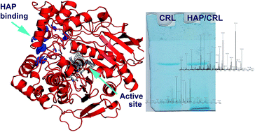
Hydroxyapatite (HAP), a calcium-phosphate bioactive ceramic, is actively employed in medical and separation sciences. Although different classes of biomacromolecules interact with this material, interactions with proteins are the most important, since they directly affect the biocompatibility of the carrier and it's industrial application. In the presented work, we thoroughly investigate and elucidate the interaction mechanism between Candida rugosa lipase (CRL) upon it's immobilization on HAP, since this immobilized enzyme showed advanced catalytic properties in previous studies. Applying elution and protein modification strategies we concluded that Ca-chelation of HAP's C-site and CRL's –COOH groups is the most probable interacting mechanism. A proteomics approach revealed that this chelation is conserved throughout all CRL isoforms, while results of molecular modelling led us to propose the involvement of a specific region of the protein surface and side chains in interactions with HAP.
Complex mechanism of protein–HAP interactions can be anticipated already from its molecular formula: Ca10(PO4)6(OH)2. Three types of functional groups are recognized in HAP: positively charged calcium ions (Ca2+), regarded as C-sites, six negatively charged oxygen ions associated with phosphate anions (PO43−), designated as P-sites and hydroxyl groups (OH). Specific topographic pattern of HAP suggests at least three distinct interaction patterns in theory: cation-exchange of –NH2 groups in basic proteins with P-sites, specific metal chelation and anion-exchange of –COOH clusters in acidic proteins with C-sites, as well as hydrogen bonds with OH-groups.12–14 Apart from these most probable interaction patterns, in our earlier work, we have observed hydrophobic interactions of lipase A from Candida antarctica (CAL A) and hydroxyapatite, which was successfully employed for one-step, high-yield purification of this enzyme.9 In reality however, protein binding is multifactorial process, governed by the characteristics of both the carrier and protein itself.15,16
Lipases are highly acclaimed catalysts in modern industry. Numerous ethical, practical and economic prerogatives have established microbial lipases as standard in production processes.17 Candida rugosa (formerly C. cylindracea) has long been utilized for its extracellular lipase production.18 Depending on the growth conditions, C. rugosa releases 5–7 extracellular lipase isoforms (LIP1–LIP7) into culture medium. These isoenzymes share some mutual characteristics: mature proteins are glycosylated monomers with about 534 amino acids in polypeptide backbone and a molecular mass of around 60 kDa. With the isoelectric point (pI) in range of 4.5–5.5, they are regarded as acidic proteins.19–21 Apart from differences in hydrophobicity, degree and position of glycosylation sites, C. rugosa lipases differ significantly in substrate specificity. High affinity towards different substrates is the key motif for widespread occurrence of CRL in industry and science.
In previous work, we have reported a high-yield and facile protocol for immobilization of C. rugosa lipase on hydroxyapatite. Our immobilization procedure has resulted in significant stabilization of catalyst, a feature directly utilized in biosynthesis of apple flavor – methyl acetate.22 Since enzyme adsorption to different materials is crucial for modulation of its biological activity and productivity, the objective of the current work was to investigate the mechanism enrolled in interactions between CRL and HAP. Obtained findings will give solid basis for further improvement of biocatalytic properties of particular immobilized enzyme, and also provide added benefit in other areas of HAP application by providing better insight into general protein–HAP interactions.
000) in the Orbitrap analyzer (mass-to-charge ratio (m/z): 400–2000), while collision-induced dissociation (CID) spectra were recorded on the five most intense ions in the LTQ traps. Raw data collected during LC-MS/MS analyses were processed into mgf format files and further searched against databanks using MASCOT Server 2.2 (Matrix Science). Protein identification was achieved by confronting mass data (MS and MS/MS spectra) against the UniProt databank (search restricted to “Fungi” taxonomy, release 09/01/2013) and Swissprot databank (unrestricted search to check the presence of keratin or trypsin contamination). One missed trypsin cleavage was set for databank searches and a mass tolerance of 0.0005%. Protein identifications were compared in the two databanks and best matches were validated. Proteins were considered when a minimum of two unique peptides were matched in their sequence (and sequence coverage is higher than 3%), with a MASCOT individual ion score of above 34 for line with soluble commercial CRL and 35 for line with bounded isoforms. The peptide scores greater than – or equal to – the threshold defined by MASCOT (p < 0.05) were considered.![]() | ||
| Fig. 1 Elution potency of different reagents for desorption of immobilized CRL. (A) Effect of Na+. (B) Effect of Ca2+. (C) Effect of PO43−. | ||
Plots from Fig. 1(a)–(c) illustrate that desorption with sodium and calcium cations is insignificant (less than 10%), while phosphate anions provide complete desorption of protein at concentration above 0.2 M. Such a trend indicates that the main factor in interactions between CRL and HAP are enzymes –COOH groups, characteristic for acidic proteins. If the interactions between CRL and HAP were solely electrostatic through protein's –NH2 groups, enzyme would be completely eluted by increase in ionic strength.34 Since, in our experiment, only 10% of CRL was eluted even when ionic strength of Na+, Cl− and Ca2+ was ≥0.5 mol L−1 in elution buffer we can eliminate this hypothesis. On the other hand, results are in very good agreement with hypothesis that main interaction points are –COOH groups of CRL and C-site of HAP. Addition of Cl− would not disturb this interaction, because dissociation constant of CaCl2 is significantly higher than dissociation constant of COO–Ca complex.13 Moreover, introducing soluble Ca2+ ions in system would result in formation of salt bridges between protein COOH groups and phosphate sites from HAP, thus even enhancing binding: HAP–PO4⋯Ca⋯OOC–protein. If, however, phosphate is added to immobilization buffer, binding of CRL on HAP is completely disabled (CRL is completely eluted for phosphate concentration in the immobilization buffer ≥ 0.3 mol L−1). Again, this can be explained through dissociation constants: dissociation constant of COO–Ca protein–HAP complex is much higher, than for the Ca–PO4 complex.13 All of data, presented so far, suggest that CRL and HAP interact through metal-coordinative bond between C-site on HAP and –COOH groups of CRL, and that other possible interactions have minor influence.
![]() | ||
| Fig. 2 Effect of different chemical treatments of CRL on its immobilization on HAP. Results are average from triplicate measurements. | ||
As shown in the Fig. 2, chemical blocking of –NH2 in CRL did not affect the binding to HAP in comparison to non-modified control regardless of type of modification reaction, corroborating our previous assumption that amino groups are not involved in CRL–HAP interactions. On the other hand, blocking of –COOH groups has strong effect on protein binding, since 64% of offered CRL activity remains in the supernatant in this experiment. Similar trend was observed when immobilization took place in the buffer containing external source of –COOH, since 87% of CRL remains unbound in the supernatant. High concentration of chelating citrate ions “mask” available Ca from HAP, thus providing direct evidence that C site of HAP is crucial for interaction with CRL. Slightly smaller effect observed in experiment with lipase obtained after –COOH blocking can be attributed to incomplete chemical modification of enzyme's –COOH groups. The plot, shown in Fig. 2 has definitely dispelled any further doubt about the binding interactions between our enzyme and the employed carrier. It is clear that –COOH play a vital role in CRL–HAP contact, which also confirms the results from previous section that the main mode of interaction is by metal-chelation through C-site. This is very significant, since protein must be in its native form in order to carry out this type of interactions.35 This fact also provides a reasonable explanation for high operational stability of immobilized CRL we have reported earlier, since the carrier we have chosen, preserves native conformation of the enzyme.
Exponentially Modified Protein Abundance Index (emPAI) was used to evaluate the relative abundance of CRL isoforms in both conditions. emPAI is an established method that estimates protein abundances from “peptide counts” (i.e. the number of MS/MS events, or “queries”, assigned to a given peptide) in a single LC-MS/MS experiment. emPAI is defined as 10PAI − 1, where PAI (Protein Abundance Index) denotes the ratio of observed to observable peptides. emPAI was first proposed by Ishihama who found that PAI is approximately proportional to the logarithm of absolute protein concentration.36 In our work, HRMS analysis of free (soluble) and immobilized CRL revealed that both preparations consist of three CRL isoforms: LIP1, LIP2 and LIP3, Table 1. In fact, according to emPAI values, the most abundant isoform in both preparations is LIP1, followed by LIP3 and LIP2. In initial CRL preparation their ratio (LIP1
:
LIP3
:
LIP2) is 18
:
9
:
1 while in immobilized CRL the ratio is of 21
:
10
:
1. The similarity of the relative abundances of the three isoforms in both situations suggests that no selective immobilization of a given lipase isoform occurred, and that interactions between HAP and lipases are mediated probably through the common amino acid sequence of the three isoforms.
| Band | Prot Ida | Uniprot best homologue name | MW | % Covb | Queries matchedc | emPAId |
|---|---|---|---|---|---|---|
| a Accession number of proteins derived from UniProt database using MASCOT search.b Percentage of sequence coverage calculated from sequence of matched amino acid/total amino acid × 100.c Number of searched queries (i.e. MS/MS spectra) matched with peptide in database.d Exponentially modified protein abundant index. It is an estimation of relative protein abundance in the mixture calculated by the number of MS/MS spectra attributed to this protein.39 | ||||||
| Initial | P20261 | Lipase 1 OS = Candida rugosa GN = LIP1 PE = 1 SV = 3 | 58 798 |
71 | 274 | 67.95 |
| P32947 | Lipase 3 OS = Candida rugosa GN = LIP3 PE = 1 SV = 1 | 59 002 |
73 | 160 | 35.84 | |
| P32946 | Lipase 2 OS = Candida rugosa GN = LIP2 PE = 1 SV = 1 | 59 169 |
45 | 33 | 3.89 | |
| Bounded | P20261 | Lipase 1 OS = Candida rugosa GN = LIP1 PE = 1 SV = 3 | 58 798 |
66 | 289 | 46.72 |
| P32947 | Lipase 3 OS = Candida rugosa GN = LIP3 PE = 1 SV = 1 | 59 002 |
65 | 147 | 23.01 | |
| P32946 | Lipase 2 OS = Candida rugosa GN = LIP2 PE = 1 SV = 1 | 59 169 |
39 | 31 | 2.19 | |
In order to find most probable part of the protein surface involved in hydroxyapatite binding, we used three-dimensional structures of LIP1, solved by X-ray diffraction in ‘open’ and in ‘closed’ form. LIP1 accounts for ∼60% of commercial preparation that we used in experiments and number of 3D structures of this isoform, in open and in closed conformations, are deposited in Protein Data Bank. PDB structures 1CRL (‘open’ form) and 1TRH (‘closed’ form) have amino-acid sequence identical to LIP1 that we used. Molecular modeling is performed with 3D structures in both open and in closed form because of obvious differences in conformation of surface loop Lb3,2. Although very good superimposition of backbone atoms of two 3D structures, apart of loop Lb3,2, during modeling we observed less-obvious differences in other part of structures, particularly in position of C-terminal part. There are 34 Asp and 18 Glu residues in CRL, which are situated along the whole 3D structure, with significant number of residues at the protein surface (see Fig. S1 in ESI†).
To estimate affinity of Ca2+ binding, we calculated interactions between Ca2+ probe and CRL structures in GRID program. This probe represent divalent Ca ion and was proved as very robust tool for estimation of the Ca2+ ion binding to proteins (see for example37,38). Results obtained from calculations, molecular interaction fields (MIFs), identify regions where chemical scaffold which GRID probe represents favorably interact with the target under consideration (CRL structures for our calculations). The whole structures of proteins are used as a target for calculations and flexibility of protein side-chains in response to probe is allowed during calculations. In this way conformational mobility of side chains are accounted, resembling situation more similar to real protein in solution, comparing to ‘static’ 3D structure obtained by X-ray diffraction. We inspected results of calculations visually, in order to find regions on the surface of proteins which favorably interact with Ca2+. As Ca2+ probe represents divalent calcium ion in solution, not ions confined by the hydroxyapatite structure, we try to find regions of MIFs having larger area situated near protein surface, on isocontour levels that represent strong interactions. Most favorable interactions between Ca2+ probe and 3D structures of CRL ‘open’ and ‘closed’ form are found in the cleft defined by residues: Pro135, Glu208, Gln240, Glu341, Asp452 and Ile453, (Fig. S2 in ESI†). This cleft is very close to the active site triad Ser209, His449 and Glu341, and is not sufficiently solvent exposed. So, it is reasonable to suppose that this part of protein is not involved in interactions with hydroxyapatite that could be attributed to binding. Next part of protein for which strong interactions with Ca2+ probe is observed is situated on the surface of the protein, defined with residues Asp167, Glu168, Asp248, Asp251, Glu257, Asp260 and C-terminal carboxyl of Val534. That region of surface is found as the most favorable in interactions with Ca2+ probe both in ‘open’ and in ‘closed’ protein form (Fig. 3). Thus, it seems that binding of CRL to HAP is similar to mechanism involved in recognition of osteocalcin to bone.39
![]() | ||
| Fig. 3 Molecular interaction field of Ca2+ probe (yellow solid representation) (A) on isocontour level of −65 kcal mol−1 near the surface of Candida rugosa lipase, isoform I, in “open” conformation (PDB entry 1CRL); (B) on isocontour level of −50 kcal mol−1 near the surface of Candida rugosa lipase, isoform I, in “closed” conformation (PDB entry 1TRH). Negatively charged residues proximal to MIF are shown in stick representation. | ||
It should be noted that interaction energies of probe with this part of protein are for some 10 kcal mol−1 stronger for ‘open’, comparing to ‘closed’ form, most probably due to somewhat different conformations of protein backbone in this region. Also, we noted different orientation of C-terminal Val534 residue. This residue is included in region with favorable interactions with Ca2+ probe in structure representing ‘open’ conformations (1CRL), but it is relatively far from the same region in structure that represents ‘closed’ form (1TRH). Due to lack of 3D structures of CRL isoform II (LIP2) in both open and closed conformations, as well as absence of isoform III (LIP3) 3D structures, from comparison of the sequence of all three isoforms it can be supposed that similar region in LIP2 and LIP3 could also be part of proteins responsible for hydroxyapatite binding. In the sequence of LIP3, residues in positions: 167, 248, 251, 257, 260 are the same as in LIP1. Exception is residue 168, where Glu is changed to Asp. In sequence of LIP3 all listed residues are ‘shifted’ by one amino-acid toward N-terminal.
This work gives for the first time insight in accurate interaction mechanism between widely used commercial enzyme and biocompatible carrier, and moreover depict “Ca binding site” on CRL. Our findings contribute to current stage of knowledge, not just as completely new data that HAP could be used for C. rugosa immobilization/purification, but also as “guide” for design strategies in CRL–HAP biotechnological application since opened and closed CRL behave differently in interaction with HAP.
Footnote |
| † Electronic supplementary information (ESI) available. See DOI: 10.1039/c6ra07521e |
| This journal is © The Royal Society of Chemistry 2016 |