Fe3+-induced bioinspired chitosan hydrogels for the sustained and controlled release of doxorubicin

Jinmao Zhanga, Xinyi Taoa, Jianwen Liub, Dongzhi Weia and Yuhong Ren*a
aState Key Laboratory of Bioreactor Engineering, New World Institute of Biotechnology, East China University of Science and Technology, Shanghai 200237, China. E-mail: yhren@ecust.edu.cn; Fax: +86 21 6425 0068; Tel: +86 21 6425 2163
bSchool of Pharmacy of East China University of Science and Technology, Shanghai 200237, China

Received 21st March 2016 , Accepted 9th May 2016

First published on 11th May 2016


Abstract

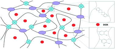
In this study, a novel Fe3+-induced bioinspired chitosan hydrogel was developed to easily deliver the anticancer drug, doxorubicin (DOX). Catechol–chitosan conjugates (CCS) and an N-acetyl cysteine–chitosan conjugate (NACCS) were synthesized and used to prepare the hydrogels. The addition of NACCS accelerated the gelation rate and improved the mechanical strength of the hydrogels, due to the Michael addition reaction between NACCS and the oxidation product of CCS. This study demonstrated that the Fe3+-induced CCS–NACCS hydrogel was a dual covalent-coordination crosslinking system under acidic conditions. Release curves for DOX were evaluated at different pH values, and the release kinetics and mechanism were also investigated. The CCS–NACCS hydrogel showed no obvious toxicity and the DOX released from the hydrogel could effectively inhibit the proliferation of several kinds of tumor cells.


Introduction

Carriers are widely used for the delivery of drugs and their sustained and controlled release in target cells or tissues. Chitosan (CS) is a linear polysaccharide that comes from chitin, which is abundant in nature.1 CS has several unique characteristics, including non-toxicity, biocompatibility, biodegradability, and high mechanical strength; therefore, it has wide applications in the biomedical field, especially for drug delivery.2–4

Several novel drug carriers have been widely used in the biomedical field, including liposomes, micelles, and nanoparticles.5,6 Compared with these carriers, injectable hydrogels that can be synthesized from CS show several obvious advantages. First, hydrogels with 3D networks can be used to load large amounts of drugs, and can release drugs in a sustained and controlled manner, which could lead to better therapeutic results.7,8 Second, hydrogels can release drugs directly to target cells or tissues, avoiding circulation of the drug in the body.8 In this manner, injectable hydrogels can quickly deliver a large dose of drugs.9 Moreover, for cancer patients whose physical conditions are not suitable for surgery, the use of injectable hydrogels could significantly reduce treatment risks.10

Various methods (enzyme-catalyzed reactions, Schiff base reactions, photocrosslinked reactions, etc.) could be used for preparing the hydrogels.10–12 Compared to these reactions, biomimetic approaches are more efficient and safer for hydrogel preparation. Marine mussels can adhere to a variety of substrate surfaces in wet conditions.13 Studies have found that an unusual amino acid, 3,4-dihydroxyphenyl-L-alanine (Dopa), is important for the formation of hydrogel-like adhesive pads.14,15 Research has also revealed that inorganic ions, especially Fe3+, are abundant in their adhesive pads.16,17 The catechol group, side chain of Dopa, is the key to mussel adhesion, and exhibits strong adhesion to various organic and inorganic surfaces.18–20 Hydrogels with catechol-containing polymers could be formed by the oxidation of catechol groups, or coordination between metal ions (e.g., Fe3+) and catechol groups, which could provide a highly biocompatible 3D matrix for drug loading.

In this study, the catechol–chitosan conjugate (CCS) and N-acetyl cysteine–chitosan conjugate (NACCS) were synthesized to prepare a novel Fe3+-induced bioinspired hydrogel for sustained and controlled release of doxorubicin (DOX). Compared to the CCS hydrogel, the novel CCS–NACCS hydrogel exhibited a faster gelation rate and a higher mechanical strength, along with an increased crosslink density due to the covalent crosslinking of the oxidized catechol of CCS with the thiol group of NACCS. The physical properties and the crosslinking mechanism of the hydrogel were characterized by evaluating gelation time, rheological properties, scanning electron microscopy (SEM) images, and Raman spectroscopy. The kinetics and mechanism of drug release were examined through the in vitro release of DOX. The cytotoxicity of the hydrogel and the viability of cells treated with the release solution from DOX-loaded hydrogels were also studied. The results demonstrated that the mussel-inspired Fe3+-induced CCS–NACCS hydrogel was a biocompatible and controllable system that would be appropriate for biomedical applications.

Experimental section

Materials and reagents

N-acetyl cysteine, chitosan (CS, viscosity: 100–200 mPa*s, degree of deacetylation ∼95%), 3,4-dihydroxy benzaldehyde, dopamine hydrochloride, 1-ethyl-3-(3-dimethylaminopropyl)-carbodiimide hydrochloride (EDC), sodium cyanoborohydride (NaBH3CN), ferric chloride hexahydrate, and sodium periodate (NaIO4) were purchased from Sigma-Aldrich (St. Louis, MO, USA). Other reagents were purchased from Sinopharm Chemical Reagent Co., Ltd. (Shanghai, China). All chemicals were used without any purification.

Synthesis of catechol–chitosan conjugates (CCSs)

Catechol–chitosan conjugates (CCSs) were synthesized by the Bosch reduction of Schiff base, according to a previous report, with some modifications (Fig. 1a).21 Briefly, 0.1 g of CS was dissolved in 20 mL of a 1% acetic acid solution. About 0.26 g of 3,4-dihydroxy benzaldehyde dissolved in 5 mL of N,N-dimethylformamide (DMF) was added to this solution. The mixture was incubated at room temperature for 7 h. Then, 0.3 g of NaBH3CN dissolved in 10 mL of H2O was added to this mixture and incubated at room temperature for another 7 h. After the reaction, the product was precipitated by the addition of 500 mL of ethanol, and washed three times with H2O and ethanol. Finally, it was dried under vacuum at room temperature for 5 hours. Formation of CCS was confirmed by nuclear magnetic resonance (1H-NMR, AVANCE III 400, Bruker, Billerica, MA, USA). The degree of substitution (DS) was determined by measuring the absorbance at 280 nm using a UV-vis spectrophotometer (U-5100, Hitachi, Tokyo, Japan), because the aromatic ring structure of the catechol group exhibits an absorption peak at this wavelength.22 A standard curve was prepared using varying concentrations of a dopamine hydrochloride solution.
image file: c6ra07369g-f1.tif
Fig. 1 Synthetic reaction of (a) catechol–chitosan conjugate (CCS) and (b) N-acetyl cysteine–chitosan conjugate (NACCS).

Synthesis of N-acetyl cysteine–chitosan conjugate (NACCS)

As shown in Fig. 1b, N-acetyl cysteine-chitosan conjugate (NACCS) was synthesized by the catalysis of EDC to couple CS and N-acetyl cysteine according to a previous report.23 Briefly, 4 g of N-acetyl cysteine was dissolved in 50 mL of H2O. 4.7 g of EDC was added to activate the carboxylic acid group of N-acetyl cysteine, and the reaction proceeded for 20 min. Then, 0.5 g of CS dissolved in 30 mL of 1% aqueous hydrochloric acid was added to the mixture. The pH was continuously monitored and maintained at pH 4–6 for 6 h. After the reaction, the solution was purified in the dark by dialysis (MWCO = 2000) for 3 d and was subsequently lyophilized, which resulted in white powder. The DS of NACCS was calculated by comparing the intensity of the C-2 proton signal of CS with that of the methylene group (2H, –CH2–S–) of N-acetyl cysteine in the 1H-NMR spectrum.

Formation of CCS–NACCS hydrogels

CCS and NACCS solution were prepared by dissolving CCS and NACCS in a 1% acetic acid solution (v/v). Predetermined amounts of FeCl3 or NaIO4 solution (10 mM phosphate buffer, pH 7.4) were added and mixed. A typical procedure for gel formation included the mixing of 500 μL of the appropriate CCS solution (catechol concentration: 49.5 mM, 33.0 mM, 16.5 mM) with 150 μL of the appropriate NACCS solution (thiol concentration: 55.0 mM, 36.7 mM, 18.3 mM) and 50 μL of the corresponding FeCl3 or NaIO4 solution (165 mM, 330 mM, 495 mM). The mixture was incubated at room temperature and gelled quickly. The gelation time was calculated by the inversion method.

Rheology of hydrogels

Fe3+- and IO4-induced CCS–NACCS bi-component hydrogels and CCS mono-component hydrogels were used for the rheology test. Fe3+- or IO4-induced CCS–NACCS bi-component hydrogels were prepared by mixing 500 μL CCS solution (catechol concentration: 49.5 mM), 150 μL NACCS solution (thiol concentration: 55.0 mM), and 50 μL FeCl3 or NaIO4 solution (165 mM). 150 μL NACCS solution was replaced by 150 μL ddH2O to prepare CCS mono-component hydrogels.

A rheometer (ARES, TA Instruments, New Castle, DE, USA) with parallel plate geometry (25 mm diameter) was used to test the mechanical properties of the hydrogels. The oscillatory shear test (as a function of frequency) was performed at 10% strain from 100 rad s−1 to 0.1 rad s−1 by recording the storage modulus (G′) and loss modulus (G′′). The modulus of the hydrogel was also measured at a time sweep mode at 10% strain and 10 rad s−1. The values were collected from three separate hydrogels and averaged.

Resonance Raman spectroscopy

An InVia microscope Raman (inVia Reflex, Renishaw, Gloucestershire, UK) was used for Raman spectroscopic studies. The lyophilized hydrogels were excited at 514 nm. For the measurements, the Laser power was maintained at 10–30 mW. The spectra were collected from three different regions and averaged.

Drug load and release

DOX, an anticancer drug, was loaded into CCS–NACCS hydrogels to evaluate the hydrogels' potential application in drug delivery. 1 mL of CCS solution (DS: 77.7%, catechol concentration: 49.5 mM) and 0.3 mL of NACCS solution (thiol concentration: 55.0 mM) was mixed, DOX solution (final concentration: 0.7 mg mL−1) was added and stirred at 4 °C. 0.1 mL of FeCl3 solution (165 mM) was added to prepare the DOX-loaded hydrogel and the gel was shaped as a cylinder. The DOX-loaded hydrogel was washed three times to obtain the free DOX solution. The amounts of DOX in the initial feed solution and in the washing solution were measured by a UV spectrophotometer at 495 nm, and the amount of the loaded DOX was calculated by subtracting the amount of DOX in the washing solution from that in the initial feed solution. The drug loading capacity (DLC) was calculated by comparing the amount of the loaded DOX with the weight of the hydrogel, and the drug loading efficiency (DLE) was calculated by comparing the amount of the loaded DOX with the amount of DOX in the initial feed solution.

The DOX-loaded hydrogels were immersed at 37 °C into 15 mL of PBS buffer at pH 5.0, 6.8, or 7.4 to mimic drug release from the hydrogels in situ. At predetermined time intervals, 5 mL of incubation solution was taken out and 5 mL of fresh PBS was added to maintain a constant volume of 15 mL. The cumulatively released amount of DOX was calculated according to a standard curve of DOX measured at an absorbance of 495 nm using a UV spectrophotometer, whose linear regression equation was A = 13.337C + 0.0314. All experiments were repeated three times.

Cell culture and cell viability study

The human lung cancer cell line NCI-H460 was cultured with RPMI-1640 medium (Gibco BRL, Gaithersburg, MD, USA) supplemented with 10% fetal bovine serum (FBS, Gibco BRL, Gaithersburg, MD, USA) at 37 °C in 5% CO2 humidified atmosphere.

Cytotoxicity was evaluated by the MTT assay to measure the viability of cells exposed to hydrogel extracts.24 Cylinder-shaped hydrogels (5 mm diameter and 2 mm thick) were incubated in RPMI-1640 medium for 24 h at 37 °C to obtain hydrogel extracts (1–100 mg mL−1). The hydrogel extracts were then filtered through a 0.22 μm sterile filter. NCI-H460 cells were seeded on 96-well plates at a density of 8000 cells/100 μL per well and incubated for 18 h. Then, the medium was replaced by 100 μL per well of hydrogel extract. Cells cultured in fresh RPMI-1640 were used as a control. After incubation for 24 h, 10 μL of MTT solution (5 mg mL−1 in PBS) was added into each well and incubated for another 4 h. Then, the solution was removed and 150 μL of DMSO was added into each well to dissolve the crystals completely. The absorbance was measured at 490 nm (n = 5) using a microplate reader (SpectraMax M5, Molecular Devices, California, USA).

Cell viability was also evaluated using a Live/Dead staining protocol. After incubation with hydrogel extracts, cells were washed with PBS and were then incubated in 100 μL of working solution containing 2 μM calcein-AM and 4 μM PI at 37 °C for 15 min. The images were taken by a fluorescence microscope (Ti–S, Nikon, Tokyo, Japan).

The potency of released DOX was also examined. DOX-loaded hydrogels and hydrogels without DOX were immersed into RPMI-1640 medium instead of PBS at 37 °C to obtain the released DOX solutions. Released DOX solutions sampled at various time points were exposed to NCI-H460 cells, and assayed for cell viability using the MTT assay. Fresh RPMI-1640 medium and the released solution from hydrogels without DOX were used as negative and positive controls, respectively.25

Other cell lines, including MCF-7, MKN45, MDA-MB-231, and A549, were also evaluated using the same method.

Cellular uptake and intercellular distribution of DOX

Cellular uptake and intercellular distribution of DOX was investigated using a confocal laser scanning microscope (CLSM, A1R, Nikon, Tokyo, Japan). The NCI-H460 cells were seeded on a culture dish at a density of 50[thin space (1/6-em)]000 cells per mL and cultured at 37 °C. After a predetermined incubation time, the dish was washed three times with PBS. The released solution from the DOX-loaded hydrogel was added to the dish for further incubation. After 2 hours, the dish was washed three times with cold PBS to remove dead cells and free DOX. Cells were fixed with 4% paraformaldehyde solution for 10 min. The sample was washed with PBS and stained with 4′,6-diamidino-2-phenylindole (DAPI) for 10 min.

Results and discussion

Synthesis and characterization of catechol–chitosan conjugates (CCSs) and an N-acetyl cysteine–chitosan conjugate (NACCS)

As shown in Fig. 1a, CCS was synthesized by Bosch reduction of a Schiff base that formed by the reaction of CS and 3,4-dihydroxy benzaldehyde. 1H-NMR analysis confirmed the conjugation of catechol to CS by showing that the peaks of aromatic protons in catechol appeared at ∼6.7 ppm (Fig. S1b).26 The UV-vis spectra of CCS exhibited an absorption peak at 280 nm (Fig. S2), confirming the successful synthesis of CCS.22,27 A series of CCS with different DS (40.7%, 62.4%, 77.7%) were synthesized as shown in Table S1.

NACCS was achieved by coupling the amino group of CS and the carboxylic acid group of N-acetyl cysteine through EDC (Fig. 1b). 1H-NMR analysis confirmed the conjugation of N-acetyl cysteine to CS by showing that the peaks of the methylene group (2H, –CH2–S–) in N-acetyl cysteine appeared at 2.8 ppm (Fig. S1c). The DS of NACCS was calculated to be 24.5%, and the yield of NACCS was 83.4%.

Hydrogel formation

The CCSs with different DSs (obtained as described in Table S1) were used for the gel experiment to investigate the impact of DS. The molar ratio of Fe3+/IO4 and catechol remained constant at 1[thin space (1/6-em)]:[thin space (1/6-em)]3, while the thiol concentration remained constant at 18.3 mM. As shown in Fig. 2a, increasing the DS of CCS from 40.7% to 77.7%, decreased the gelation time of Fe3+-induced CCS–NACCS hydrogels from 1874 s to 45 s. This was also observed for the gelation time of IO4-induced hydrogels, which decreased from 848 s to 97 s (Fig. 2b). These results suggest that CCSs with a higher DS gel faster for both Fe3+-induced and IO4-induced hydrogels. The degree of crosslinking included in the system was proportional to the concentration of catechol groups included, because higher concentrations of catechol groups were more conducive to forming chemical interactions (oxidation and coordination).
image file: c6ra07369g-f2.tif
Fig. 2 Influence of DS (a and b), the molar ratio of Fe3+/IO4 and catechol (c and d), and thiol concentration (e and f) on the gelation time of Fe3+-induced hydrogels (left) and IO4-induced hydrogels (right).

Fig. 2c & d showed the impact of the molar ratio of Fe3+/IO4 and catechol on gelation time. The effect of catechol concentration was investigated at a constant DS of 77.7%, thiol concentration of 18.3 mM, and Fe3+/IO4 concentration of 11.8 mM. When the catechol concentration included in the system was 11.8 mM, it remained liquid, possibly because the low catechol concentration could not sufficiently crosslink. When the catechol concentration was increased from 23.6 mM to 35.4 mM, the gelation time of Fe3+-induced hydrogels decreased from 432 s to 45 s. This was also confirmed by an increased absorption peak at 420 nm (Fig. S3b), which was changed from the peak of quinone at 400 nm by the addition of NACCS (Fig. S3a & d).28,29 IO4-induced hydrogels exhibited similar results; as the catechol concentration increased from 23.6 mM to 35.4 mM, the gelation time decreased from 216 s to 97 s. Next, the effect of Fe3+/IO4 concentration was investigated at a constant DS of 77.7%, thiol concentration of 18.3 mM, and catechol concentration of 35.4 mM. When the Fe3+/IO4 concentration was increased from 11.8 mM to 35.4 mM, the gelation time of Fe3+-induced hydrogels increased from 45 s to 165 s, which is contrary to the gelation time of IO4-induced hydrogels, which decreased from 97 s to 54 s. A higher concentration of oxidant (NaIO4) is favorable for covalent crosslinking; thus, the gelation rate of IO4-induced hydrogels was increased. However, Fe3+-induced hydrogels were not a covalent-dominated system like IO4-induced hydrogels, but were possibly a dual covalent-coordination crosslinking system under acidic condition (pH ∼ 3). This was confirmed by the appearance of absorption peaks at 420 nm (due to covalent interactions) and between 500 and 700 nm (due to coordination) in its UV-vis spectra (Fig. S3).29 The colors of the hydrogels also showed differences between Fe3+-induced and IO4-induced hydrogels. The Fe3+-induced hydrogel was black, while the IO4-induced hydrogel was brown (Fig. S4).

The effect of thiol concentration was investigated at a constant DS of 77.7% and a constant Fe3+/IO4-catechol molar ratio of 1[thin space (1/6-em)]:[thin space (1/6-em)]3. As show in Fig. 2e & f, the gelation time decreased from 53 s to 12 s for Fe3+-induced hydrogels and from 116 s to 72 s for IO4-induced hydrogels when the thiol concentration was increased from 0 to 55.0 mM. This is mainly because the Michael addition reaction has occurred between the thiol group of NACCS and the highly reactive quinone, the oxidation product of catechol, which results in faster formation of the hydrogel. The absorption peak of the UV-vis spectrum shifted from 400 nm to 420 nm (Fig. S3d), which also confirmed the Michael addition reaction.

Hydrogel characterization

The mechanism of IO4-induced and Fe3+-induced CCS–NACCS hydrogels were illustrated in Scheme S1. The rheology test was used to elucidate the mechanical properties of the hydrogels. As shown in Fig. 3a and Fig. 3c, the G′ and G′′ of IO4-induced CCS and CCS–NACCS hydrogels do not change significantly with the change in oscillation frequency, and G′≫G′′, suggesting that the IO4-induced hydrogel fits the Hookean elastic solid model.21 This was consistent with the properties of other NaIO4-induced covalently crosslinked hydrogels prepared with catechol-modified polymers.30,31 It is believed that the plateau G′ can be used to represent the mechanical strength of the hydrogel.11 The G′ values of CCS–NACCS bi-component hydrogels and CCS mono-component hydrogels were 609.1 ± 25.83 Pa and 527.2 ± 25.91 Pa, respectively, which represented that the mechanical strength of hydrogel was improved by 15.5% with the addition of NACCS. This is mainly because the Michael addition reaction has occurred between the thiol group of NACCS and the oxidation product of CCS, which results in an increase of crosslinking density.
image file: c6ra07369g-f3.tif
Fig. 3 Rheological properties of IO4-induced hydrogels (a and c) and Fe3+-induced hydrogels (b and d).

For Fe3+-induced hydrogels, the G′ and G′′ of CCS–NACCS bi-component hydrogels were higher than that of CCS mono-component hydrogels, which was consistent with the higher crosslinking density caused by the Michael addition reaction. The pore sizes of freeze-dried hydrogels were analyzed by SEM (S-3400N, Hitachi, Tokyo, Japan). As shown in Fig. S5, the average pore size of CCS–NACCS hydrogels was 48 ± 8.1 μm, which was smaller than that of CCS hydrogel (65 ± 20.8 μm), further supporting that the addition of NACCS increased the crosslinking density of the system. From Fig. 3b and d, we can see that the CCS–NACCS bi-component hydrogels and the CCS mono-component hydrogels exhibited similar rheological properties, which fit neither the Hookean elastic solid model nor the Maxwell viscoelasticity model; rather, they are neither a simple covalently crosslinked system nor a simple coordinately crosslinked system, but a dual covalently-coordinately crosslinked system.29,30 In general, the mechanical strength of Fe3+-induced hydrogels was weaker than that of IO4-induced hydrogels. G′ values of Fe3+-induced and IO4-induced CCS–NACCS were 197.1 ± 17.04 Pa and 609.1 ± 25.83 Pa, respectively. G′ values of Fe3+-induced and IO4-induced CCS hydrogel were 181.5 ± 11.80 Pa and 527.2 ± 25.91 Pa, respectively. This is because the presence of mechanically functional Fe3+-catechol coordination bonds endowed the hydrogels with a new viscoelastic behavior that dissipate energy under applied force.21

Raman spectroscopy was also used to characterize Fe3+-induced CCS mono-component hydrogels and CCS–NACCS bi-component hydrogels (Fig. 4). Compared with the CCS spectra, the CCS–NACCS spectra showed an obvious peak at around 690 cm−1 that represented C–S bond vibrations, and when the thiol concentration of NACCS was increased from 18.3 to 55.0 mM, the intensity of this peak was increased from 61.0 ± 18.12 to 105.5 ± 15.23 a.u., which confirmed that increasing the amount of NACCS could improve the Michael addition reaction of the oxidized CCS and NACCS. The addition of NACCS also increased the intensity ratio of the peaks at 1487 cm−1 (C–H bond vibrations) and 1571 cm−1 (C[double bond, length as m-dash]N bond vibrations), because that catechols in Fe3+-induced CCS mono-component hydrogel were oxidized to covalently crosslinked with amino groups, and to coordinately crosslinked with Fe3+, while in CCS–NACCS bi-component hydrogel, catechols were oxidized to covalently crosslinked with both amino groups and thiol groups, which decreased the intensity of C[double bond, length as m-dash]N bond vibrations. Peaks at 533 cm−1, 582 cm−1, and 630 cm−1 represented Fe–O bond vibrations according to a previous report.29 Specifically, the peak at 533 cm−1 indicated the charge transfer resonance energy of the chelate, and the intensity of this peak represented the degree of Fe–O coordination.21


image file: c6ra07369g-f4.tif
Fig. 4 Resonance Raman spectroscopy of Fe3+-induced CCS–NACCS bi-component hydrogels (thiol concentration: 55.0 mM, 36.7 mM, 18.3 mM) and CCS mono-component hydrogel.

In vitro DOX release from hydrogels

DOX was loaded into hydrogels to evaluate their in vitro release properties. As shown in Table S2, the drug loading capacity (DLC) and drug loading efficiency (DLE) at pH 7.4 were 0.998 mg DOX/g hydrogel and 99.5%, respectively. A continuous and controllable release of DOX was clearly observed (Fig. 5). The hydrogels quickly released DOX for the first 12 h, because of the large concentration gradient between the hydrogel and PBS. The release rate then slowed over a period of 12–36 h, and then plateaued. There was a cumulative release of ∼44.9% of the loaded DOX within 60 h in PBS at pH 7.4. When the pH of PBS was decreased to 5.0, the cumulative release of DOX increased to 61.1%, because the solubility of DOX at pH 5.0 is higher than that at pH 6.8 or 7.4.
image file: c6ra07369g-f5.tif
Fig. 5 Release curves for DOX from CCS–NACCS hydrogels in PBS buffers with different pHs (n = 3).

The amount of cumulatively released DOX was fitted to the Ritger-Peppas model (eqn (1)) to describe the drug release mechanism:

 
Mt/M = ktn (1)

It has been reported that when samples were cylindrical and the diffusion exponent, n, was tested to be less than 0.45, the release mechanism could be described by Fickian diffusion.32,33 In the present study, the n values for release in PBS at pH 7.4, 6.8, and 5.0 were 0.1326, 0.1492, and 0.2426, respectively (Table S2). Thus, they also conformed to the Fickian diffusion model, indicating that DOX is released from Fe3+-induced CCS–NACCS hydrogels by diffusion.

Cellular uptake and intercellular distribution of DOX

Cellular uptake and intercellular distribution of DOX was investigated by CLSM. The nuclei of NCI-H460 cells were stained blue with DAPI (Fig. 6a), DOX is shown as red fluorescence (Fig. 6b), and Fig. 6c is a merged image of Fig. 6a and b. Intensive red fluorescence appeared in the nucleus, which indicated that DOX had released from the hydrogel and had accumulated in the cell nuclei. This is consistent with the report that DOX had direct interactions with DNA in the cell nuclei and inhibited the proliferation of tumor cells by inhibiting the replication of DNA.34 As shown in Fig. 6c, most DOX stayed in the nuclei, and only a small amount of DOX was located in the cytoplasm, which also indicated that DOX had released from the hydrogel and was taken up by cells.
image file: c6ra07369g-f6.tif
Fig. 6 CLSM images of NCI-H460 cells incubated with released solution from a DOX-loaded hydrogel. (a) Blue indicates the nucleus, which was stained with DAPI; (b) red indicates DOX fluorescence; (c) a merged image of (a) and (b); scale bars are 50 μm for all images.

Cell viability study

The cytotoxicity of CCS–NACCS hydrogels was examined to determine their biocompatibility. The half maximal inhibitory concentration (IC50) was measured for different kinds of cells, including NCI-H460, MCF-7, MKN45, MDA-MB-231, A549, Hela, and SKOV-3. The IC50 of free DOX for NCI-H460 cells was the lowest (Table S3), suggesting that NCI-H460 cells were more sensitive to DOX than the other cells. For NCI-H460 cells, the IC50 of DOX-loaded hydrogels was 11.5 mg mL−1 (5.1 μg DOX/mL), which was close to that of free DOX (4.8 μg mL−1). As shown in Fig. 7a, the viability of NCI-H460 cells that were exposed to different concentrations (1–100 mg mL−1) of hydrogel extracts was higher than 75%, suggesting no serious cytotoxicity.35 This was also confirmed for the other cell lines (Fig. S6). These results indicate that the CCS–NACCS hydrogel was biocompatible, and that it could be used in the biomedical field.
image file: c6ra07369g-f7.tif
Fig. 7 Cytotoxicity test of an Fe3+-induced CCS–NACCS hydrogel (a), the viability of NCI-H460 cells incubated with released solutions from DOX-loaded hydrogels and hydrogels without DOX at various release time points (b), fluorescence microscope images of NCI-H460 cells cultured with released solutions from hydrogels without DOX (c) and DOX-loaded hydrogels (d). Green indicates live cells, red indicates both dead cells and DOX; scale bars are 200 μm for both images.

The viability of NCI-H460 cells cultured with the DOX released solutions from DOX-loaded CCS–NACCS hydrogels and CCS–NACCS hydrogels without DOX was tested at different release time points. As shown in Fig. 7b, with increasing release time from 2 h to 1 d, the viability of NCI-H460 cells decreased from 90.4% to 56.8% due to the fast release of DOX. Cell viability was 38.0% after a release time of 7 d. The other cell lines exhibited similar results (Fig. S7). These results indicate that DOX was released in a sustained and controlled manner from CCS–NACCS hydrogels, and that the potency of DOX was maintained during the whole process.

Viability of cells cultured with DOX releasing solutions from CCS–NACCS hydrogels without DOX and from DOX-loaded CCS–NACCS hydrogels was also evaluated by live/dead staining. As shown in Fig. 7c, almost all cells are alive (green) when cultured with the released solution from hydrogels without DOX, with a viability >95%. This is consistent with the experimental results of the cytotoxicity test described above, further supporting that the hydrogel is biocompatible. However, in Fig. 7d, a significant number of NCI-H460 cells cultured with the released solution from DOX-loaded hydrogels were dead (red). For example, in response to the 44.5% of DOX released from the DOX-loaded hydrogel within 24 h at pH7.4 (Fig. 5), large amounts of cells were dead. This also supports that DOX was released from the hydrogels, and that it maintained its potency, which is consistent with the experimental results of Fig. 7b.

Conclusions

In this study, we used two chitosan derivatives, CCS and NACCS, to develop a novel and injectable Fe3+-induced CCS–NACCS hydrogel for the delivery of DOX. This crosslinked hydrogel was formed by the covalent and coordinate interactions of catechol and Fe3+. The Michael addition reaction between NACCS and the oxidation product of CCS also accelerated the gelation rate and improved the mechanical strength of the hydrogel. The hydrogel can be used for the sustained and controlled release of DOX, which was confirmed by the release curves for DOX, the MTT assay, fluorescence microscope images, and CLSM images. NCI-H460 cells incubated with hydrogel extracts maintained high viability, demonstrating the biocompatibility of the hydrogel.

Acknowledgements

This work was funded by the National Special Fund for the State Key Laboratory of Bioreactor Engineering (2060204) and the National major science and technology projects of China (No. 2012ZX09304009).

Notes and references

  1. J. Y. Kim, J. K. Lee, T. S. Lee and W. H. Park, Int. J. Biol. Macromol., 2000, 32, 23–27 CrossRef.
  2. C. E. Orrego, N. Salgado, J. S. Valencia, G. I. Giraldo, O. H. Giraldo and C. A. Cardona, Carbohydr. Polym., 2010, 79, 9–16 CrossRef CAS.
  3. O. Felt, P. Buri and R. Gurny, Drug Dev. Ind. Pharm., 1998, 24, 979–993 CrossRef CAS PubMed.
  4. N. Bhattarai, J. Gunn and M. Zhang, Adv. Drug Delivery Rev., 2010, 62, 83–99 CrossRef CAS PubMed.
  5. J. A. Champion and S. Mitragotri, Pharm. Res., 2009, 26, 244–249 CrossRef CAS PubMed.
  6. M. A. C. Stuart, W. T. S. Huck, J. Genzer, M. Muller, C. Ober, M. Stamm, G. B. Sukhorukov, I. Szleifer, V. V. Tsukruk, M. Urban, F. Winnik, S. Zauscher, I. Luzinov and S. Minko, Nat. Mater., 2010, 9, 101–113 CrossRef PubMed.
  7. J. K. Cho, K. Y. Hong, J. W. Park, H. K. Yang and S. C. Song, J. Drug Targeting, 2011, 19, 270–280 CrossRef CAS PubMed.
  8. S. J. Lee, Y. Bae, K. Kataoka, D. Kim, D. S. Lee and S. C. Kim, Polym. J., 2007, 40, 171–176 CrossRef.
  9. J. P. Smith, S. Kanekal, M. B. Patawaran, J. Y. Chen, R. E. Jones, E. K. Orenberg and N. Y. Yu, Cancer Chemother. Pharmacol., 1999, 44, 267–274 CrossRef CAS PubMed.
  10. J. Shi, W. Guobao, H. Chen, W. Zhong, X. Qiu and M. M. Q. Xing, Polym. Chem., 2014, 5, 6180–6189 RSC.
  11. J. Hou, C. Li, Y. Guan, Y. Zhang and X. X. Zhu, Polym. Chem., 2015, 6, 2204–2213 RSC.
  12. Y. F. Poon, Y. B. Zhu, J. Y. Shen, M. B. Chan-Park and S. C. Ng, Adv. Funct. Mater., 2007, 17, 2139–2150 CrossRef CAS.
  13. H. Lee, B. P. Lee and P. B. Messersmith, Nature, 2007, 448, 338–341 CrossRef CAS PubMed.
  14. J. H. Waite, J. Biol. Chem., 1983, 258, 2911–2915 CAS.
  15. B. P. Lee, P. B. Messersmith, J. N. Israelachvili and J. H. Waite, Annu. Rev. Mater. Res., 2011, 41, 99–132 CrossRef CAS PubMed.
  16. S. W. Taylor, G. W. Luther and J. H. Waite, Inorg. Chem., 1994, 33, 5819–5824 CrossRef CAS.
  17. M. J. Harrington, A. Masic, N. Holten-Andersen, J. H. Waite and P. Fratzl, Science, 2010, 328, 216–220 CrossRef CAS PubMed.
  18. J. H. Waite, N. H. Andersen, S. Jewhurst and C. Sun, J. Adhes., 2005, 81, 297–317 CrossRef CAS.
  19. L. Ninan, R. L. Stroshine, J. J. Wilker and R. Shi, Acta Biomater., 2007, 3, 687–694 CrossRef CAS PubMed.
  20. H. Lee, N. F. Scherer and P. B. Messersmith, Proc. Natl. Acad. Sci. U. S. A., 2006, 103, 12999–13003 CrossRef CAS PubMed.
  21. Z. Guo, K. Ni, D. Wei and Y. Ren, RSC Adv., 2015, 5, 37377–37384 RSC.
  22. J. H. Ryu, Y. Lee, W. H. Kong, T. G. Kim, T. G. Park and H. Lee, Biomacromolecules, 2011, 12, 2653–2659 CrossRef CAS PubMed.
  23. T. Schmitz, V. Grabovac, T. F. Palmberger, M. H. Hoffer and A. Bernkop-Schnürcha, Int. J. Pharm., 2008, 347, 79–85 CrossRef CAS PubMed.
  24. M. Mehdizadeh, H. Weng, D. Gyawali, L. Tang and J. Yang, Biomaterials, 2012, 33, 7972–7983 CrossRef CAS PubMed.
  25. R. Rastogi, N. Gulati, R. K. Kotnala, U. Sharma, R. Jayasundar and V. Koul, Colloids Surf., B, 2011, 82, 160–167 CrossRef CAS PubMed.
  26. S. Hong, K. Yang, B. Kang, C. Lee, I. T. Song, E. Byun, K. I. Park, S. Cho and H. Lee, Adv. Funct. Mater., 2013, 23, 1774–1780 CrossRef CAS.
  27. C. Lee, J. Shin, J. S. Lee, E. Byun, J. H. Ryu, S. H. Um, D. Kim, H. Lee and S. Cho, Biomacromolecules, 2013, 14, 2004–2013 CrossRef CAS PubMed.
  28. E. Mentasi, E. Pelizzetti and G. Saini, J. Chem. Soc., Dalton Trans., 1973, 23, 2609–2614 RSC.
  29. N. Holten-Andersen, M. J. Harrington, H. Birkedal, B. P. Lee, P. B. Messersmith, K. Y. C. Lee and J. H. Waite, Proc. Natl. Acad. Sci. U. S. A., 2011, 108, 2651–2655 CrossRef CAS PubMed.
  30. B. P. Lee, J. L. Dalsin and P. B. Messersmith, Biomacromolecules, 2002, 3, 1038–1047 CrossRef CAS PubMed.
  31. D. G. Barrett, D. E. Fullenkamp, L. He, N. Holten-Andersen, K. Y. C. Lee and P. B. Messersmith, Adv. Funct. Mater., 2013, 23, 1111–1119 CrossRef CAS PubMed.
  32. P. L. Ritger and N. A. Peppas, J. Controlled Release, 1987, 5, 37–42 CrossRef CAS.
  33. P. L. Ritger and N. A. Peppas, J. Controlled Release, 1987, 5, 23–36 CrossRef CAS.
  34. S. Goto, Y. Ihara, Y. Urata, S. Izumi, K. Abe, T. Koji and T. Kondo, FASEB J., 2001, 15, 2702–2714 CrossRef CAS PubMed.
  35. Y. Liu, H. Meng, S. Konst, R. Sarmiento, R. Rajachar and B. P. Lee, ACS Appl. Mater. Interfaces, 2014, 6, 16982–16992 CAS.

Footnote

Electronic supplementary information (ESI) available. See DOI: 10.1039/c6ra07369g

This journal is © The Royal Society of Chemistry 2016
Click here to see how this site uses Cookies. View our privacy policy here.