Graphene oxide coated Fe3O4@mSiO2 NPs for magnetic controlled bioimaging

Aihua Liac, Hongjing Maab and Jingquan Liu*ab
aShandong Provincial Key Laboratory of Detection Technology for Tumor Markers, College of Chemistry and Chemical Engineering, Linyi University, Shuangling Road, Linyi, China. E-mail: jliu@qdu.edu.cn
bCollege of Materials Science and Engineering, Qingdao University, No. 308, Ningxia Road, Qingdao, China
cKey Laboratory of Special Functional Aggregated Materials, Key Laboratory of Colloid and Interface Chemistry (Shandong University) Ministry of Education, School of Chemistry and Chemical Engineering, Shandong University, No. 27, South Shanda Road, Jinan, China

Received 21st March 2016 , Accepted 18th June 2016

First published on 22nd June 2016


Abstract

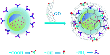
A novel GO coated porous magnetic NPs (Fe3O4@mSiO2@GO) for bioimaging was synthesized. First, Fe3O4 magnetic NPs were prepared through a solvothermal reaction. Second, through a surfactant-templating approach with cetyltrimethylammonium bromide (CTAB) as a template, a mesostructured CTAB/SiO2 composite was deposited on Fe3O4 NPs surfaces. Third, CTAB templates were removed to form a mesoporous SiO2 shell, resulting in mesoporous magnetic (Fe3O4@mSiO2) NPs. Forth, the magnetic NPs surfaces were functionalized with amino groups by (3-aminopropyl)triethoxysilane (APTES). Finally, the dye rhodamine B (RB) was loaded inside porous magnetic NPs, and the magnetic NPs surfaces were then wrapped with graphene oxide (GO) nanosheets. SEM analysis revealed that GO sheets have been successfully coated on magnetic NPs surfaces. The hysteresis loops analysis indicated that these magnetic NPs still retained strongly magnetic property. Furthermore, Fe3O4@mSiO2@GO NPs could efficiently protect the loaded dye from releasing. In addition, MTT assay revealed that the RB loaded Fe3O4@mSiO2@GO NPs exhibited insignificant cytotoxicity at moderate concentrations and the RB loaded Fe3O4@mSiO2@GO NPs could undergo target-directed move under the action of a magnetic field. The noncytotoxic magnetic hybrids presented significant potential for applications in cell imaging.


Introduction

Magnetic iron oxide NPs have attracted extensive research interest due to their unique physicochemical properties and great potential for various biomedical applications. In recent decades, magnetic NPs, especially Fe3O4 (magnetite) and Fe2O3 (maghemite), have been applied in various fields, particularly in biotechnology and biomedicine, such as enzyme-immobilization,1 separation and purification,2 biosensing and bioelectrocatalysis,3 magnetic resonance imaging,4 drug delivery,5 and tumor therapy.6 The requirements for any biomedical application of magnetic NPs include the chemical stability, biocompatibility and strong magnetization of the dispersed magnetic NPs. However, these magnetic particles are sensitive and unstable, especially under acidic conditions. In order to tackle these problems Fe3O4 particles are usually protected with proper shells to form core–shell structures such as Fe3O4@Al2O3 (ref. 7) and Fe3O4@SiO2 (ref. 8) and so on. For example, the silica shell can not only prevent the aggregation of the Fe3O4 particles but also protect the inner magnetite core from oxidation, it also provides numerous surface Si–OH groups for further modification,9 especially mesoporous silica.

Mesoporous materials, which possess easily modified surface, ordered pores, proper pore volume and large surface area, are widely applied in various fields.10–14 Since the first discovery in 1992,15,16 synthesis and applications of mesoporous silica microspheres have attracted great interest due to their outstanding properties.11,17 Up to now, mesoporous silica NPs have been well studied and employed as drug delivery platforms.14,18 For example, Zhang19 et al. studied polymeric prodrug grafted hollow mesoporous silica nanoparticles for drug release. Liu20 et al. prepared gold nanorods coated with mesoporous silica shell as drug delivery system, in this paper 1-tetradecanol molecules were also utilized as gatekeepers to regulate the drug release. However, most research work just focused on conventional constant, rather than controlling the drug release to the targeting sites. Undoubtedly, the combination of mesoporous silica and magnetic property can generate a new class of materials with great application potential.21–26 The core–shell magnetic mesoporous silica NPs have strong magnetic responsivity, high dispersibility and orientated and accessible mesopores, which is highly valuable. On the other hand, the integration of mesoporous silica NPs and graphite oxide (GO) via electrostatic adsorption properties have been applied as an avenue for biological applications such as fluorescence imaging.27 For example, Yang28 et al. recently reported the fabrication of GO-encapsulated metal oxide NPs as high-performance anode materials through the co-assembly of GO sheets and positively charged oxide NPs. Zhao27 et al. studied graphene oxide wrapping on squaraine-loaded mesoporous silica nanoparticles for bioimaging.

GO with its unique structure provides a broad range of applications, including DNA detection,29,30 drug delivery,31,32 and biological imaging.33–36 GO produced by the oxidative treatment of graphite is two-dimensional and full of hydrophilic groups around the edges and on the planar surfaces, therefore, it has been an attractive choice as the substrate for preparation of nanocomposites due to its large surface area, novel optical, electronic, mechanical, and catalytic properties. The hydroxyl groups and carboxyl groups are responsible for the negative charges on the GO sheets in aqueous suspensions.37,38 These functional groups will benefit for the preparation of GO composites with other materials either via electrostatic interactions27,39,40 or covalent bonding.39,41–43

Herein we synthesized a novel Fe3O4@mSiO2@GO magnetic NPs with loaded RB for bioimaging. The magnetic Fe3O4 particles were prepared through a solvothermal reaction,44 followed by the surface deposition of mesostructured CTAB/SiO2 composite through a surfactant-templating approach with cetyltrimethylammonium bromide (CTAB) as template. The CTAB template was removed and then the magnetic NPs were aminated by APTES. The Fe3O4@mSiO2 NPs were loaded with the dye RB, followed by wrapping of GO around the surfaces of Fe3O4@mSiO2 NPs via electrostatic interaction. This novel magnetic hybrid material exhibited remarkable stability and could efficiently protect the loaded dye from releasing and could undergo target-directed move under the action of a magnetic field. Therefore, the mesoporous magnetic hybrids with superior properties present potential for application in cell imaging.

Experimental

Materials

Ferric chloride hexahydrate (FeCl3·6H2O, 99%) and ethylene glycol (AR) were purchased from Adamas-beta. Sodium citrate (99%), sodium acetate trihydrate (NaAc, 99%) and ethanol (AR) were purchased from Aldrich. Ammonia water (NH3·H2O, 28 wt%), hexadecyl trimethyl ammonium bromide (CTAB, 99%), tetraethyl orthosilicate (TEOS, AR), (3-aminopropyl)triethoxysilane (APTES, 99%), ammonium nitrate (99%) and rhodamine B (RB, 99%) were purchased from Aladdin. GO was prepared by a modified Hummer's method.45,46

Instrumentation

The TEM images of the magnetic NPs were taken on a JEOL JEM-1200EX microscope at an accelerating voltage of 100 kV. The particles were dispersed in water (1 mg mL−1) and deposited onto holey carbon grids (200 mesh copper holey carbon) and allowed to air-dry at room temperature. Fourier-transform infrared (FT-IR) spectroscopy was obtained using Tensor 27 produced by Bruker Corporation. Each sample was analyzed using 128 scans. The X-ray photoelectron spectroscopy (XPS) measurements were performed using a Kratos Axis Ultra DLD spectrometer employing a monochromated Al Kα X-ray source (hγ = 1486.7 eV), hybrid (magnetic/electrostatic) optics, and a multi-channel plate, and delay line detector. Spectra were recorded at a takeoff angle of 35 (angle between the plane of the sample surface and the entrance lens of the analyzer) with pass energy of 150 eV. UV spectra were recorded on a UV/V-16/18 UV spectrophotometer (Shanghai Mapada), which was equipped with a double beam proportion monitoring optical system with a wavelength detection range from 200 to 800 nm. The images of Fe3O4@mSiO2@GO were measured using SEM analysis (Zeiss Auriga). Zeta potential was measured by Malvern Nanosizer. The magnetic property was measured at room temperature using VSM (VSM7407, Lakeshore, USA). Polarization microscopic image of the magnetic nanohybrids was obtained using a Nikon Eclipse E600 microscope equipped with a Nikon DXN1200 digital camera and a Nikon Plan Fluor 50× objective (Nikon, Tokyo, Japan). The excitation laser was reflected by a polarized mirror to a high numerical aperture (NA) oil objective (50×) and focused to a diffraction limited spot (∼300 nm) on the sample surface.

Synthesis of Fe3O4 NPs

The magnetic particles were synthesized through a solvothermal reaction according to the reported method.44 FeCl3·6H2O (1.087 g) was dissolved in ethylene glycol (40 mL) to form a clear solution, NaAc (3.6 g) was added in five minutes and stirred to dissolve, followed by the addition of sodium citrate (0.225 g). The resulting mixture was stirred vigorously for 30 min and then sealed in a Teflon-lined stainless-steel autoclave (50 mL capacity). The autoclave was heated to 200 °C and maintained for 10 h, and then allowed to cool to room temperature. The black products were washed several times with ethanol and water and then dried.

Preparation of Fe3O4@mSiO2

Magnetic mesoporous silica NPs with CTAB (Fe3O4@CTAB/SiO2) were synthesized referring to the previously published method.47 Fe3O4 (0.1 g) NPs was homogeneously dispersed in a mixture of CTAB (0.3 g), ethanol (60 mL), deionized water (80 mL) and NH3·H2O solution (1 g). The obtained mixture was then homogenized by vigorously stirring for 30 min. Finally, TEOS (0.4 g) was added to the mixture with continuous stirring at room temperature. After reacting for 8 h, the product was collected with a magnet and washed several times with ethanol and deionized water and then dried. The Fe3O4@CTAB/SiO2 (0.101 g) NPs was then dispersed in a mixture of isopropyl alcohol (100 mL) and APTES (1 mL) and then refluxed for 5 h to functionalize the magnetic NPs surfaces with amino groups. The purified product was dispersed in ethyl alcohol (120 mL), followed by the addition of ammonium nitrate (1.15 g) and refluxed for 1 h to remove the template CTAB. The Fe3O4@mSiO2 NPs were washed several times with ethanol and deionized water and then dried for subsequent use.

Preparation of Fe3O4@mSiO2@GO

Fe3O4@mSiO2 (40 mg) NPs were homogeneously dispersed in a mixed solvent of ethanol (25 mL) and deionized water (30 mL), followed by the addition of RB (0.1 g) and stirred for 10 h at room temperature. To the resulting mixture was added GO suspension (5 mL, 0.5 mg mL−1) under stirring for 12 h at room temperature. Finally, the product was washed several times with ethanol and deionized water and then dispersed in deionzied water for further use. In addition, the loading efficiency of RB was determined by UV-vis spectra.

Cytotoxicity and image study of magnetic NPs

Human corneal stromal cells were seeded in Dulbecco's Modified Eagle Medium (DMEM) supplemented with 10% fetal bovine serum (FBS) and L-glutamine under 5% CO2 atmosphere at 37 °C for 24 h prior to the cytotoxicity study. Cell density in a 96-well microplate was 105 per well. Magnetic NPs were pretreated under high temperature and high pressure before introduction to the cell culture. After incubation, the viability was chosen as cytotoxicity parameter and determined using the 3-(4,5-dimethylthiazol-2-yl)-2,5-diphenyl tetrazolium bromide (MTT) assay.48 MTT solution (5 mg mL−1) was added to each well and further incubation for 4 h. The medium was then discarded and HeLa cells were washed with phosphate buffered saline (PBS). The relative cell viability (%) was calculated based on the following formula: (Aof treated cells/Aof untreated cells) × 100%.

HeLa cells were seeded in complete DMEM medium for 24 h before treated with RB loaded Fe3O4@mSiO2@GO. Then RB loaded Fe3O4@mSiO2@GO was added, after 12 h incubation, the medium was removed. Further incubation of cells for another 4 h, 8 h and 12 h in original medium without fluorescent nanoparticles was carried out. The free RB loaded cells were also imaged as the contrast group. HeLa cells were washed with phosphate buffered solution (PBS) (pH = 7.4) for three times to remove the free NPs in the culture dish. The cells treated with RB loaded magnetic NPs were observed under a florescence microscope.

Results and discussion

Preparation of Fe3O4, Fe3O4@CTAB/SiO2, Fe3O4@mSiO2 and Fe3O4@mSiO2@GO magnetic NPs

The procedure to prepare Fe3O4@mSiO2@GO NPs is shown in Scheme 1. First, the Fe3O4 magnetite particles were prepared by a solvothermal reduction method at 200 °C.44 Second, through a surfactant-templating approach with CTAB as a template, mesoporous CTAB/SiO2 shell was deposited on the Fe3O4 microsphere. Third, the template CTAB was easily removed by ammonium nitrate ethanol solution to form a mesoporous SiO2 shell on Fe3O4 (Fe3O4@mSiO2). Fourth, the Fe3O4@mSiO2 NPs were functionalized with amino groups by covalent attachment of APTES. Subsequently, the Fe3O4@mSiO2 NPs were loaded with RB, followed by wrapping of GO around the surface of Fe3O4@mSiO2 NPs via electrostatic interaction, resulting in well-dispersed Fe3O4@mSiO2@GO NPs.
image file: c6ra07336k-s1.tif
Scheme 1 Schematic illustration of the synthesis of RB loaded Fe3O4@mSiO2@GO NPs.

Characterization of the Fe3O4, Fe3O4@CTAB/SiO2, Fe3O4@mSiO2 and Fe3O4@mSiO2@GO magnetic NPs

The particle size and morphology of Fe3O4, Fe3O4@mSiO2, Fe3O4@mSiO2@GO NPs are shown in Fig. 1. It can be observed that the Fe3O4 NPs were of well spherical structure with good mono-dispersibility and the average diameter was about 200 nm as shown in Fig. 1a. The surfactant templating process resulted in CTAB/SiO2 shell coated on the Fe3O4 NPs surfaces. The subsequent treatment with ammonium nitrate by refluxing could remove CTAB templates, resulting in magnetic mesoporous NPs. As shown in Fig. 1b, the thick mesoporous SiO2 shell was clearly seen to be uniformly coated on Fe3O4 NPs surfaces. GO was prepared from purified natural graphite and the average size of GO sheets was about 500 nm as shown in Fig. 1c. The SEM image of Fe3O4@mSiO2@GO NPs recorded in gentle beam mode revealed the morphologies of GO loaded Fe3O4@mSiO2, which confirmed the successful coating of GO on the surface of Fe3O4@mSiO2 (Fig. 1d).
image file: c6ra07336k-f1.tif
Fig. 1 TEM images of Fe3O4 (a), Fe3O4@mSiO2 (b) and GO sheets (c), SEM image of Fe3O4@mSiO2@GO NPs (d).

In order to further confirm the composition and structure of Fe3O4, Fe3O4@SiO2 and Fe3O4@mSiO2 NPs, FT-IR spectra were recorded (Fig. 2). From FT-IR spectrum of Fe3O4, the strong absorption peak at about 582 cm−1 should be attributed to Fe–O vibration. The characteristic peaks at about 1635 and 1403 cm−1 represent the O–H bending vibration and C–H stretching vibration, respectively. The typical peak at 1072 cm−1 can be ascribed to Si–O asymmetric stretching vibration, which suggested that silica shell was successfully coated onto the surfaces of Fe3O4 NPs. The characteristic peaks of Fe3O4@CTAB/SiO2 at 929 and 2851 cm−1 should be contributed to C–H stretching vibration from CTAB. The peak at 3425 cm−1 was resulted from the stretching vibration of hydroxyl groups. Moreover, the disappearance of C–H stretching vibration peaks in the FT-IR spectrum of Fe3O4@mSiO2 suggested the complete removal of CTAB.


image file: c6ra07336k-f2.tif
Fig. 2 FT-IR spectra of Fe3O4, Fe3O4@CTAB/SiO2, Fe3O4@mSiO2 NPs.

The surface charges of Fe3O4 NPs, GO sheets, Fe3O4@CTAB/SiO2, Fe3O4@mSiO2–NH2 and Fe3O4@mSiO2@GO NPs were examined using ζ potential measurement in deionized water, as shown in Table 1. Fe3O4 NPs showed negative ζ of −26.3 ± 3.6 because of the presence of hydroxy groups on the surfaces of Fe3O4 NPs, while CTAB/SiO2 modified Fe3O4 NPs exhibited a positive ζ potential of 39.5 ± 4.6 mV due to the electro-negativity of the CTAB template, which testified the silica template has been successfully coated on Fe3O4 NPs. Fe3O4@mSiO2–NH2 showed a positive ζ potential of 38.1 ± 1.7 mV due to the introduction of amine groups on the Fe3O4@mSiO2 NPs surfaces. Thus, the strong electrostatic interactions between the negative charged GO sheets and positively charged Fe3O4@mSiO2–NH2 led to the formation of Fe3O4@mSiO2@GO NPs. In addition, the Fe3O4@mSiO2@GO NPs showed a positive ζ potential of −13.0 ± 0.6 mV, confirming that GO sheets have been successfully coated on the surfaces of Fe3O4@mSiO2–NH2 NPs.

Table 1 The ζ potentials of Fe3O4 NPs, GO sheets, Fe3O4@CTAB/SiO2 NPs, Fe3O4@mSiO2–NH2 NPs and Fe3O4@mSiO2@GO NPs in deionized H2O
Samples Zeta (ζ) potential/mV
Fe3O4 −26.3 ± 3.6
GO −79.1 ± 2.1
Fe3O4@CTAB/SiO2 39.5 ± 4.6
Fe3O4@mSiO2–NH2 38.1 ± 1.7
Fe3O4@mSiO2@GO −13.0 ± 0.6


In addition to FT-IR spectra analysis, the Fe3O4 and Fe3O4@CTAB/SiO2 magnetic NPs were also characterized by XPS. The spectrum exhibited elemental signals at 294.32, 532.05 and 980.07 eV, which confirmed the presence of C1s, O1s and CkLL respectively (Fig. 3a). For Fe3O4 NPs, the characteristic peaks at 711.82 and 724.89 eV represented Fe2p3/2 and Fe2p1/2 (Fig. 3c) from iron oxide. The data is consistent with the reported values of pure Fe3O4.49 However, the characteristic peak of Fe2p3/2 and Fe2p1/2 disappeared in the spectrum of Fe3O4@CTAB/SiO2 NPs as shown in Fig. 3b. The peaks at 102.65, 149.26 and 532.05 eV can be assigned to Si2p, Si2s (Fig. 3d) and O1s, respectively, indicating that silica shell was successfully formed on the Fe3O4 NPs surfaces.


image file: c6ra07336k-f3.tif
Fig. 3 XPS broad scan spectra of Fe3O4 NPs (a) and Fe3O4@CTAB/SiO2 NPs (b). XPS narrow scan spectra of Fe (2p) (c) and Si (2s, 2p) (d).

Characterization of RB loaded magnetic NPs

The Fe3O4@mSiO2@GO and RB loaded Fe3O4@mSiO2@GO NPs were uniformly dispersed in deionized water, and then the UV/vis spectra were measured. As shown in Fig. 4a the absorption spectrum of RB loaded Fe3O4@mSiO2@GO NPs showed an absorbance peak at about 554 nm corresponding to the RB dye. Nevertheless, the Fe3O4@mSiO2@GO NPs without RB did not show any obvious absorbance peak at around 554 nm under the same condition. The result indicated that the dye RB has been successfully loaded into the magnetic mesoporous NPs. The successful loading of RB into the Fe3O4@mSiO2@GO NPs was also evidenced by polarization microscopy. As shown in Fig. 4b the red particles were clearly observed by polarization microscopy, further evidencing the RB loading. It is noteworthy that the polarization microscopy cannot visualize the single nano-sized particles and the observed are mostly particle aggregates.
image file: c6ra07336k-f4.tif
Fig. 4 UV/vis spectra of Fe3O4@mSiO2@GO NPs (black) and RB loaded Fe3O4@mSiO2@GO NPs (red) in aqueous medium (a) and polarized light microscopy image of RB loaded Fe3O4@mSiO2@GO NPs (b).

Magnetic characterization using a magnetometer indicated that the Fe3O4@mSiO2 NPs had a magnetization saturation value of 39.5 emu g−1, which was lower than that of Fe3O4 NPs (70.2 emu g−1) as shown in Fig. 5a. The phenomenon might be related to the shielding of Fe3O4 NPs by the magnetic inactive layer, that is the mesoporous silica shell. However, the decrease of magnetization saturation value did not seriously affect the magnetic. The Fe3O4@mSiO2 NPs could be accumulated within 8 s in aqueous solution when the magnetic field was applied and redispersed quickly once the magnetic field was removed, which suggested that these magnetic NPs possessed excellent magnetic and can be used for varied applications. UV/vis absorbance spectroscopy was also used to monitor the release of dye molecules from RB loaded Fe3O4@mSiO2@GO NPs and RB loaded Fe3O4@mSiO2 NPs. RB loaded Fe3O4@mSiO2@GO NPs and RB loaded Fe3O4@mSiO2 NPs were uniformly dispersed into deionized water and subject to dialysis using a 2000 cut-off membrane. The outer aqueous solution of the dialysis membrane was measured by UV/vis absorbance spectroscopy. As shown in Fig. 5b a marked reduction of the dye release was observed when GO sheets were coated on RB loaded Fe3O4@mSiO2 NPs surfaces, compared with GO-free Fe3O4@mSiO2 NPs. The result proved that the surface covered GO sheets could effectively protect the dye release from the mesoporous NPs. Hence, these kinds of magnetic hybrids could envision great potential for application in fluorescence imaging.


image file: c6ra07336k-f5.tif
Fig. 5 The hysteresis loops of Fe3O4 NPs and Fe3O4@mSiO2 NPs (inserted images were the photograph of magnetic response of Fe3O4@mSiO2 to an external magnetic field at 298 K) (a) and the dye RB release from RB loaded Fe3O4@mSiO2 NPs (black) and RB loaded Fe3O4@mSiO2@GO NPs (red) in aqueous solution at each time point (b).

The surface area and porous nature of Fe3O4@mSiO2 NPs were characterized by N2 adsorption/desorption measurements as shown in Fig. 6a. A typical type-IV isotherm indicative of mesoporous structures was observed, with a Brunauer–Emmett–Teller (BET) surface area of 721.4 m2 g−1. The pore size distribution curve (Fig. 6a, inset) derived from the adsorption branch reveals that the sample has a mesopore size of 2.41 nm. As shown in Fig. 6b the absorption spectrum of RB loaded Fe3O4@mSiO2@GO NPs and the free RB solution showed an absorbance peak at about 554 nm, corresponding to the RB dye. According to the UV/vis absorption of RB at 554 nm, the encapsulation efficiency of RB was calculated to be 21.08%.


image file: c6ra07336k-f6.tif
Fig. 6 Nitrogen adsorption–desorption isotherms and pore size distribution profile (inset) of Fe3O4@mSiO2 NPs (a) and fluorescence spectroscopy analysis of the immobilization of RB loaded porous magnetic NPs. The UV/vis spectra of the RB solutions separated from the mixture of porous magnetic NPs with RB and the free RB solution at the same concentration (b).

Cell test of RB loaded magnetic NPs

To examine the potential of RB loaded Fe3O4@mSiO2@GO NPs for biological application, the cytotoxic effect of RB loaded Fe3O4@mSiO2@GO NPs was evaluated using MTT viability assay with human corneal stromal cells for an incubation period of 48 h. Fig. 7a showed the cell viabilities of HeLa cells treated with RB loaded Fe3O4@mSiO2@GO NPs for 12, 36 and 48 h at the same dye concentration range (0.005–50 μg mL−1). For an incubation period of 36 h, the cell viability was about 90%. All samples showed high viability of more than 80% after 48 h treatment. These results indicated RB loaded Fe3O4@mSiO2@GO NPs showed insignificant cytotoxicity at moderate concentrations. The fluorescence imaging studies were performed with HeLa cells. Fig. 7b–e showed the fluorescence microscopy images of HeLa cells treated with RB loaded Fe3O4@mSiO2@GO NPs after incubation periods of 4 h, 8 h, 12 h and 24 h. It can be seen that the cells maintained their normal morphology and the red fluorescent intensity of the cells increased with the increasing incubation time. However, when the incubation time was 24 h, the red fluorescent intensity of the cells was the same to that after 12 h incubation, which indicated the cells completely swallowed Fe3O4@mSiO2@GO NPs in 12 h.
image file: c6ra07336k-f7.tif
Fig. 7 Cytotoxicity (a) and fluorescence microscopy images of cells incubated with RB loaded Fe3O4@mSiO2@GO NPs for 4 h (b), 8 h (c), 12 h (d) and 24 h (e).

The fluorescence microscopy images of cells incubated with RB loaded Fe3O4@mSiO2@GO NPs after 12 h incubation for another 4 h (a), 8 h (b) and 12 h (c) were investigated. As shown in Fig. 8a–c, the fluorescent intensity of cells was almost the same, which proved that the surface immobilized GO sheets could effectively prevent the dye releasing from the mesoporous NPs. However, the fluorescence microscopy images of the free RB loaded cells indicated that the free RB intruded into the nucleus, which further proved the surface immobilized GO sheets could effectively protect the dye in the mesoporous NPs. In a word, our investigations demonstrated the great potential of the novel magnetic hybrid for bioimaging applications.


image file: c6ra07336k-f8.tif
Fig. 8 Fluorescence microscopy images of cells incubated with RB loaded Fe3O4@mSiO2@GO NPs after 12 h incubation, followed by the removal of RB loaded Fe3O4@mSiO2@GO NPs and then incubated with the normal medium for another 4 h (a), 8 h (b) and 12 h (c) and cells after 12 h incubation with free RB and then in normal medium for another 4 h (d).

Conclusions

We have successfully prepared novel Fe3O4@mSiO2@GO NPs that can be utilized for magnetic controlled bioimaging. The preparation of Fe3O4@mSiO2@GO NPs started from the synthesis of Fe3O4 NPs via a solvothermal method, followed by the formation of the porous silica shells from the hydrolysis and condensation reaction of TEOS in the presence of template CTAB. After the removal of CTAB and loading of RB inside porous magnetic NPs, the magnetic NPs surfaces were wrapped with graphene oxide (GO) sheets to prevent the self-releasing of the loaded stuff. The hysteresis loops indicated that these magnetic NPs still remained strong magnetism. Most importantly, these Fe3O4@mSiO2@GO NPs could efficiently protect the loaded dye from releasing, which can be used as stable imaging reagents. In addition, the RB loaded Fe3O4@mSiO2@GO NPs exhibited insignificant cytotoxicity at moderate concentrations and could undergo target-directed move under the action of a magnetic field. These novel magnetic hybrids are expected to serve as a platform for a variety of biological applications including cellular imaging.

Acknowledgements

This work was supported by Qingdao Innovation Leading Talent Program, Natural Science Foundation of Qingdao (12-1-4-2-2-jch) and Taishan Scholars Program.

Notes and references

  1. J. R. McCarthy and R. Weissleder, Adv. Drug Delivery Rev., 2008, 60, 1241–1251 CrossRef CAS PubMed.
  2. H.-Y. Park, M. J. Schadt, L. Y. Wang, I. I. S. Lim, P. N. Njoki, S. H. Kim, M.-Y. Jang, J. Luo and C.-J. Zhong, Langmuir, 2007, 23, 9050–9056 CrossRef CAS PubMed.
  3. R. De Palma, C. Liu, F. Barbagini, G. Reekmans, K. Bonroy, W. Laureyn, G. Borghs and G. Maes, J. Phys. Chem. C, 2007, 111, 12227–12235 CAS.
  4. C. Corot, P. Robert, J.-M. Idée and M. Port, Adv. Drug Delivery Rev., 2006, 58, 1471–1504 CrossRef CAS PubMed.
  5. B. Chertok, A. E. David, B. A. Moffat and V. C. Yang, Biomaterials, 2009, 30, 6780–6787 CrossRef CAS PubMed.
  6. Z. Liu, Y. Liu, K. Yao, Z. Ding, J. Tao and X. Wang, J. Mater. Synth. Process., 2002, 10, 83–87 CrossRef CAS.
  7. A. Ballesteros-Gómez and S. Rubio, Anal. Chem., 2009, 81, 9012–9020 CrossRef PubMed.
  8. Y.-H. Deng, C.-C. Wang, J.-H. Hu, W.-L. Yang and S.-K. Fu, Colloids Surf., A, 2005, 262, 87–93 CrossRef CAS.
  9. A.-L. Morel, S. I. Nikitenko, K. Gionnet, A. Wattiaux, J. Lai-Kee-Him, C. Labrugere, B. Chevalier, G. Deleris, C. Petibois and A. Brisson, ACS Nano, 2008, 2, 847–856 CrossRef CAS PubMed.
  10. J. Y. Ying, C. P. Mehnert and M. S. Wong, Angew. Chem., Int. Ed., 1999, 38, 56–77 CrossRef CAS.
  11. M. Vallet-Regí, F. Balas and D. Arcos, Angew. Chem., Int. Ed., 2007, 46, 7548–7558 CrossRef PubMed.
  12. L. Giraldo, B. López, L. Pérez, S. Urrego, L. Sierra and M. Mesa, Macromol. Symp., 2007, 258, 129–141 CrossRef CAS.
  13. Z. Wu and D. Zhao, Chem. Commun., 2011, 47, 3332–3338 RSC.
  14. A. Popat, S. B. Hartono, F. Stahr, J. Liu, S. Z. Qiao and G. Q. M. Lu, Nanoscale, 2011, 3, 2801–2818 RSC.
  15. J. Beck, J. Vartuli, W. Roth, M. Leonowicz, C. Kresge, K. Schmitt, C. Chu, D. H. Olson and E. Sheppard, J. Am. Chem. Soc., 1992, 114, 10834–10843 CrossRef CAS.
  16. C. Kresge, M. Leonowicz, W. Roth, J. Vartuli and J. Beck, Nature, 1992, 359, 710–712 CrossRef CAS.
  17. I. I. Slowing, B. G. Trewyn, S. Giri and V. Y. Lin, Adv. Funct. Mater., 2007, 17, 1225–1236 CrossRef CAS.
  18. M. Manzano and M. Vallet-Regí, J. Mater. Chem., 2010, 20, 5593–5604 RSC.
  19. Y. Zhang, C. Y. Ang, M. Li, S. Y. Tan, Q. Qu and Y. Zhao, ACS Appl. Mater. Interfaces, 2016, 8, 6869–6879 CAS.
  20. J. Liu, C. Detrembleur, D. Pauw-Gillet, S. Mornet, C. Jérôme and E. Duguet, Small, 2015, 11, 2323–2332 CrossRef CAS PubMed.
  21. I. S. Ibarra, J. A. Rodriguez, J. M. Miranda, M. Vega and E. Barrado, J. Chromatogr. A, 2011, 1218, 2196–2202 CrossRef CAS PubMed.
  22. S. Liu, H. Chen, X. Lu, C. Deng, X. Zhang and P. Yang, Angew. Chem., 2010, 122, 7719–7723 CrossRef.
  23. Q. Gao, D. Luo, J. Ding and Y.-Q. Feng, J. Chromatogr. A, 2010, 1217, 5602–5609 CrossRef CAS PubMed.
  24. J.-H. Wu, X.-S. Li, Y. Zhao, Q. Gao, L. Guo and Y.-Q. Feng, Chem. Commun., 2010, 46, 9031–9033 RSC.
  25. P. Wu, J. Zhu and Z. Xu, Adv. Funct. Mater., 2004, 14, 345–351 CrossRef CAS.
  26. L. Zhang, S. Qiao, Y. Jin, Z. Chen, H. Gu and G. Q. Lu, Adv. Mater., 2008, 20, 805–809 CrossRef CAS.
  27. S. Sreejith, X. Ma and Y. Zhao, J. Am. Chem. Soc., 2012, 134, 17346–17349 CrossRef CAS PubMed.
  28. S. Yang, X. Feng, S. Ivanovici and K. Müllen, Angew. Chem., Int. Ed., 2010, 49, 8408–8411 CrossRef CAS PubMed.
  29. S. Bi, T. Zhao and B. Luo, Chem. Commun., 2012, 48, 106–108 RSC.
  30. M. Luo, X. Chen, G. Zhou, X. Xiang, L. Chen, X. Ji and Z. He, Chem. Commun., 2012, 48, 1126–1128 RSC.
  31. D. Depan, J. Shah and R. D. K. Misra, Mater. Sci. Eng., C, 2011, 31, 1305–1312 CrossRef CAS.
  32. H. Shen, L. Zhang, M. Liu and Z. Zhang, Theranostics, 2012, 2, 283 CrossRef CAS PubMed.
  33. Y. Wang, Z. Li, J. Wang, J. Li and Y. Lin, Trends Biotechnol., 2011, 29, 205–212 CrossRef CAS PubMed.
  34. L. Feng, L. Wu and X. Qu, Adv. Mater., 2013, 25, 168–186 CrossRef CAS PubMed.
  35. G. Gonçalves, M. Vila, M. T. Portolés, M. Vallet-Regi, J. Gracio and P. A. A. Marques, Adv. Healthcare Mater., 2013, 2, 1072–1090 CrossRef PubMed.
  36. K. Yang, L. Feng, X. Shi and Z. Liu, Chem. Soc. Rev., 2013, 42, 530–547 RSC.
  37. W. Scholz and H. Boehm, Z. Anorg. Allg. Chem., 1969, 369, 327–340 CrossRef CAS.
  38. A. Lerf, H. He, M. Forster and J. Klinowski, J. Phys. Chem. B, 1998, 102, 4477–4482 CrossRef CAS.
  39. Z. Liu, J. Liu, D. Li, P. S. Francis, N. W. Barnett, C. J. Barrow and W. Yang, Chem. Commun., 2015, 51, 10969–10972 RSC.
  40. H. Yan, X. Tao, Z. Yang, K. Li, H. Yang, A. Li and R. Cheng, J. Hazard. Mater., 2014, 268, 191–198 CrossRef CAS PubMed.
  41. N. Kong, J. Liu, Q. Kong, R. Wang, C. J. Barrow and W. Yang, Sens. Actuators, B, 2013, 178, 426–433 CrossRef CAS.
  42. J. Liu, N. Kong, A. Li, X. Luo, L. Cui, R. Wang and S. Feng, Analyst, 2013, 138, 2567–2575 RSC.
  43. Y. Shan, X. Y. Xu, K. Z. Chen and L. Gao, Adv. Mater. Res., 2013, 774–776, 532–535 CrossRef CAS.
  44. J. Liu, Z. Sun, Y. Deng, Y. Zou, C. Li, X. Guo, L. Xiong, Y. Gao, F. Li and D. Zhao, Angew. Chem., Int. Ed., 2009, 48, 5875–5879 CrossRef CAS PubMed.
  45. D. A. Dikin, S. Stankovich, E. J. Zimney, R. D. Piner, G. H. B. Dommett, G. Evmenenko, S. T. Nguyen and R. S. Ruoff, Nature, 2007, 448, 457–460 CrossRef CAS PubMed.
  46. W. S. Hummers and R. E. Offeman, J. Am. Chem. Soc., 1958, 80, 1339 CrossRef CAS.
  47. Y. Deng, D. Qi, C. Deng, X. Zhang and D. Zhao, J. Am. Chem. Soc., 2008, 130, 28–29 CrossRef CAS PubMed.
  48. P. Cerrai, G. Guerra, L. Lelli, M. Tricoli, R. S. Del Guerra, M. Cascone and P. Giusti, J. Mater. Sci.: Mater. Med., 1994, 5, 33–39 CrossRef CAS.
  49. G. Du, Z. Liu, X. Xia, Q. Chu and S. Zhang, J. Sol-Gel Sci. Technol., 2006, 39, 285–291 CrossRef CAS.

This journal is © The Royal Society of Chemistry 2016
Click here to see how this site uses Cookies. View our privacy policy here.