Facile synthesis of highly porous N-doped CNTs/Fe3C and its electrochemical properties

Yanzhong Wang*a, Guoxiang Zhanga, Guiwu Liub, Wei Liua, Huiyu Chen*a and Jinlong Yangac
aSchool of Materials Science and Engineering, North University of China, Taiyuan 030051, P. R. China. E-mail: wyzletter@nuc.edu.cn; hychen09@sina.com; Fax: +86-351-3557519; Tel: +86-351-3557519
bSchool of Materials Science and Engineering, Jiangsu University, Zhenjiang 030051, P. R. China
cState Key Lab of New Ceramics and Fine Processing, Department of Materials Science and Engineering, Tsinghua University, Beijing 100084, P. R. China

Received 17th March 2016 , Accepted 28th April 2016

First published on 29th April 2016


Abstract

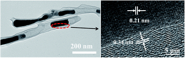
Highly porous N-doped CNTs/Fe3C was synthesized by a facile and one-pot method. In this method, melamine was used as the carbon precursor; iron chloride was employed as a catalyst of graphic carbon and iron carbon source, and zinc chloride as a chemical activator to obtain highly porous structure. Fe3C nanorods were in situ formed and embedded into the inner structure of CNTs. The pore structure analysis shows that N-doped CNTs/Fe3C possesses the large specific surface area up to 1021.26 m2 g−1. The N-doped CNTs/Fe3C electrode exhibits a high specific capacitance of 181 F g−1 at the current density of 0.1 A g−1, and excellent capacitance rate and cycling stability.


1 Introduction

Carbon nanotubes (CNTs) have attracted intensive attention due to their unique structure and properties, such as large pore size, high electric conductivity and chemical stability.1–3 These features allow CNTs to be widely used as ideal electrode materials for supercapacitors,4–7 lithium ion batteries,8–10 and fuel cells.7,11,12 However, CNTs generally possess a low specific surface area (below 200 m2 g−1), which leads to the low specific capacitance (20–80 F g−1) for electric double-layer capacitors (EDLCs) electrode materials.3,13

In order to solve this problem, KOH activation is regarded as an effective method to create lots of micropores by the strong chemical etching carbon of CNTs, dramatically increasing their surface area, and therefore its specific capacitance.14 The specific surface area of CNTs via KOH activation are about 1000 m2 g−1, and their specific capacitances are in the range of 80–100 F g−1 in an aqueous electrolyte.13,15–18 It is much lower than those of activated carbons for EDLCs electrode materials.

The heteroatoms doping has been shown to improve the specific capacitance of CNTs by introducing the additional pseudocapacitance.7,19–21 For example, Denisa et al. reported that N enriched nonporous carbon materials exhibited a specific capacitance of 198 F g−1 in aqueous electrolyte.22 Yun et al. reported that the specific capacitance of N-doped CNTs was 190.8 F g−1.21 The surface modification is generally used to incorporate electroactive heteroatoms on CNTs.12,23 Unfortunately, this technology is complicated and costly.

Recently, melamine has been employed to effectively prepare N-doped CNTs,7,24 such as S. Chen et al.7 reported that Fe–N co-doped CNTs/graphene were prepared by calcining the mixture of melamine, FeSO4, and graphene at 700–1000 °C, and it showed a superior electrocatalytic activity. L. Chen et al.24 prepared N-doped CNTs with bamboo-like structure by the pyrolysis of melamine and FeCl3 at 700 °C, possessing the enhanced hydrogen storage capacity. Wang et al.25 reported that the morphologies of N-doped CNTs can be tuned by the molar ratio of melamine and FeCl3 and the calcined temperature. When the molar ratio of melamine and FeCl3 was 6, the specific surface area of as-prepared N-doped CNTs reaches 294.4 m2 g−1. However, the specific capacitance is only 67 F g−1, which is not sufficient for the EDLCs electrode materials.

Iron carbide (Fe3C) is attractive in the field of catalysis due to its favorable magnetic properties and good air stability.26,27 Recently, C/Fe3C was reported as an anode material for lithium ion batteries and a counter electrode for dye-sensitized solar cell.28–31 However, to our knowledge, there are no reports of N-doped CNTs/Fe3C for EDLCs electrode materials. In this study, based on our previous reported results,25 highly porous N-doped CNTs/Fe3C were one-pot prepared by the pyrolysis of melamine with FeCl3 and ZnCl2 additions. In this method, FeCl3 was used as a catalyst, and ZnCl2 as a chemical activator. The results show that the specific surface area of N-doped CNTs/Fe3C reaches 1021.26 m2 g−1, and the specific capacitance was 181 F g−1 in 6 M KOH.

2 Experimental

2.1 Preparation of porous N-doped CNTs/Fe3C

All chemicals were in analytical grade and used without further purification. 3 g melamine, 3 g ZnCl2 and different FeCl3·6H2O contents were added into 30 mL ethanol under magnetic stirring (the molar ratio of melamine and FeCl3·6H2O is 6[thin space (1/6-em)]:[thin space (1/6-em)]1, 4[thin space (1/6-em)]:[thin space (1/6-em)]1, and 2[thin space (1/6-em)]:[thin space (1/6-em)]1, denoted as CNT/Fe3C-6, CNT/Fe3C-4, and CNT/Fe3C-2 in the following). Then the mixture was dried at 80 °C for 10 h. Subsequently, the obtained powders were heated to 573 K and 773 K (5 K min−1) for 2 h, respectively, and then heated to 973 K (10 K min−1) for 2 h in the flow of Ar gas. Finally, the collected product was treated with 1 M hydrochloric acid for 24 h to remove the catalyst residues, washed with deionized water several times, and dried at 100 °C for 10 h.

2.2 Structural characterization

The microstructures of the obtained products were investigated using a field emission scanning electron microscope (FE-SEM, JSM-7001F). Transmission electron microscopy (TEM) images were recorded on a JEOL JEM 2100F field emission transmission electron microscope. X-ray diffraction (XRD) patterns were obtained on a Bruker D8 Advance diffractometer with Cu Kα radiation operating at 40 kV and 20 mA. Raman spectra were collected with a confocal microprobe Raman system (Thermo Nicolet Almega XR Raman Microscope). The thermal analysis was carried out by using the thermogravimetric-differential scanning calorimetry analysis (TG-DSC). Nitrogen adsorption–desorption isotherm measurements were performed on a Micromeritics ASAP 2020 volumetric adsorption analyzer at 77 K. The specific surface area of the materials was calculated by the Brunauer–Emmett–Teller (BET) theory. The micropore size distribution was calculated according to the Horvath–Kawazoe (HK) method.

2.3 Electrochemical measurements

The working electrode was prepared by loading the mixture of 95 wt% of active material and 5 wt% of polytetrafluoroethylene (PTFE) on the nickel foam. The assembled electrodes were dried at 110 °C for 12 h. The electrochemical measurements were conducted in a three-electrode system in 6 M KOH under ambient conditions. A platinum foil and saturated calomel electrode (SCE) were used as the counter electrode and reference electrode, respectively. The capacitive performance was investigated by the cyclic voltammeter (CV), galvanostatic charge–discharge (GCD), and electrochemical impedance spectroscopy (EIS) techniques using an electrochemical workstation (Bio-logic SP 200). EIS was performed in a frequency range from 0.01 Hz to 100 kHz at the open circuit potential of 5 mV amplitude. The working voltage windows of CV and GCD were between −0.8 and 0 V. The specific capacitance was calculated using the following eqn (1):
 
C = IΔt(mΔV) (1)
where C is the specific capacitance (F g−1), I is the discharge current (A), Δt is the discharge time (s), m is the mass of the active material in the electrode (g), and ΔV is the potential change in discharge (V).

3 Results and discussion

The XRD pattern of the prepared sample without HCl treatment is shown in Fig. s1. It indicates that many phases are observed for the sample without a HCl treatment. Besides the diffraction peaks of C, Fe3C and Fe, the diffraction peaks of Zn-based compounds can be identified as ZnO, ZnFe2O4 and Zn3FeC0.5. After the treatment with 1 M HCl for 24 h, the diffraction peaks of Fe and Zn-based compounds disappear (Fig. 1a), implying the Fe and Zn-based compounds had been completely removed. The strong diffraction peaks can be indexed as the orthorhombic phase of Fe3C (JCPDS 35-772) and graphitic carbon. A diffraction peak centered at about 26° indicates the existence of graphitic carbon. This graphitization of carbon has been further confirmed by a Raman spectroscopy measurement. A weak D peak and a strong G peak in the Raman spectrum of N-doped CNTs/Fe3C are shown in Fig. 1b. Two characteristic peaks around 1373 cm−1 and 1602 cm−1 could be ascribed to D and G band, respectively. The IG/ID values increase with increasing FeCl3 contents because FeCl3 is a good catalyst for the graphitization, and their values of all samples are greater than 1, implying a crystalline structure of the obtained N-doped CNTs/Fe3C.
image file: c6ra07101e-f1.tif
Fig. 1 (a) XRD patterns, and (b) Raman spectroscopy of N-doped CNTs/Fe3C.

The microstructures of N-doped CNTs/Fe3C were investigated by SEM, and shown in Fig. s2. It can be seen that the nanotubular structure was observed for all the samples regardless of FeCl3 contents. The nanotubes were tangled together into clusters with a length of more than 10 μm. A magnification SEM image of CNT/Fe3C-6 in Fig. 2a demonstrates that the diameter of CNTs is about 100 nm, and the cracks were observed in the surface of CNTs. The internal structure and components of CNT/Fe3C-6 were further investigated by TEM images. As shown in Fig. 2b and c, the wall thickness of CNTs is about 5–10 nm. Fe3C nanorods were encapsulated by graphitic carbons, embedded into the inner of CNTs, leading to be difficult to remove them by HCl treatment. HRTEM images of CNT/Fe3C-6 are shown in Fig. 2d. It can be seen that the well-crystallized structures with lattice fringes of about 0.34 and 0.21 nm, corresponding to an interplanar spacing of graphite (002) and Fe3C (211) crystal plane, respectively.30


image file: c6ra07101e-f2.tif
Fig. 2 (a) SEM images, (b) and (c) TEM, and (d) HRTEM images of CNT/Fe3C-6.

In order to define the quality of Fe3C in CNT/Fe3C-6, the thermal analysis was carried out by using TG-DSC. The strongest endothermal peak appears in the temperature range of 450–660 °C, and the weight of the sample decreased rapidly (Fig. 3a). It indicates that the oxidation of carbon and Fe3C in this temperature range at the same time. Based on the chemical equations: C + O2 = CO2, 2Fe3C + 13/2O2 = 3Fe2O3 + 2CO2,31 the weight percentage of Fe3C in the composites is about 13.2 wt%.


image file: c6ra07101e-f3.tif
Fig. 3 (a) TG-DSC thermograms for heating CNT/Fe3C-6, (b) nitrogen adsorption–desorption isotherms of CNT/Fe3C-6, and the inset shows the pore size distributions, which are calculated by HK method.

Nitrogen adsorption–desorption isotherms of N-doped CNTs/Fe3C are shown in Fig. s3. They exhibit a typical I isotherm, which is indicative of microporous structures in the sample. The specific surface area of samples increases with decreasing FeCl3 contents. CNT/Fe3C-6 exhibits the maximum specific surface area up to 1026.23 m2 g−1, and the pore volume is 0.57 cm3 g−1. The pore size distributions calculated by HK method, as shown in inset in Fig. 3b, is indicative of a quite uniform pore size distribution centered in micropore ranges of about 0.68 nm and 0.78 nm. In our previous report,25 the specific surface area of N-doped CNTs was only 294.4 m2 g−1 without a ZnCl2 activator. It suggests that ZnCl2 is an effective chemical activator to improve the high specific surface area of N-doped CNTs/Fe3C, which is much higher than the specific surface area of CNTs activated by a KOH chemical activator.13 Therefore, it is expected that CNT/Fe3C-6 with high specific surface area will possess superior capacitive performance.

The X-ray photoelectron spectroscopy (XPS) of CNT/Fe3C-6 is shown in Fig. 4. The wide XPS spectrum (Fig. 4a) confirms that CNT/Fe3C-6 contains nitrogen (398.2 eV), oxygen (531.1 eV), and Fe (711.9 eV). The surface N, O and Fe contents in the composite was 12.83 at%, 15.05 at% and 1.3 at%, respectively. The high-resolution N1s XPS spectrum of CNT/Fe3C-6 (Fig. 4b) shows three peaks with binding energies at 398.3 eV, 399.8 eV, and 400.9 eV, assigned to pyridinic-, pyrrolic-, and graphitic-like nitrogen species, respectively.12 The high N and O contents are considered to contribute to additional pseudocapacitance and improved wettability.32


image file: c6ra07101e-f4.tif
Fig. 4 (a) XPS spectrum of a wide survey scan, and (b) high-resolution N1s XPS spectra of CNT/Fe3C-6.

The electrochemical performances of samples were evaluated by CV, GCD, and EIS. Fig. s4a and 5a show the CV curves of all samples at a scan rate of 100 mV s−1 and CNT/Fe3C-6 at scan rates ranging from 5 to 300 mV s−1, respectively. It shows that the curves maintain the quasi-rectangular shape with little distortion at the scan rate up to 300 mV s−1, indicating the favorable accessibility of electrolyte ions into the pores and superior capacitive behavior.32 The GCD curves of N-doped CNT/Fe3C electrode are presented in Fig. s4b and 5b. The curves show symmetric triangular shapes between 0 and −0.8 V vs. SCE electrode, indicating the good capacitive properties. The curves still remain a typical triangle shape at a high current density of 20 A g−1, implying excellent columbic efficiency. The specific capacitances of N-doped CNT/Fe3C electrodes were calculated according to the eqn (1), and the relationship between the specific capacitance and current densities is shown in Fig. 5c. It exhibits that the specific capacitances of N-doped CNT/Fe3C increase with decreasing FeCl3 contents, which is in accordance with the specific surface area. CNT/Fe3C-6 shows the maximum specific capacitance of 181 F g−1 at a current density of 0.1 A g−1. The specific capacitance retention of CNT/Fe3C-6 electrode is about 51% for the current densities ranging from 0.5 to 20 A g−1, which exhibits the good rate capability.


image file: c6ra07101e-f5.tif
Fig. 5 (a) CV curves at different scan rates, (b) GCD curves at various current densities of CNT/Fe3C-6, (c) the relationship between the specific capacitances and current densities, and (d) Nyquist plots of N doped CNTs/Fe3C.

Nyquist plots of N-doped CNT/Fe3C electrodes are shown in Fig. 5d. The curves are composed of a semicircle in the high frequency region and a nearly vertical line in the low frequency region indicating a typical capacitive behavior of EDLCs electrodes. The semicircle shape represents the charge-transfer resistance (Rct) at the electrode/electrolyte interface. The obtained values of Rct are 0.43, 1.17, and 3.32 Ω for CNT/Fe3C-2, CNT/Fe3C-4 and CNT/Fe3C-6, respectively. The Rct of CNT/Fe3C-6 is much higher than that of the previous reported CNTs,13,25 which is assigned to the high microposity and Fe3C nanorods of N-doped CNT/Fe3C-6. However, the equivalent series resistances (ESR) are only 0.26 Ω calculated from x-intercepts with the real axis of the Nyquist plots in high frequency part due to the high graphitization, which is close to the values of CNTs activated by KOH activation.13 The lower ESR of N-doped CNT/Fe3C electrodes is beneficial to enhance the electrochemical performance.

The cycling stability plays an important role for the practical application of supercapacitors. Fig. 6 shows that after 5000 successive charging–discharging cycles at 1 A g−1, the specific capacitance of CNT/Fe3C-6 remains 135.6 F g−1, which is 93.6% of its initial value (144.8 F g−1), indicating the high electrochemical stability. No significant electrochemical changes were observed after long-term process, as shown in the inset of Fig. 6. It further proves the excellent cycling stability of CNT/Fe3C-6.


image file: c6ra07101e-f6.tif
Fig. 6 The cycling performance of CNT/Fe3C-6 at the current density of 1 A g−1, and the inset shows the charge/discharge curves of the 10 cycles of the electrode.

4 Conclusions

In summary, a highly porous N-doped CNTs/Fe3C was facilely synthesized through one-step pyrolysis of melamine with a FeCl3 catalyst and ZnCl2 activator. The as-prepared CNT/Fe3C-6 possess the characteristics of large specific surface area (1021.26 m2 g−1), and Fe3C nanorods were embedded into the inner of N-doped CNTs. As a result, CNT/Fe3C-6 electrode exhibits the outstanding capacitive behavior (181 F g−1 at 0.1 A g−1), rate capacity and cycling stability in 6 M KOH aqueous electrolyte, which is promising for applications as EDLCs electrode materials.

Acknowledgements

The work was financially supported by the National Natural Science Foundation of China (No. 51102216 and 51572140), the Program for the Innovative Talents of Higher Learning Institutions of Shanxi, and Scientific and Technological Innovation Programs of Higher Education Institutions in Shanxi.

References

  1. S. Iijima, Nature, 1991, 354, 56–58 CrossRef CAS.
  2. E. Frackowiak and F. Béguin, Carbon, 2002, 40, 1775–1787 CrossRef CAS.
  3. G. Lota, K. Fic and E. Frackowiak, Energy Environ. Sci., 2011, 4, 1592–1605 CAS.
  4. H. Pan, J. Li and Y. Feng, Nanoscale Res. Lett., 2010, 5, 654–668 CrossRef CAS PubMed.
  5. R. A. Fisher, M. R. Watt and W. Jud Ready, ECS J. Solid State Sci. Technol., 2013, 2, M3170–M3177 CrossRef CAS.
  6. P. Bondavalli, D. Pribat, J. P. Schnell, C. Delfaure, L. Gorintin, P. Legagneux, L. Baraton and C. Galindo, Eur. Phys. J.: Appl. Phys., 2012, 60, 10401–10414 CrossRef.
  7. S. Zhang, H. Zhang, Q. Liu and S. Chen, J. Mater. Chem. A, 2013, 1, 3302–3308 CAS.
  8. H. Zhang, G. Cao and Y. Yang, Energy Environ. Sci., 2009, 2, 932–943 CAS.
  9. J. Wang, L. Li, C. L. Wong and S. Madhavi, Nanotechnology, 2012, 23, 495401 CrossRef PubMed.
  10. W. D. Zhang, B. Xu and L. C. Jiang, J. Mater. Chem., 2010, 20, 6383–6391 RSC.
  11. X. Xie, M. Ye, L. Hu, N. Liu, J. R. McDonough, W. Chen, H. Alshareef, C. S. Criddle and Y. Cui, Energy Environ. Sci., 2012, 5, 5265–5270 CAS.
  12. Z. Wang, R. Jia, J. Zheng, J. Zhao, L. Li, J. Song and Z. Zhu, ACS Nano, 2011, 5, 1677–1684 CrossRef CAS PubMed.
  13. B. Xu, F. Wu, Y. Su, G. Cao, S. Chen, Z. Zhou and Y. Yang, Electrochim. Acta, 2008, 53, 7730–7735 CrossRef CAS.
  14. J. Wang and S. Kaskel, J. Mater. Chem., 2012, 22, 23710–23725 RSC.
  15. E. Frackowiak, S. Delpeux, K. Jurewicz, K. Szostak, D. Cazorla-Amoros and F. Béguin, Chem. Phys. Lett., 2002, 361, 35–41 CrossRef CAS.
  16. C. Emmenegger, P. Mauron, P. Sudan, P. Wenger, V. Hermann, R. Gallay and A. Züttel, J. Power Sources, 2003, 124, 321–329 CrossRef CAS.
  17. Q. Jiang, Y. Zhao, X. Y. Lu, X. T. Zhu, G. Q. Yang, L. J. Song, Y. D. Cai, X. M. Ren and L. Qian, Chem. Phys. Lett., 2005, 410, 307–311 CrossRef CAS.
  18. E. Raymundo-Piñero, P. Azaïs, T. Cacciaguerra, D. Cazorla-Amorós, A. Linares-Solano and F. Béguin, Carbon, 2005, 43, 786–795 CrossRef.
  19. G. Wang, R. Liang, L. Liu and B. Zhong, Electrochim. Acta, 2014, 115, 183–188 CrossRef CAS.
  20. L. X. Li, J. Tao, X. Geng and B. G. An, Acta Phys.-Chim. Sin., 2013, 29, 111–116 Search PubMed.
  21. Y. S. Yun, H. H. Park and H. J. Jin, Materials, 2012, 5, 1258–1266 CrossRef CAS.
  22. D. Hulicova-Jurcakova, M. Kodama, S. Shiraishi, H. Hatori, Z. H. Zhu and G. Q. Lu, Adv. Funct. Mater., 2009, 19, 1800–1809 CrossRef CAS.
  23. P. Ayala, R. Arenal, M. Rümmeli, A. Rubio and T. Pichler, Carbon, 2010, 48, 575–586 CrossRef CAS.
  24. L. Chen, K. Xia, L. Huang, L. Li, L. Pei and S. Fei, Int. J. Hydrogen Energy, 2013, 38, 3297–3303 CrossRef CAS.
  25. Y. Wang, Y. Liu, W. Liu, H. Chen, G. Zhang and J. Wang, Mater. Lett., 2015, 154, 64–67 CrossRef CAS.
  26. A. Kraupner, A. Markus, R. Palkovits, K. Schlicht and C. Giordano, J. Mater. Chem., 2010, 20, 6019–6022 RSC.
  27. Z. Wen, S. Ci, F. Zhang, X. Feng, S. Cui, S. Mao, S. Luo, Z. He and J. Chen, Adv. Mater., 2012, 24, 1399–1404 CrossRef CAS PubMed.
  28. L. Su, Z. Zhou and P. Shen, Electrochim. Acta, 2013, 87, 180–185 CrossRef CAS.
  29. Y. Tan, K. Zhu, D. Li, F. Bai, Y. Wei and P. Zhang, Chem. Eng. J., 2014, 258, 93–100 CrossRef CAS.
  30. Y. Liao, K. Pan, L. Wang, Q. Pan, W. Zhou, X. Miao, B. Jiang, C. Tian, G. Tian, G. Wang and H. Fu, ACS Appl. Mater. Interfaces, 2013, 5, 3663–3670 CAS.
  31. X. Zhao, D. Xia, J. Yue and S. Liu, Electrochim. Acta, 2014, 116, 292–299 CrossRef CAS.
  32. Y. Zhao, M. Liu, X. Deng, L. Miao, P. K. Tripathi, X. Ma, D. Zhu, Z. Xu, Z. Hao and L. Gan, Electrochim. Acta, 2015, 153, 448–455 CrossRef CAS.

Footnote

Electronic supplementary information (ESI) available. See DOI: 10.1039/c6ra07101e

This journal is © The Royal Society of Chemistry 2016
Click here to see how this site uses Cookies. View our privacy policy here.