Ruiqi Zhang
,
Fanyong Yan
*,
Yicun Huang
,
Depeng Kong
,
Qianghua Ye
,
Jinxia Xu
and
Li Chen
State Key Laboratory of Hollow Fiber Membrane Materials and Processes, Key Lab of Fiber Modification and Functional Fiber of Tianjin, College of Environmental and Chemical Engineering, Tianjin Polytechnic University, 300387 Tianjin, People's Republic of China. E-mail: yanfanyong@tjpu.edu.cn; Tel: +86 22 83955766
First published on 11th May 2016
Ratiometric fluorescent probes allow the simultaneous measurement of two fluorescence signals at different wavelengths followed by calculation of their intensity ratio, which can provide more precise measurement results than intensity-based fluorescent probes. Excitation energy transfer is widely used in the design of ratiometric fluorescent probes. Rhodamine is a convenient platform for the construction of “OFF–ON” ratiometric chemosensors. Rhodamine-based ratiometric fluorescent probes based on the excitation energy transfer mechanism can be constructed by conjugated or non-conjugated connections with other chromophores. In this review, we summarized the recent advances regarding rhodamine-based ratiometric fluorescent probes based on excitation energy transfer. We reviewed these probes according to the classification of “through-space” and “through-bond” probes; we focused on the contributions of different donor fluorophores and the types of connections between the energy donors and acceptors.
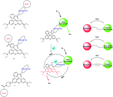
Rhodamine fluorophores have excellent photophysical properties, such as large absorption extinction coefficients, sharp fluorescence emissions and high fluorescence quantum yields. Moreover, they can undergo great fluorescence enhancements via a structural change from the spirocyclic state (non-fluorescent) to the ring-open (fluorescent) state, induced by metal ions, inorganic anions,10–13 etc. Their absorption and emission spectra appear at a longer wavelength (ca. 580 nm); thus, in the FRET system, the rhodamine fluorophore often acts as the energy acceptor.8 A conjugated or non-conjugated spacer moiety is chosen as the linker, and other fluorophores such as naphthalimide14 and coumarin15 are chosen as energy donors; rhodamine is a convenient platform to construct “OFF–ON” ratiometric chemosensors. Currently, many novel rhodamine-based energy transfer probes are being successfully designed, synthesized and applied.
In this review, we mainly summarized the recent advances in EET systems with rhodamine skeletons, including rhodamine–naphthalimide, rhodamine–coumarin, rhodamine–dansyl, rhodamine–quinoline, and rhodamine and other organic dyes; meanwhile, rhodamine nanoparticles, quantum dots, carbon dots and micelles have also been described.
Many of these probes are based on analyte promoted spiro-opening of rhodamine, while some other chemosensors exploit special reactions with the FRET process.
Using an ethylenediamine moiety as the linker, Luxami et al. synthesized a reversible naphthalimide–rhodamine-based fluorescent sensor, F1.17 F1 could act as a ratiometric sensor for Hg2+ based on FRET (Fig. 1). F1 displayed a blue fluorescence centered at 485 nm when excited at 410 nm in CH3CN–HEPES buffer (1:
1), pH = 7.12 ± 0.1. Upon addition of Hg2+ to the solution, the intensity of the fluorescent peak at 485 nm gradually decreased and that of a new fluorescent band centered at 585 nm gradually increased; thus, the FRET process occurred. The ratio of the fluorescence intensities at 585 nm and 485 nm (I585/I485) exhibited a drastic change from 0.11 to 15.6, a 141-fold variation in the emission ratio. Fang et al. synthesized a similar ratiometric Hg2+ probe, F2, based on FRET by connecting 4-propylamine-1,8-naphthalimide with rhodamine 6G ethylenediamine; the limit of detection was found to be 7.91 × 10⁻⁷ M (Fig. 2).18 Through connecting a pyrrolidine-1,8-naphthalimide directly with rhodamine B hydrazine, Mahato et al. synthesized a ratiometric Hg2+/Cr3+ probe, F3, based on FRET. The limits of detection for Hg2+ and Cr3+ were 0.35 and 0.14 ppb, respectively (Fig. 3).19
Through the introduction of different electron-donating groups by C-4 substitution, naphthalimide–rhodamine fluorescent sensors can show different properties.
Wang et al. synthesized a series of FRET-based fluorescent ratiometric chemosensors, F4a–f (Fig. 4).20 In terms of the degree of separation of Fe3+ and Cr3+ (FFe3+/FCr3+), F4b had the best selectivity toward Fe3+. In ethanol/Tris–HCl buffer (9:
1, v/v) solution; when excited at 420 nm, it only showed an emission band at 520 nm. In the presence of Fe3+ ions, F4b showed an emission band at 577 nm, which is the region of rhodamine. The fluorescence ratio at I577/I520 showed a 17-fold increase in the case of Fe3+ complexation, with a detection limit of 0.418 ppm.
Li et al. synthesized a series of fluorescence probes, F5a–c, based on naphthalimide–rhodamine compounds (Fig. 5).21 For compound F1b, the small distance between two fluorophores and the lower number of binding sites made it difficult to bind Al3+. Meanwhile, Al3+ was difficult to bind to F5c; thus, the FRET process was forbidden. The diethylenetriamine linker offers a suitable distance between the energy donor (naphthalimide) and acceptor (rhodamine) and the binding sites. Therefore, the FRET process could occur in F1a. In the absence of Al3+ ions, the system of F5a was FRET-OFF; when it was excited with 420 nm in H2O/EtOH (99:
1, v/v) buffered (Tris–HNO3, pH 7.0) solution, an emission at 530 nm appeared. Meanwhile, along with the addition of Al3+, a new emission of rhodamine at 580 nm increased; the steady decrease in emission intensity at 530 nm showed that the FRET process occurred. The ratio of the fluorescence intensities at 580 nm and 530 nm (I580/I530) gradually increased with increasing Al3+ concentration and varied from 0.42 to 4.88, an 11.5-fold variation in the emission ratios. The limit of detection was found to be 1.0 × 10⁻⁷ M. Furthermore, confocal laser microscopy studies confirmed that F5a could be used as an imaging probe for detecting Al3+ in HeLa cells.
Tang et al. synthesized a naphthalimide–rhodamine compound, F6, as a ratiometric fluorescent probe for adenosine triphosphate (ATP) detection (Fig. 6).22 The interaction of ATP with F6 through hydrogen bonds and unique π–π stacking interactions triggered the opening of the spirolactam of rhodamine. In the absence of ATP, the system F6 was FRET-OFF; when excited at 420 nm, an emission from naphthalimide at 530 nm appeared, while along with the addition of ATP (FRET-OPEN), a new emission of rhodamine at 580 nm increased and a steady decrease in emission intensity was observed at 530 nm. The emission intensity ratio, I580/I530, gradually increased with increasing ATP concentration and varied from 0.34 to 10.8, with the concentration of ATP ranging from 0 to 50 μM; the limit of detection was found to be 10 μM. Furthermore, confocal laser microscopy studies confirmed that the probe F6 could also be used as an imaging probe for detecting ATP in HeLa cells.
Based on the pH dependent spirolactam to ring-opening amide equilibrium of the rhodamine core,23–26 Georgiev et al. reported a pH probe (F7, Fig. 7).27 Due to the photoinduced electron transfer (PET) quenching process in peripheral 1,8-naphthalimides and the non-fluorescent spirolactam form of rhodamine, in alkaline solution, F7 did not emit light. PET was not feasible at pH < 6.5, and the rhodamine was in the ring-opened form at pH 2 to 6. At pH 6.5, when excited with 400 nm in water/DMF (4:
1, v/v) solution, an emission band at 520 nm was observed; at pH 2, the emission band at 520 nm decreased while the observed emission shifted to 561 nm, which could be attributed to the energy transfer from 1,8-naphthalimide to the ring opened form of the rhodamine. The energy transfer efficiency was 91%.
Fan et al. reported a naphthalimide–rhodamine FRET probe, F8, as a ratiometric and intracellular pH probe, in which 1,2,3-triazole was identified as an ideal bridge and biocompatible moiety (Fig. 8).28 At pH > 6.2, F8 showed green fluorescence and a yellow color which was mainly ascribed to the optical properties of 1,8-naphthalimide. When the pH changed from 6.20 to 2.00, the intensity of the green fluorescent band at 538 nm gradually decreased and a new pink fluorescent band at 580 nm gradually increased (excitation wavelength at 430 nm). The FRET efficiency was 72%. Furthermore, F8 was highly membrane permeable and could localize in lysosomes.
Yu et al. reported a FRET fluorescent probe, F9, based on the spirolactam of rhodamine through induced hydrazide and oxalaldehyde (Fig. 9).29 In the absence of Cu2+, F9 exhibited emission at 525 nm, attributed to the 1,8-naphthalimide donor, when excited at 355 nm in ethanol/water (1:
9, v/v, 50 mM HEPES, pH 7.4) solution (FRET-OFF). Upon addition of Cu2+ to the solution, the intensity of the fluorescent peak at 525 nm gradually decreased and a new fluorescent band at 580 nm gradually increased, indicating that the spirolactam ring was open and the FRET process occurred. The limit of detection was found to be 0.25 μM. At the same time, F9 could also be used as an imaging probe for detecting Cu2+ in RAW cells.
Based on the Hg2+-promoted ring opening of the spirolactam of the rhodamine moiety, leading to a cyclization reaction of a thiourea moiety, Song et al. designed a FRET probe (F10, Fig. 10).30 When excited in MeOH/water (7:
3, v/v) solution with 420 nm, an emission peak at 543 nm was observed, which could be assigned to naphthalimide. Upon addition of Hg2+ to the solution, the emission peak at 543 nm decreased, and the emission peak of rhodamine at 589 nm appeared and increased remarkably, which showed that the FRET process occurred. The ratio of the fluorescence intensities at 589 nm and 543 nm (I589/I543) exhibited a great change, from 0.35 to 11.49. The detection limit of probe for Hg2+ was 2.11 × 10⁻⁸ M.
F11 was synthesized as a ratiometric Au3+ probe. The reaction of rhodamine with N-(2-ethynlphenyl) increased steric strain around the lactam moiety, which increased the ground state energy of the probe and thus accelerated the gold ion-promoted ring-opening process (Fig. 11).31 When Au3+ was added to excited F11 at 420 nm, it showed an emission band at approximately 587 nm, which is the region of rhodamine. Selectivity and competition experiments showed that this system displayed high sensitivity and excellent selectivity over other metal ions. The detection limit for Au3+ was 0.5 ppb. Furthermore, F11 enables fluorescent imaging of gold species in N2A cells.
Cao et al. synthesized a ratiometric fluorescent chemosensor, F12, containing a coumarin moiety bound to rhodamine hydrazide (Fig. 12).35,36 F12 can respond to Al3+ with two different sets of fluorescence signals. F12–Ca2+ complex gave a green emission with a peak centered at 490 nm. When excited at 377 nm, F12–Al3+ showed an emission band of rhodamine centred at 580 nm. Al3+ ions could displace the Ca2+ ions in the F12–Ca2+ complex. In the absence of Al3+ ions, the system of F12–Ca2+ was FRET-OFF, while upon excitation at 377 nm, an emission band from the donor at 490 nm appeared; meanwhile, upon addition of Al3+, the FRET process occurred, a new emission band of rhodamine centred at 580 nm increased, and a steady decrease in the emission intensity at 490 nm was observed. The detection limit of Al3+ was 9.4 × 10⁻⁹ M. At the same time, F12 could also be used as an imaging probe for detecting Al3+ in hepatoma cells.
The measurement of pH values inside cells is of great significance. Shen et al. designed a ratiometric pH probe, F13, based on FRET and PET (Fig. 13).37 The coumarin moiety served as a FRET donor with the rhodamine moiety as a FRET acceptor; a 2-aminoethanol moiety was chosen as the linker. The pKa of F13 was 3.21, suggesting that F13 was suitable for studying acidic environments (pH < 4). At weakly basic pH, the PET process of the N atom of the rhodamine moiety partly quenched the coumarin emission. The PET process was gradually inhibited upon acidification; meanwhile, the rhodamine spirolactam changed to the ring-open form, followed by the FRET process between coumarin and rhodamine. Based on the ratio of the fluorescence intensities at 583 nm and 470 nm (I583/I470), F13 could be used for the ratiometric detection of pH in the range of 2.20 to 4.20 in buffer (1:
1, Britton–Robinson buffer/EtOH, v/v) solution. Furthermore, confocal laser microscopy studies confirmed that F13 could also be used as an imaging probe for detecting pH in bacteria. Zhang et al. synthesized a coumarin–rhodamine based chemosensor as a ratiometric pH probe.38 An N-ethylamino piperazine moiety was chosen as the linker. This probe (pKa = 4.98) could be used in the ratiometric detection of pH in the range of 4.20 to 6.00 in Britton–Robinson buffer solution. At the same time, this probe has been successfully applied in fluorescence imaging in HeLa cells.
Wang et al. reported a coumarin–rhodamine based FRET fluorescent probe, F14, with a thiourea moiety as the receptor unit and m-phenylenediamine as a linker (Fig. 14).39 Upon gradual addition of Hg2+, the emission peak at 478 nm decreased, while a new emission band centered at 587 nm appeared and gradually increased when excited at 440 nm. The ratio of the fluorescence intensities at 587 nm and 478 nm (I587/I478) exhibited a drastic change from 0.11 to 15.6, which represents a 994-fold variation in the emission ratios. The FRET efficiency was calculated to be 90%, while the detection limit of Hg2+ was 3.2 × 10⁻⁹ M. Furthermore, probe F14 was successfully used for recognition of Hg2+ in HeLa cells.
Based on the fact that the energy difference of the HOMO and LUMO for the coumarin moiety is lower than that of an azido-capped rhodamine moiety, but significantly higher than that of the rhodamine moiety, Wei et al. took advantage of the unique reduction of an azide moiety with H2S to synthesize a coumarin–rhodamine-based dyad, F15, for H2S detection (Fig. 15).40 In the absence of S2− ions, the F15 system was FRET-OFF, while upon excitation at 400 nm, an emission from the donor at 465 nm appeared; meanwhile, upon the addition of S2−, a new emission of rhodamine at 525 nm increased and showed a steady decrease in emission intensity at 465 nm, indicating that the FRET process occurred. Selectivity and competition experiments showed that this system featured high sensitivity and excellent selectivity over other ions. Furthermore, the probe F15 could detect endogenous production of H2S in living HEK293 cells.
Although partial success has been obtained in the design of first-generation and second-generation rhodamine-based FRET probes, further improvements in terms of versatility, sensitivity, and synthetic accessibility are required. To address these issues with first-generation and second-generation rhodamine-based FRET probes, third-generation rhodamine-based FRET probes have been designed and synthesized. The design of third-generation rhodamine-based FRET probes involves the utilization of 3-(piperazin-1-yl)-phenol as a raw material, because the piperazine can act as a linker. This approach only yields a single FRET probe; meanwhile, the energy donor is distant from the reaction site.
Guan et al. designed a series of rhodamine–coumarin-based ratiometric fluorescent probes, F16a–c, based on FRET (Fig. 16).41 F16a, whose energy donor is linked to the interaction site, was not suitable to detect the analyte. In F16b, the interaction site is far away from the donor, unlike F16a; thus, the sensing sensitivity should not be affected. However, F16b was very difficult to synthetize and separate. To solve this problem, F16c was designed by utilization of 3-(piperazin-1-yl)-phenol as a raw material; the piperazine acted as a linker, and the energy donor (coumarin) was distant from the reaction site of Cu2+. Thus, the coumarin donor would not affect the interactions between the reaction site and Cu2+. This strategy was suitable for the target analytes. In the presence of Cu2+ ions, a fluorescence emission band appeared from 481 nm to 581 nm when excited at 410 nm in HEPES buffer solution (pH 7.0, containing 20% CH3CN as a cosolvent). The ratio of the fluorescence intensities at 581 nm and 481 nm (I581/I481) exhibited a drastic change from 0.059 to 1.63, a 27.6-fold variation in the emission ratio. At the same time, the chemosensor F16c could also be used as an imaging probe for detecting Cu2+ ions in MCF-7 cells. Furthermore, this new FRET technology has great potential for the development of a wide variety of ratiometric fluorescent probes.
Zhang et al. reported two ratiometric HOCl probes, F17a–b, based on FRET. The coumarin moiety acted as a FRET donor, the rhodamine moiety acted as a FRET acceptor, and a piperazine moiety was chosen as the linker (Fig. 17).42 F17a took advantage of diacylhydrazine as a detection group, while F17b utilized a rhodamine acid as a detection group. Probe F17a could respond to OCl− under alkaline conditions. In a range of concentrations of OCl−, a new emission occurs at 580 nm, and the intensity increases with increasing hypochlorite dose; meanwhile, a steady decrease in emission intensity at 470 nm was observed, indicating that the FRET process occurred. Probe F17a showed an excellent linearity between the intensity ratio (I580/I470) and the concentration of OCl− from 170 to 230 μM. Probe F17b could rapidly respond to HOCl at pH < 6.0. Probe F17b showed a trend of the change in fluorescence intensity that was opposite to that of probe F17a. Due to the chlorination-induced cyclization properties of rhodamine acid, the fluorescence intensity increased markedly at 470 nm, and a great decrease in emission intensity was observed at 580 nm with increasing hypochlorous acid dose (FRET-OFF). Probe F17b shows an excellent linearity between the intensity ratio (I470/I580) and the concentration of HOCl from 60 to 160 μM. Hou et al. introduced a triphenylphosphonium moiety to realize the OCl− mitochondrial target ability.43
F19, a FRET ratiometric fluorescence probe for Cd2+ ions, was synthesized from a rhodamine spirolactam with a quinolone–benzothiazole conjugated dyad (Fig. 19).46 In MeOH/H2O (1/4, v/v, 1 mM HEPES buffer) solution, when excited with 360 nm, an emission from the quinoline dyad at 470 nm appeared; meanwhile, along with the addition of Cd2+, the FRET process occurred, a new emission of rhodamine at 585 nm increased, and a steady decrease in emission intensity was observed at 470 nm. The ratio of the emissions at the two wavelengths (I585/I470) increased from 0.054 to 2.07 with increasing Cd2+ concentration; the FRET efficiency was calculated to be 48%, while the detection limit of Cd2+ was 2.7 × 10⁻⁷ M.
Through connecting an 8-aminoquinoline derivative with rhodamine spirolactam, Zhou et al. reported a FRET-based ratiometric chemosensor, F20, for Hg2+ ion detection (Fig. 20).47 An ethanediamine moiety was chosen as the linker. F20 could respond to Hg2+ with two different sets of fluorescence signals. In the presence of Hg2+ ions, a fluorescence resonance energy transfer phenomenon occurred in F20–Zn2+ by exciting the quinolone–zinc complex moiety at 420 nm; upon gradual addition of Hg2+ into F20–Zn2+ solution, the emission peak of the sensor at 497 nm decreased with concomitant formation of a new peak at 562 nm. Furthermore, the ratiometric change of the fluorescence spectra became evident with a clear isoemission point at 547 nm. The detection limit of Hg2+ was 2.95 × 10⁻⁸ M. Sen et al. synthesized a similar ratiometric chemosensor for Hg2+ bearing a rhodamine and 8-aminoquinoline derivative moiety; a hydrazine moiety was chosen as the linker.48 The FRET efficiency was calculated to be 97.4%. The detection limit was determined and was found to be 90 ppb.
With an ethylenediamine moiety as the linker, through connecting dansyl with rhodamine 101, F21 was synthesized as a ratiometric sensor for Fe3+ based on the resonance energy transfer process (Fig. 21).50 The fluorescence intensity ratio at 605 nm and 515 nm (I605/I515) exhibited a great change from 0.29 to 62.2, about a 214-fold emission ratio increase due to FRET modulation. The efficiency of the energy transfer was calculated to be 84%; the detection limit of Fe3+ was 0.64 μM in CH3CN–Tris (9:
1, v/v) buffer solution. Choosing a diethylenetriamine moiety as the linker, Xie et al. synthesized a dansyl–rhodamine based probe for Hg2+ based on FRET.51 The efficiency of energy transfer was calculated to be 65%; the detection limit was 7.7 × 10⁻⁸ M.
Hu et al. connected the dansyl donor and rhodamine acceptor by the click reaction and introduced an ion ligand to report a FRET Cu2+ sensor F22 (Fig. 22).52 In CH3CN–H2O (1:
1, v/v) buffered solution, a Cu2+ titration experiment showed that upon excitation at 420 nm, the fluorescence intensity around 540 nm decreases along with the incremental addition of Cu2+; simultaneously, a new emission band around 568 nm gradually increases. The ratio of the emission intensities at 568 and 540 nm (I568/I540) exhibited a drastic change from 0.6 to 16.8, a 28-fold variation in the case of Cu2+ complexation. The detection limit of the sensor was determined to be 0.12 μM. The F22 could also be used as an imaging probe for detecting Cu2+ ions in HeLa cells.
The emission of BODIPY was at 510 nm in CH3CN–H2O (60:
40) buffer solution, which overlaps with the absorption of rhodamine. Yu et al. employed an o-phenylenediamine as a NO trapper to synthesize a ratiometric NO sensor F23 (Fig. 23).53 In the absence of NO, F23 exhibited an intense emission band with maxima at 510 nm, which was typical for BODIPY; upon addition of NO solution, the emission peak at 510 nm gradually decreased and a new emission peak at 590 nm of rhodamine gradually increased, which reflected that an efficient FRET process was turned on. The detection limit of NO was determined to be 26 nM. Furthermore, the probe F23 could detect endogenous production of NO in living MCF-7 cells.
Sui et al. reported a ratiometric chemosensor, F24, for Pd2+ ion detection (Fig. 24).54 F24 was synthesized by connecting 3-amino-2-naphthoic acid directly with rhodamine B ethylenediamine. In EtOH/H2O (1:
1, v/v) solution, with increasing Pd2+ concentration, the emission of the naphthylamine moiety at 490 nm gradually decreased and a new emission peak of rhodamine appeared at 590 nm. The fluorescence intensity ratios (I590/I490) of F24 exhibited a linear relationship in the concentration range from 0 μM to 2 μM of Pd2+ ions. The detection limit of Pd2+ was 45.9 nM. In addition, this probe was successfully applied for detecting Pd2+ ions in live mice.
Using an imidazole derivative of the phenanthrene moiety as an energy donor and a rhodamine 6G moiety as an energy acceptor, Reddy et al. synthesized a ratiometric fluorescence sensor, F25, for pH detection based on FRET (Fig. 25).55 In PBS buffer–DMSO (98:
2, v/v) solution, at pH 7.0, F25 was present exclusively in the cyclic lactam form, FRET-OFF, while upon excitation at 400 nm, an emission from the donor at 493 nm appeared; meanwhile, the acyclic xanthene form of F26–H+ prevailed at pH 3.5 (FRET-OPEN), the emission peak at 493 nm decreased and the emission peak of rhodamine at 552 nm appeared. The energy transfer efficiency was calculated to be 42%. Furthermore, F25 could also be used as an imaging probe for detecting pH in Hct116 cells.
Kar et al. synthesized an indole functionalized rhodamine derivative, F26, which could specifically bind to Cu2+ in the presence of large excesses of other competing ions (Fig. 26).56 In the presence of Cu2+ ions, a fluorescence emission band appeared from 490 nm to 587 nm upon excitation at 340 nm in CH3CN/aqueous HEPES buffer (1 mM, pH 7.3; 1:
4 v/v) medium. The FRET efficiency was calculated to be 51.5%. The detection limit was found to be 3.6 ppb. At the same time, this fluorescent probe could also be used as a fluorescence microscopic imaging probe for Cu2+ detection in HeLa cells.
Liu et al. reported a FRET ratiometric fluorescence probe F27 for Hg2+ detection which comprised an indole–triazole conjugate moiety donor and a rhodamine acceptor, linked by a hydrazine moiety (Fig. 27).57 Compound F27 displayed a large shift (from 490 to 590 nm) in its emission spectrum with increasing Hg2+ concentration in EtOH–MOPS mixed solvent. The detection limit of Hg2+ was 11 nM. Furthermore, F27 could also be used as an imaging probe for Hg2+ detection in MCF-7 cells.
F28 is a ratiometric fluorescent Hg2+ probe consisting of a phenothiazine donor and a rhodamine acceptor (Fig. 28).58 In the presence of Hg2+ ions, a fluorescence emission band appears from 520 nm to 580 nm when excited at 415 nm in acetonitrile. The intramolecular energy transfer efficiency from the phenothiazine donor to the rhodamine acceptor was calculated to be 76.8%. The detection limit of Hg2+ was 9.2 × 10⁻⁹ M.
F29 is a FRET ratiometric fluorescence mercury probe with a 7-nitrobenz-2-oxa-1,3-diazole (NBD) fluorophore as the energy donor and rhodamine B as an energy acceptor; a 1-(2-aminoethyl) piperazine moiety was chosen as the linker (Fig. 29).59 In the presence of Hg2+ ions, a fluorescence emission peak appeared from 525 nm to 580 nm upon excitation at 420 nm in CH3CN–H2O (9:
1 v/v) mixture medium. The FRET efficiency was calculated to be 86.5%. The detection limit was found to be 5.7 × 10⁻⁹ M.
Using per-acetyl glycosyl as an energy donor and rhodamine B as an energy acceptor, F30 was synthesized through a click reaction. Fluorescent probe F30 could detect Hg2+ based on FRET (Fig. 30).60 In the absence of Hg2+ ions, the F30 system was FRET-OFF, while upon excitation at 315 nm, an emission from the donor at 470 nm appeared; meanwhile, along with the addition of Hg2+, the FRET process occurred, a new emission of rhodamine at 584 nm increased, and a steady decrease in emission intensity at 470 nm was observed in aqueous solution (H2O/CH3CN = 4:
1, v/v). The detection limit of Hg2+ was 7.6 × 10⁻⁸ M.
In addition, double ion detection probes and two-photon technology have opened up new prospects for traditional energy transfer systems.
Fan et al. reported the design of ratiometric chemosensor F31 for Al3+ ions by connecting a 6-ethoxychromone-3-carbaldehyde chromone with rhodamine 6G hydrazide. Based on the spiro-ring of rhodamine, F31 could respond to Al3+ with two different sets of fluorescence signals (Fig. 31).61 The prepared zinc complex of the sensor could detect Al3+ on the basis of the fluorescence resonance energy transfer mechanism, and the detection limit for Al3+ was 1.83 × 10⁻⁷ M.
The emission peak of fluorescein is at 523 nm, which overlaps perfectly with the absorption of rhodamine B; at the same time, based on the spiro-ring of fluorescein and rhodamine, a fluorescein–rhodamine dyad probe could detect two different ions. Based on Hg2+-promoted desulfation followed by a cyclization reaction of thiourea and fluoride ion-selective cleavage of the Si–O linkage, Chereddy et al. reported a fluorescein–rhodamine dyad probe, F32 (Fig. 32).62 F32 permitted ratiometric detection of Hg2+ and F− ions, individually and collectively, with different fluorescence outputs.
Wang et al. developed a bichromophoric two-photon excited FRET-based ratiometric fluorescent probe, F33 (Fig. 33).63 F33 had a large two-photon absorption cross-section in the 735.8 nm region (359.9 GM). F33 was capable of undergoing efficient FRET following either two-photon (IR laser) or single-photon (UV-visible) excitation. For probe F33, using a 735.8 nm laser as the two-photon (TP) excitation light, the probe was excited and the fluorescence emitted from the energy donor was observed (FRET-OFF); meanwhile, along with the addition of Cu2+, the spirolactam ring of rhodamine opened (FRET-OPEN), and the fluorescence emission band of the acceptor could be observed. The energy transfer efficiency was nearly perfect (>99%). Furthermore, F33 could produce a larger Stokes shift and prevent photodamage to biological samples.
Mesoporous silica is an excellent host material with good optical transparency in the visible-light region and good biocompatibility. Wu et al. reported a FRET ratiometric fluorescence sensor, F34, through thioRB-esters connected with mesoporous silica nanoparticles (MSN) functionalized with fluorescein isothiocyanate (FITC) (Fig. 34).72 In the absence of HOCl, F34 was FRET-OFF, while upon excitation at 488 nm in Na2HPO4–citrate buffer solution, an emission from the donor at 525 nm appeared; upon the addition of HOCl, the analyte triggered oxidative opening of the intramolecular thioether, a new emission of rhodamine at 590 nm increased, and a steady decrease in the emission intensity at 525 nm showed that the FRET process occurred. The detection limit was 5 μM. At the same time, F34 could also be used as a fluorescence image probe for detecting HOCl in L929 cells.
Liu et al. developed a FRET ratiometric silica core–shell nanoparticle fluorescent probe F35 based on 7-nitro-benzo-furazan (NBD) and a rhodamine B derivative for Hg2+ detection (Fig. 35).64 The fluorophores were linked to silica core–shell nanoparticles by γ-aminopropyl triethoxysilane. With increasing concentration of Hg2+, the ratio of the emission intensity at 578 nm to that at 526 nm (I578/I526) increased steadily upon excitation at 420 nm in water. The detection limit was 100 nM.
Lu et al. developed a FRET fluorescent probe, F36, for ratiometric sensing of Hg2+ in water with periodic mesoporous organosilica (PMO) nanoparticles (NPs) as the scaffold (Fig. 36).73 Silylated rhodamine 6G with a spiro-ring as the energy acceptor was covalently attached on the pore walls of anthracene encapsulated PMO NPs, which played the role of energy donor. In the absence of Hg2+ ion, the system of F38 was FRET-OFF; upon excitation at 370 nm in HEPES buffer solution, an emission from anthracene at 450 nm appeared, and a significant increase of the I550/I430 value was observed upon the addition of Hg2+ ion. The detection limit was 6.0 × 10⁻⁹ M.
Graphene quantum dots exhibit an excitation-dependent photoluminescence emission; the emission peak shifts from ca. 440 nm to ca. 550 nm, while the excitation wavelength changes from 280 to 460 nm. Graphene quantum dots could be prepared from graphite powder by a modified Hummers' method. Liu et al. synthesized a FRET ratiometric probe, F37, based on graphene quantum dots (GQDs) and vulcanized rhodamine B derivative moiety (Fig. 37).74 An ethanediamine moiety was chosen as the rigid linker. In the absence of Hg2+ ions, F37 was FRET-OFF, while upon excitation at 400 nm in HEPES buffered (pH 7.4) water–ethanol (8:
1, v/v), an emission from graphene quantum dots at 500 nm appeared; meanwhile, upon the addition of Hg2+, a new emission of rhodamine at 585 nm increased, and a steady decrease in emission intensity at 500 nm was observed, which suggested that FRET process occurred. The detection limit was 0.23 μM. Furthermore, F37 could be used as a fluorescence microscopic image probe for detecting Hg2+ in HeLa cells.
Choosing CdSe (CdZnS) quantum dots as the energy donor, Moquin et al. described a nanosensor, F38, for caspase-1 enzymatic activity ratiometric measurements. F38 consisted of a rhodamine-labeled, caspase-1 cleavable peptide and quantum dots (QDs) (Fig. 38).75 In the absence of caspase-1, when excited at 355 nm, they transfer their energy to the rhodamine moiety, and emission was observed at 590 nm. After enzymatic cleavage of the peptide molecules, the acceptor molecules were detached from the QDs, FRET could no longer occur, and the emission of the QDs at 550 nm appeared. Furthermore, F38 could function as a sensitive assay to measure caspase-1 enzymatic activity in microglia cells.
Carbon dots (Cds), a new species of fluorescent material, possess great dispersibility in water and good biocompatibility and have received much attention. Kim et al. developed Cds by microwave irradiation of acrylic acid and 1,2-ethylenediamine. The carbon dots exhibited an excitation-dependent photoluminescence emission; when excited at 350 nm, a fluorescence emission band at 410 nm appeared. Kim et al. developed a FRET ratiometric fluorescent probe, F39, with rhodamine 6G and carbon dots (Fig. 39).76 In the presence of Al3+ ions, the fluorescence emission band shifted from 410 nm to 560 nm when excited at 350 nm in aqueous solution. The detection limit was determined and was found to be 3.8 × 10⁻⁵ M. Furthermore, F39 could be embedded to fabricate a paper-based sensor strip, with which the detection of Al3+ was performed in aqueous solution.
Polymers with well-designed fluorescent probes have been widely explored in recent decades. Georgiev et al. reported a pH sensitive probe, F40, based on highly water-soluble fluorescent micelles (Fig. 40).77 F40 was synthesized by grafting a 1,8-naphthalimide–rhodamine bichromophoric FRET system (RNI) to the PMMA block of a well-defined amphiphilic diblock copolymer, poly(methyl methacrylate)-b-poly(methacrylic acid) (PMMA48-b-PMAA27). The RNI-PMMA48-b-PMAA27 adduct was capable of self-assembling into micelles with a hydrophobic PMMA core, containing the anchored fluorescent probe and a hydrophilic shell composed of PMAA blocks. F40 had greater photostability in comparison with the pure organic dye label and could serve as a highly sensitive pH probe in water. At the same time, this fluorescent probe could be used as a fluorescence image probe for detecting pH in HeLa cells and HEK cells.
Due to its excellent solubility in pure aqueous solution and the good match of its fluorescence emission spectrum with the absorption spectrum of rhodamine 6G, poly[p(phenylene ethynylene)-alt-(thienylene ethyn-ylene)] (PPETE), whose fluorescence is insensitive to environmental factors, has been chosen as an energy donor. Wu et al. reported a Fe3+ sensitive FRET-based ratiometric fluorescent probe, F41 (Fig. 41).78 In the presence of Fe3+ ions, the fluorescence emission band shifted from 442 nm to 538 nm when excited at 400 nm in aqueous solution; the ratio of the emissions at the two wavelengths (I538/I442) increased from 0.18 to 3.7, and the FRET efficiency was calculated to be 61.8%. The detection limit was determined and was found to be 3.0 × 10⁻⁷ M. Furthermore, confocal laser microscopy studies confirmed that the reagent F41 could also be used as an imaging probe for detecting Fe3+ in Hela cells.
Based on the Hg2+-promoted ring opening of the spirolactam of a rhodamine moiety, leading to a cyclization reaction of a thiourea moiety, Gong et al. synthesized a TBET probe, T1. T1 used vulcanized rhodamine B derivative as the signaling moiety, with a spirolactam structure (nonfluorescent); coumarin moiety acted as the energy donor (Fig. 42).79 On excitation of T1 at 420 nm in a buffered (0.01 M HEPES, pH 7.2) H2O/THF (1:
1, v/v) solution, an emission band at 470 nm was observed, which could be assigned to the coumarin moiety. Upon gradual addition of Hg2+ to the solution of T1, the emission at 470 nm decreased and the emission of rhodamine at 580 nm appeared and increased remarkably, which showed that the TBET process occurred. The ratio of the emission intensities at 589 nm and 543 nm (I589/I543) exhibited a great change from 0.0133 to 9.280, a 697.7-fold increase. The TBET efficiency was calculated to be 97.4%, and the detection limit of Hg2+ was 7 nM. Furthermore, T1 has been preliminarily used for ratiometric imaging of Hg2+ in Hela cells.
Bhalla et al. synthesized a ratiometric fluorescence probe, T2, which contained a hexaphenylbenzene derivative as the energy donor and rhodamine B as the energy acceptor (Fig. 43).80 T2 was the first report where a donor–acceptor system displayed solvent dependent switching of its energy transfer mechanism in the presence of Hg2+ ions. The FRET mechanism was operative in THF and CH3CN, whereas in protic solvents such as MeOH, TBET was operative. Chopra et al. developed a similar TBET fluorescence probe which could sense picric acid in pure methanol.81
Through the conjugated connection of a naphthalimine derivative with rhodamine spirolactam, T3 was synthesized as a ratiometric sensor for Cu2+ detection based on TBET (Fig. 44).82 Cu2+ acts as not only a selective recognizing guest but also as a hydrolytic promoter. In the absence of Cu2+, upon excitation at 420 nm, a emission peak of naphthalimine derivative appeared at 537 nm; there is no TBET in the free T3. The addition of Cu2+ caused a significant decrease in the emission intensity at 535 nm, while simultaneously, a new emission band at around 577 nm gradually increased; thus, the TBET process occurred. The detection limit of T3 was 3.88 × 10⁻⁷ M. Furthermore, confocal laser microscopy studies confirmed that the reagent T3 could also be used as an imaging probe for detecting Cu2+ in MCF-7 cells.
Wang et al. developed a TBET ratiometric fluorescence probe, T4, using a naphthalimine derivative moiety as the energy donor and rhodamine B as the energy acceptor (Fig. 45).83 A p-phenylenediamine moiety was chosen as the rigid linker. In the presence of Fe3+ ions, the fluorescence emission band shifted from 535 nm to 586 nm; when excited at 420 nm in a CH3OH–H2O (4:
6, v/v) solution, the ratio of the emissions at the two wavelengths (I586/I535) exhibited a 30-fold enhancement. The detection limit was found to be 0.105 μM.
Based on the fact that cerium(IV) ammonium nitrate (CAN) specially promotes an irreversible oxidative cyclization of N-acylhydrazones into 1,3,4-oxadiazoles under mild conditions, Goswami et al. developed a TBET ratiometric fluorescence probe (T5) which uses carbazole moiety as the energy donor and rhodamine B as the energy acceptor (Fig. 46).84 On excitation of T5 at 450 nm in CH3–CN:
H2O (7
:
3, v/v), a weak emission band at 483 nm was observed, which could be assigned to the carbazole moiety. Upon gradual addition of Ce4+, the emission at 483 nm decreased, while the emission of rhodamine at 585 nm appeared and increased remarkably, which showed that the TBET process occurred. The TBET efficiency was calculated to be 98%, and the detection limit was 0.685 μM. Yang et al. developed a similar TBET fluorescence probe which could detect Hg2+.85 The energy transfer efficiency reaches 99.6%; the detection limit was determined to be 3 ppb.
The donor fluorophore was connected directly to the rhodamine by a conjugated linker. Zhou et al. synthesized naphthalene derivatives with an appended rhodamine based fluorescent chemosensor, T6 (Fig. 47).86 The fluorescence spectrum of compound T6 exhibited an emission at 475 nm, attributed to the naphthalene moiety, when excited at 420 nm in Tris–HCl/CH3CH2OH (9:
1, v/v, 10 mM) aqueous solution. After addition of Cu2+, fluorescence appeared at 575 nm. The efficiency of energy transfer was calculated to be 93.7%; the detection limit of Cu2+ was 3.0 × 10⁻⁷ M. Furthermore, T6 is a two-photon probe which had a two-photon active absorption cross-section of 115 GM at 475 nm upon excitation at 780 nm. At the same time, T6 could be used as an imaging probe for detecting Cu2+ ions in HeLa cells. Furthermore, Zhou et al. synthesized a similar naphthalene derivative appended rhodamine based TBET fluorescent chemosensor for Pd2+ with an anthracene appended rhodamine spirolactam as the signaling moiety.87 The detection limit was estimated to be 2.3 × 10⁻⁷ M, and the energy transfer efficiency was calculated to be 90%.
This journal is © The Royal Society of Chemistry 2016 |