Electronically excited state of neutral/protonated, indole/5-hydroxyinodole–water clusters: a theoretical study

Reza Omidyan*, Masoud Omidyan and Atefeh Mohammadzadeh
Department of Chemistry, University of Isfahan, 81746-73441, Isfahan, Iran. E-mail: r.omidyan@sci.ui.ac.ir; reza.omidyan@u-psud.fr; Fax: +98 311 6689732

Received 14th March 2016 , Accepted 25th March 2016

First published on 29th March 2016


Abstract

The second-order approximate coupled-cluster (RI-CC2) method was employed to investigate photoinduced hydrogen-bond weakening or strengthening in neutral and protonated indole–, 5-hydroxyindole–water clusters. In addition to the protonation effect on the electronic structure of 5-hydroxyindole, the intermolecular H-bond weakening or strengthening of selected systems in the S1 excited state has been investigated. According to our calculated results, it has been predicted that the electronic excitation effect on the hydrogen-bond strength in protonated clusters is essentially more pronounced than neutral analogues. Also, a charge transfer character of the excited state over the chromophore moiety can be suggested for interpreting the excited state dynamics of H-bonds in protonated complexes. Moreover, it has been predicted that protonation is accompanied by a strong red-shift effect (∼1.10 eV) on the S1–S0 transition energy of 5-hydroxyindole.

1. Introduction

Water is the most abundant substance in living systems, making up 70% or more of the weight of most organisms. The first living organisms certainly arose in an aqueous environment, and the course of evolution has been shaped by the properties of the aqueous medium in which life began.1 The special characteristics of water are directly related to its hydrogen-bond networks in liquid or its solid states. Protein folding in water environments and the special features of DNA and RNA molecules are essentially connected with H-bonds.1–4 Therefore, hydrogen bonding is a modern research interest in a wide range of scientific fields nowadays.2,5,6

So far, the hydrogen bonding has been studied in the ground state by many different experimental and theoretical methods.2,4,7–10 However, little is known about electronic excited-state characters of hydrogen bonding. Fortunately, based on the new advances in computational methods during last two decades, it is now possible to investigate the physical properties of hydrogen-bond complexes in their excited electronic states accurately.11–15 According to pioneer studies of Zhao et al.,4,6,16–19 it has been established that photoexcitation may induce essential alterations on hydrogen bond characters.20–23 In this regard, Zhao and co-workers have demonstrated that intermolecular hydrogen bonds can be significantly strengthened or weakened in electronically excited states.23–25 In addition, the intermolecular hydrogen bonding interactions in electronically excited state was investigated by Liu et al.26 It has been clarified that intermolecular hydrogen bonds of C[double bond, length as m-dash]O⋯H–O and N–H⋯O–H are significantly strengthened upon photoexcitation to electronically excited states. Additionally, Chudoba et al.20 studied the excited-state infrared (IR) spectra of coumarin 102 dye in hydrogen-donating solvents.20 They proposed that intermolecular hydrogen bond C[double bond, length as m-dash]O⋯H–C forming between C102 and chloroform solvent can be broken following electronic excitation. However, in contrast to the vast numbers of reports dealing with H-bond characters in neutral clusters, rare reports has been dedicated to this subject in ionic states.27,28

Protonated aromatic molecules29 are short lives species which are important in the wide range of science from astrochemistry,30 environment,31 jet engine gas exhaust and hydrocarbon plasmas.32 They are also intermediates in electrophilic aromatic substitution reactions (EAS),29 one of the most commonly reaction mechanisms in organic chemistry. The effects of protonation on aromatic biomolecular systems is an interesting issue for models rationalizing the UV photostability of biological macromolecules, such as proteins and DNA.30,33

In the present study, our results on protonation and excited state hydrogen-bond characters of indole34–42/5-hydroxyindole37,43,44 with one water molecule will be presented. In addition to excited state dynamics of hydrogen bonding in neutral clusters, we have investigated the corresponding parameters of protonated systems. Thus, the geometry and electronic structures, electronic transition energies and oscillator strength of neutral/protonated clusters of indole, 5-hydroxyindole and water, will be accurately addressed. Then, the weakening and strengthening of H-bond following photoexcitation will be explored and discussed.

2. Computational details

The second-order approximate coupled cluster (RI-CC2),45 which is a valuable tool in electronic structure theory, is the method of choice in this study because it gives reasonable results on excited state properties and dynamics of medium size organic molecules, not only in isolated form but also in cluster structures and hydrogen bond systems (see ref. 47–52, and references there in). All of calculations have been performed with the TURBOMOLE program suit46 making use of the resolution-of-identity (RI) approximation for the evaluation of the electron-repulsion integrals. The equilibrium geometry of individual and cluster forms of selected systems at the ground electronic states (S0) have been determined at the RI-MP2 (Möller–Plesset second order perturbation theory) level.53,54 Excitation energies of the lowest lying singlet states and equilibrium geometry of the lowest excited state (S1) have been determined at the RI-CC2 method.47,55,56 The electronic transition energies have been determined using two basis functions of correlation-consistent polarized valence double-zeta (cc-pVDZ)57,58 and the augmented cc-pVDZ basis set, while the relevant parameters to hydrogen bonding (binding energies and geometry parameters), have been determined by the larger basis function of aug-cc-pVDZ. The vibrational frequencies and zero point vibrational energies (ZPE) have been determined based on the cc-pVDZ basis function. Moreover, the full counterpoise (CP)59 method was used to correct the interaction energy from the inherent basis set superposition error (BSSE), associated with different electronic states.

The abbreviations of “H-bond, In, InH+, 5-HIn, 5-HIn–H+ and W” have been used instead of hydrogen bond, indole, protonated indole, 5-hydroxyindole, protonated 5-hydroxyindole and water, respectively.

3. Results and discussions

3.1 Geometry and electronic structures

3.1.1 Monohydrated indole and protonated indole. In the first step of this study, the most stable structures for complexation of selected monomers with water have been investigated. In this regard, the preferred positions of water molecule around the neutral and protonated indole chromophore were considered.

Therefore, the N–H and C–H of pyrrole ring, in addition to C–H of benzene and O–H⋯π interaction have been studied. It has been found that H-bond formation between the N–H side of indole and water molecule produces the most stable structure for indole–water complex (Fig. 1(a1)). As shown in Fig. 1, the structure is stabilized by a strong N–H⋯OH hydrogen bond.


image file: c6ra06716f-f1.tif
Fig. 1 Optimized structures of neutral and protonated In–W clusters calculated at RI-MP2/aug-cc-pVDZ level of theory. In each panel, the values (in kJ mol−1) indicate to the relative stabilities of clusters, compared to the first isomer of each row. Also, the ground state hydrogen bond binding energy (EHb, corrected with BSSE), has been presented for the most stable isomers of neutral and protonated systems in kJ mol−1.

Also, when additional water molecule connects to indole, by formation a hydrogen bond between pyrrole's C–H and H2O, the b1 complex is produced. This cluster has been found to be 24 kJ mol−1 less stable than a1 (at the RI-MP2/aug-cc-pVDZ level of theory). Also, adding a water molecule between pyrrole and benzene rings, obtains another complex (c1, see Fig. 1), with 6.8 kJ mol−1 less stability than a1 complex. The CH⋯OH hydrogen bond between water and benzene ring obtains another structure which is less stable than other CH⋯OH and NH⋯OH hydrogen bond clusters. Finally, we have considered a OH⋯π interaction between water and indole chromophore. The optimized structure is depicted in Fig. 1(d1), involving the H(water)⋯π hydrogen bond. That is also more stable than CH⋯OH hydrogen bounded systems; nevertheless, it is 12.5 kJ mol−1 less stable than a1, (the most stable structure). Thus, from inspection of relative stabilities in Fig. 1, the decreasing pattern of H-bond interactions in indole–water clusters can be presented as: NH⋯OH > H(water)⋯π > CH⋯OH.

The protonation effect on electronic structure and spectroscopic properties of indole has been extensively investigated by Alata et al.60 Based on their report, the pyrrole ring provides the most attractive site of protonation. The relevant protonated isomer has been reported to be ∼37 kJ mol−1 more stable than others. Thus, we have selected this isomer60 and investigated its mono-hydration from different sites. As the same as neutral indole, we have considered three type complexes, with N–H, C–H and also OH⋯π interaction. However, no local minimum has been found involving the OH⋯π interaction. As shown in Fig. 1(a2–d2), the NH⋯OH hydrogen bounded is the most stable complex, being ∼37.0 kJ mol−1 more stable than other clusters. Comparing the stability of different clusters of protonated indole–water, it is seen that the NH⋯OH hydrogen bond cluster of protonated indole–water is substantially more stable (∼41.5 kJ mol−1) than all of CH⋯OH clusters. This stabilization is arising from increasing the polarity of N–H bond, due to presence of additional positive charge density on pyrrole ring.

In addition, one may compare the H-bond strength between neutral and protonated indole, by considering the hydrogen bond binding energies (EHb), of the ground state for the most stable isomer of neutral and protonated indole–water complexes (Fig. 1(a1 and a2)).

The ground state EHb has been calculated by comparing the ground state optimized energy of complex and individual monomers:61

 
EHb(S0) = [EGs]complex − ([EGs]Ch + [EGs]H2O) (1)
where EGs stands to ground state energy and the Ch term, refers to chromophore.

As shown in Fig. 1, the absolute value of ground state binding energies of protonated indole–water clusters have been determined to be 29 kJ mol−1 larger than those of neutral homologues, indicating to stronger interactions between protonated chromophores and water molecule.

Regarding the transition energies and electronic structures, for the most stable isomers of monohydrated systems (a1 and a2 in Fig. 1), the first and second singlet transition energies have been calculated, using the RI-CC2 method and the cc-pVDZ/aug-cc-pVDZ basis functions.

The results have been tabulated in Table 1. According to the RI-CC2 results, it has been found that the S1–S0 transition in neutral system is corresponding to the HOMO–LUMO single electron transition (80%) and it is arising from HOMO−1–LUMO in protonated system (70%). We have depicted few selected MOs of the most stable isomers of our clustered systems in Fig. 2. As shown, both HOMO and HOMO−1 for neutral and protonated indole–water clusters are of the π character and the LUMO orbital of both systems are of the π* nature. Thus the S1–S0 transition can be assigned as 1ππ* nature in both systems. Additionally, from inspection of Fig. 2, it can be seen that both of the HOMO−1 and LUMO locate over the neutral or protonated chromophores, and no contribution of water molecule is presented on the S1–S0 transition. Thus the S1–S0 transition of neutral/protonated clusters of indole–water can be identified as local transitions.

Table 1 Calculated electronic transition energies (in eV) and corresponding oscillator strengths of two low-lying singlet excited states for neutral/protonated indole in their individual and monohydrated forms. The values in parentheses represent the ΔZPE-corrected values for S1–S0 transition at the CC2/cc-pVDZ level of theory
  Electronic state Vertical transition energy/eV Adiabatic transition energy/eV
aug-cc-pVDZ Oscillator strength cc-pVDZ aug-cc-pVDZ cc-pVDZ
a The experimental S1(0–0) band of indole has been adopted from ref. 62.b The experimental S1(0–0) band of protonated indole has been adopted from ref. 60.c The experimental S1(0–0) band of indole–water cluster has been adopted from ref. 63.
Indole (In) S1(ππ*) 4.83 0.0022 4.89 4.66 4.71 (4.50)
4.37a
S2(ππ*) 5.29 0.0053 5.20    
Protonated indole (In–H+) S1(ππ*) 3.97 0.0419 4.08 3.43 3.68 (3.46)
3.45b
S2(ππ*) 4.12 0.157 4.24    
In–W S1(ππ*) 4.70 0.0328 4.86 4.37 4.67 (4.60)
4.31c
S2(ππ*) 5.51 0.0930 5.08
[In–W]H+ S1(ππ*) 4.18 0.0351 4.28 3.56 3.68 (3.52)
S2(ππ*) 4.31 0.1713 4.43


image file: c6ra06716f-f2.tif
Fig. 2 The HOMO, HOMO−1 and LUMO, of neutral and protonated cluster systems, having the most important contributions on the S1–S0 electronic transitions.

The electronic spectrum of indole and indole–water cluster has been reported by several groups.34,35,37,41,42,44 Recently, Alata et al.,60 reported the electronic spectrum of protonated indole in the jet-cold molecular beam. In order to evaluate our method and basis sets, we have recalculated the S1–S0 adiabatic transition energies of neutral and protonated individual indole, in addition to indole–water cluster. The gas-phase experimental S1(0–0) band of indole, indole–water complex and protonated indole have been reported to be 4.37 eV (35[thin space (1/6-em)]232 cm−1),62,64 4.31 eV (34[thin space (1/6-em)]782 cm−1)63 and 3.45 eV (27[thin space (1/6-em)]826 cm−1)60 respectively. The calculated results are comparable with corresponding experimental results. The best agreement is related to protonated indole, which has been determined to be 3.45 eV at the RI-CC2/aug-cc-pVDZ level and 3.46 eV at the RI-CC2/cc-pVDZ; (corrected by the difference between the zero point vibrational energy of the ground and S1 excited state, ΔZPE = −0.22 eV). Nevertheless, the adiabatic S1–S0 electronic transition energy of indole and indole–water cluster are comparable with experimental values by small errors (ΔE < 0.30 eV). As clarified by Aquino et al.,48,65 this error is mostly related to over-estimation occurred by CC2 method.

3.1.2 Excited state hydrogen bond dynamics (ESHBD). Following photo-excitation and redistribution of electron densities, the geometry reorganization leads the excited systems to the minimum of excited state potential energy (PE) surface. This process in hydrogen bond complexes can be accompanied by alterations in H-bond length and strength. This process has been known as the excited state hydrogen bonding dynamics (ESHBD).22,28 In order to investigate the excitation effect on hydrogen bond character of neutral, protonated indole–water clusters, we have tabulated several selected parameters relevant to intermolecular hydrogen bonding in Table 2. The selected parameters of the ground and S1 excited states have been determined on the basis of ground and S1 optimized geometries respectively. The HN⋯OH bond length in neutral and protonated indole–water clusters have been determined to be 1.946 Å and 1.693 Å respectively. Following photoexcitation of clustered systems to S1 excited-state, the HN⋯OH bond-lengths of neutral system decreases to 1.511 Å and the corresponding bond of protonated system increases to 1.951 Å. The shortening and lengthening of H-bond lengths in neutral and protonated systems deal with strengthening and weakening of H-bond following excitation respectively. These strengthening and weakening can be confirmed by inspection of binding energies (EHb). The H-bond binding energy for ground state has been determined based on eqn (1), while for the S1 excited state, it can be determined by the energy of H-bond complex in the S1 state minus the energy of isolated chromophore in its S1 state and the energy of water in ground state:61
 
EHb(S1) = [EGs + Eex]complex − ([EGs + Eex]Ch + [EGs]H2O) (2)
the EGs in eqn (2) refers to the ground-state energy, determined using the RI-MP2 method, Eex is the adiabatic S1 ← S0 transition energy determined using the RI-CC2 method and Ch, denotes to the neutral or protonated chromophores.
Table 2 H-bond parameters of neutral, protonated indole–water clusters at ground (S0) and S1 excited states. The E(S0,c) and E(S1,c) denote to corrected H-bond binding energies of ground and excited states with BSSE
  Electronic state In–H2O [In–W]H+

image file: c6ra06716f-u1.tif

image file: c6ra06716f-u2.tif

a The experimental values for the S0 and S1 vibrational frequencies of N–H bond stretching have been adopted from ref. 12.
l(O⋯H) S0 1.946 1.693
l(N–H) 1.019 1.047
l(O⋯H) S1 1.511 1.951
l(N–H) 1.093 1.026
vN–H/cm−1 S0 3545 3067
3436a
S1 3446 3425
3387a
EHb (kJ mol−1) E(S0) −28.0 −73.0
BSSE (3.9) (5.3)
E(S0, c) −24.1 −67.7
E(S1) −56.0 −60.4
BSSE (9.7) (6.8)
E(S1, c) −46.3 −53.6


As shown in Table 2, the H-bond binding-energy of neutral and protonated indole–water clusters has been determined to be 24.1 kJ mol−1 and 67.7 kJ mol−1 respectively. The corresponding values of binding energies for the S1 excited-state have been determined to be 46.3 and 53.6 kJ mol−1 respectively, (the BSSE corrections have been considered). Thus, it can be concluded that photoexcitation of neutral and protonated indole–water clusters are accompanied by 22.2 kJ mol−1 increasing and 14.2 kJ mol−1 decreasing in the H-bond binding energies respectively.

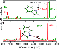
According to pioneer works of Nibbering,66 Zhao and Han,67 it has been established that hydrogen-bonding dynamics can be investigated by monitoring the vibrational absorption spectra of H-bonded groups at the ground and excited states. In Fig. 3, we have given the stretching vibrational frequency of N–H bond in different electronic states of neutral and protonated indole–water clusters. As shown the ground state N–H vibrational frequency of neutral and protonated indole–water clusters have been determined to be 3550 and 3067 cm−1 respectively (see blue curves of Fig. 3 and also see Table 2), while the corresponding values of S1 excited states have been determined to 3446 cm−1 and 3425 cm−1 (red spectra in Fig. 3). Thus, the N–H vibrational frequency in neutral indole–water cluster moves to the red by 105 cm−1, and the corresponding frequency of protonated system moves to the blue by 342 cm−1. The red and blue shifts on frequencies of N–H bond-stretching in neutral and protonated clusters following photoexcitation are in accord with increasing and decreasing of N–H bond-length respectively in neutral and protonated systems.


image file: c6ra06716f-f3.tif
Fig. 3 Selected part of vibrational absorption spectra of the neutral and protonated indole with water clusters in different electronic states. The blue colour represents the vibrational spectrum on the ground state and the red one is related to the S1 excited state. The spectra have been calculated with the RI-MP2 (S0) and RI-CC2 (S1) methods.

It is noteworthy that experimental vibrational frequencies, corresponding to the ground and S1 excited states of N–H stretching mode of indole–water cluster, have been reported respectively to be 3436 and 3387 cm−1 by Zwier's group.12 As seen, our theoretical values of 3545 and 3446 cm−1 (respectively for the ground and excited states), can be compared with corresponding experimental values of Zwier et al.12 by applying the scaling factors of 0.97 and 0.98 on the respective S0 and S1 simulated spectra.

3.2 Neutral/protonated 5-hydroxyindole with water clusters

3.2.1 Neutral and protonated monomers: 5-HIn and 5-HInH+. 5-Hydroxyindole (abbreviated by 5-HIn) is the chromophore of serotonin,68,69 which is a neurotransmitter in biologic systems. The neutral form of 5-hydroxyindole has been studied extensively by Oeltermann71 and others,43,70,71 thus, we disregarded to present further details on its geometry and electronic properties. In contrary to its importance, rarely reports are dedicated to photophysical characters 5-HIn38,43,44,72 in its neutral state. Nevertheless, on best of our knowledge, no report is dedicated on protonation and mono-hydration of 5-hydroxyindole. Thus we have determined the geometry, electronic structures and oscillator strengths of 5-hydroxyindole in its neutral and protonated states.

One may expect several protonated isomers for 5-HIn, corresponding to its different carbon sites. We have determined the optimized structures and consequently the relative stability of these isomers at the RI-MP2/cc-pVDZ level of calculation (see Fig. 4 and its inset for numbering). As shown, protonation of C2 carbon site, leads to the most stable isomer. Also, the C4 carbon atom is the second attractive-position for protonation, producing another protonated isomer with 15.4 kJ mol−1 (0.15 eV) less stability than C2 isomer. With exception of C4 isomer, other protonated isomers of 5-HIn, have been predicted to contain higher internal-energy of 43 kJ mol−1, in respect to C2, the most stable protonated isomer. Moreover, protonation of C3, C8 and oxygen atoms, obtain other high-energy isomers (ΔE > 100 kJ mol−1). Because of the low possibility for populating these isomers in a gas phase study, we ignored to present further details of these isomers. Therefore, in the next sections, we will only consider the most stable structure of protonated 5-HIn (i.e. C2 isomer) for further calculations.


image file: c6ra06716f-f4.tif
Fig. 4 Comparison the stability of different protonated isomers of 5-hydroxyindole, computed at the RI-MP2/cc-pVDZ level of theory. The inset, represent the numbering pattern of carbon sites.
3.2.2 Neutral and protonated 5-HIn–water clusters: 5-HIn–W and [5-HIn–W]H+. An extensive search on the neutral and protonated [5-HIn–W] complexes, resulted in few low energy configurations, based on the position of water in respect to neutral/protonated chromophore. In Fig. 5, four local minima for neutral and protonated systems have been presented. These isomers are stabilized either by HN⋯OH, OH⋯OH, CH⋯OH or π⋯H hydrogen bonds. However, for neutral 5-HIn–W, the isomer involving a strong H-bond between chromophore's hydroxyl-(OH) and water molecule (OH(5-HIn)⋯OH(W) bond), is found as the most stable isomer. Also, a slightly less-stable isomer of 5-HIn–W; (with relative energy of 3.00 kJ mol−1), is obtained by adding a water molecule to N–H bond of 5-HIn, leading to the structure involving NH(5-HIn)⋯OH(W) H-bond. In addition, our attempts on optimization of another complex of 5-HIn–W, by adding a water molecule to the C–H side of 5-HIn chromophore converged to another structure, for which the water molecule is shared between two C–H bonds (Fig. 5(c1)). As shown, this structure is significantly less stable than a1, (18 kJ mol−1). In addition, the latest structure has been determined by adding the water molecule, interacting with π-aromatic by O–H bond; (i.e. O–H⋯π interaction). This cluster (Fig. 5(d1)), is found to be more stable than c1, nevertheless that is 14 kJ mol−1 less stable than a1 cluster. However, from inspection of relative stabilities in Fig. 5, the decreasing pattern of H-bond interactions in 5-hydroxyindole–water clusters can be presented as:
OH(5-HIn)⋯OH(W) > NH(5-HIn)⋯OH(W) > OH(water)⋯π(5-HIn) > CH(5-HIn)⋯OH(W).

image file: c6ra06716f-f5.tif
Fig. 5 Optimized structures along with relative stabilities (in kJ mol−1), of neutral and protonated 5-HIn–W clusters calculated at RI-MP2/aug-cc-pVDZ level of theory. For a1, a2/b1, b2 isomers, the H-bond binding energies, have been presented in parenthesis.

For mono-hydration of protonated 5-HIn, four different isomers, based on the O–H, N–H, C–H and H⋯π interactions have been considered. The results have been presented in Fig. 5(a2–d2). Comparison the total binding energies in neutral and protonated clusters (a1, a2 and b1, b2), the H-bond interaction in protonated species has been found to be substantially stronger than neutral homologues. Also, in contrast to neutral clusters, for protonated 5-HIn–W, the most stable configuration arises from the NH⋯OH H-bond formation. The large charge-density on pyrrole ring strongly affects the NH⋯OH hydrogen bond interaction.

3.2.3 Electronic structures and transition energies. In Table 3, the vertical and adiabatic electronic transition energies for individual and monohydrated forms of the neutral/protonated 5-HIn are presented. The electronic spectrum of 5-HIn has been recorded by Arnold73 and Huang.71 The band origin of the S1–S0 transition has been reported to be 4.05 eV. The adiabatic S1–S0 transition energy of 5-HIn have been determined to be 4.13 and 4.29 eV respectively at the RI-CC2/aug-cc-pVDZ and cc-pVDZ level of theory.
Table 3 Calculated electronic transition energies (in eV) and corresponding oscillator strengths (in parenthesis) of low-lying singlet electronically excited states for neutral/protonated 5-HIn accompanied by their water clusters. The values in parentheses represent the ΔZPE-corrected values for S1–S0 transition at the CC2/cc-pVDZ level of theory
  Electronic state Vertical transition energy/eV Adiabatic transition energy/eV
aug-cc-pVDZ Oscillator strength cc-pVDZ aug-cc-PVDZ cc-pVDZ
a The experimental band origin of 5-HIn has been adopted from ref. 73.
5-HIn S1(ππ*) 4.33 0.0580 4.50 4.13 4.29 (4.11)
4.05a
S2(ππ*) 4.64 0.0002 5.18
5-HIn–H+ S1(ππ*) 3.43 0.0240 3.49 3.05 3.12 (2.96)
S2(ππ*) 4.11 0.0053 4.28
5-HIn(OH)–W S1(ππ*) 4.28 0.0786 4.45 4.06 4.24 (4.04)
S2(ππ*) 4.42 0.1182 5.24
5-HIn(NH)–W S1(ππ*) 4.31 0.0570 4.46 4.10 4.26 (4.07)
S2(ππ*) 4.49 0.0005 5.08
[5-HIn(NH)–W]H+ S1(ππ*) 3.63 0.2487 3.71 3.11 3.21 (3.01)
S2(ππ*) 4.33 0.0048 4.43
[5-HIn(OH)–W]H+ S1(ππ*) 3.19 0.2776 3.23 2.71 2.88 (3.92)
S2(ππ*) 4.11 0.0056 4.19


The former result (4.13 eV) is comparable with experiment while the later is far. However, when the difference between the zero point vibrational energy of the ground and S1 excited state (ΔZPE = −0.19 eV at the RI-CC2/cc-pVDZ level of theory), is taken to account, the corresponding transition energy (4.10 eV) will be in well agreement with experiment.

In addition, the adiabatic S1–S0 transition of protonated 5-HIn has been determined to be 3.05 and 2.96 eV (corrected by ΔZPE = −0.16 eV), at the RI-CC2/aug-cc-pVDZ and cc-pVDZ level respectively. Thus, the S1–S0 transition of protonated 5-HIn is predicted to be strongly red shifted in comparison with its neutral homologue; (by around 1.00 eV).

In order to investigate the H-bond characters, we have considered two isomers for monohydrating of neutral and protonated 5-HIn, based on the connection of water to N–H or O–H groups, obtaining the 5-HIn(NH)–W/[5-HIn(NH)–W]H+ and 5-HIn(OH)–W/[5-HIn(OH)–W]H+ clusters. The results have been presented in Table 3. As shown, mono-hydration dose not significantly affect the S1–S0 transition energies.

Concerning the nature of electronic transitions, the RI-CC2 calculated results, predicted that S1–S0 and S2–S0 for bare 5-HIn, [5-HIn]H+ and their monohydrated systems arise from HOMO–LUMO and HOMO−1–LUMO transitions respectively. The contribution of HOMO and LUMO for the first electronic transition is more than 90% while the second electronic transition is mostly arising from HOMO−1–LUMO single electron transition (75%). In Fig. 2, the selected frontier MOs of the bare and hydrated systems are presented. As shown, both HOMO, HOMO−1 have π character and LUMO is a π* orbital. Thus the S1 and S2 states can be assigned as 1ππ* nature. In addition, in ESI file further details on electronic transitions and configurations of our studied systems have been presented.

3.2.4 Hydrogen bonding dynamics. In order to investigate the excitation effect on the H-bond dynamics of neutral and protonated 5-HIn–water clusters, two types of H-bond clusters; (OH(5-HIn)⋯OH(W) and NH(5-HIn)⋯OH(W)) have been studied. In Table 4, selected H-bond parameters of neutral and protonated 5-HIn–W, consisting the OH(5-HIn)⋯OH(W) bond have been presented. The ground state OH⋯OH bond-length for neutral and protonated clusters is determined to be 1.888 and 1.743 Å respectively. The corresponding values for S1 excited state also have been determined to be 1.743 and 1.600 Å. Thus for neutral and protonated clusters, excitation is accompanied by shortening of OH⋯OH bond length. The H-bond binding energies for both cases at the ground state have been determined to be −25.9 kJ mol−1 and −53.7 kJ mol−1 respectively (the BSSE corrections have been considered). However, following photoexcitation, the binding energies move to 30.7 kJ mol−1 and 84.1 kJ mol−1 respectively for neutral and protonated clusters. Thus, photoexcitation of both systems are along with strengthening of OH⋯OH bond in neutral and protonated systems.
Table 4 The H-bond parameters of neutral, protonated 5-HIn(OH)–W clusters at ground, (S0) and S1 excited states calculated at RI-MP2/RI-CC2 level of theory. The EHb(S0,c) and EHb(S1,c) denote to corrected binding energies of ground and excited states with BSSE
  State 5-HIn(OH)–W [5-HIn(OH)–W]H+

image file: c6ra06716f-u3.tif

image file: c6ra06716f-u4.tif

l(O⋯H) S0 1.888 1.743
l(O–H) 0.975 0.987
l(O⋯H) S1 1.789 1.600
l(O–H) 0.976 1.015
vO–H/cm−1 S0 3675 3441
S1 3445 2915
EHb (kJ mol−1) S0 −30.2 −58.8
BSSE (4.3) (5.1)
E(S0,c) −25.9 −53.7
S1 −37.2 −91.8
BSSE (6.5) (7.7)
E(S1,c) −30.7 −84.1


As mentioned before, there is another possibility for adding water molecule to the N–H bond of neutral and protonated 5-HIn, constructing the NH(5-HIn)⋯OH(W) H-bond. The results of NH⋯OH H-bond parameters have been presented in Table 5. As shown, the NH⋯OH bond lengths in the ground state of neutral and protonated 5-HIn(NH)–W have been determined to be 1.953 and 1.699 Å respectively. The substantially shorter H-bond of protonated system indicates to stronger hydrogen bond in protonated cases. Also, the corresponding value of NH⋯OH bond lengths for the S1 excited state have been determined to be 1.918 and 1.931 Å in neutral and protonated systems respectively. Thus, photoexcitation of neutral 5-HIn(NH)–W is accompanied by only 1.7% shortening of NH⋯OH bond length, and 15% lengthening of NH⋯OH bond in protonated system.

Table 5 H-Bond parameters of neutral, protonated 5-HIn–(NH)–water clusters at ground (S0) and S1 excited states calculated at RI-MP2, RI-CC2/aug-cc-pVDZ level of theory. The E(S0,c) and E(S1,c) denote to corrected binding energies of ground and excited states with BSSE
  State 5-HIn(NH)–W [5-HIn(NH)–W]H+

image file: c6ra06716f-u5.tif

image file: c6ra06716f-u6.tif

l(O⋯H) S0 1.953 1.699
l(N–H) 1.018 1.046
l(O⋯H) S1 1.918 1.931
l(N–H) 1.027 1.030
vN–H/cm−1 S0 3558 3092
S1 3449 3387
EHb (kJ mol−1) E(S0) −28.0 −71.8
BSSE (3.8) (5.3)
E(S0,c) −24.1 −66.5
E(S1) −30.6 −65.8
BSSE (6.1) (7.1)
E(S1,c) −24.5 −58.7


In addition, from inspection of hydrogen-bond binding energies of NH⋯OH for ground and excited states of neutral and protonated systems, it is seen that NH⋯OH bond in neutral stays roughly unchanged following photoexcitation; (based on the small alteration of EHb from −24.1 to −24.5 kJ mol−1 respectively from ground to the S1 excited state).

Also, for protonated system, photoexcitation is accompanied by slightly strengthening of NH⋯OH hydrogen bond; (increasing the absolute value of EHb by 7.8 kJ mol−1 after excitation).

The increasing in the absolute value of binding energy after excitation for protonated systems is in accord with NH⋯OH H-bond shortening, and indicating to strengthening of NH⋯OH bond after excitation.

Additionally, we have determined the ground and S1 state IR spectra of 5-HIn–W clusters in neutral and protonated cases. A selected part for IR spectra has been presented in Fig. 6. Considering the vibrational frequencies of normal modes and IR spectra of Fig. 6, it is seen that in neutral and protonated systems, involving OH⋯OH H-bond, the O–H bond stretching of chromophore, shows red-shift in the S1 excited state, by decreasing with 230 and 526 cm−1 respectively in neutral and protonated systems. This decreasing pattern of O–H vibrational frequency is accompanied by increasing of the O–H bond lengths of chromophore and also decreasing of OH⋯OH bond lengths, following photoexcitation of both clustered systems, indicating to H-bond strengthening.


image file: c6ra06716f-f6.tif
Fig. 6 Selected part of vibrational absorption spectra of neutral and protonated 5-HIn with water clusters in different electronic states. The blue colour represents the vibrational spectrum on the ground state and the red one is related to the S1 excited state. The spectra have been calculated at the RI-MP2 (S0) and RI-CC2 (S1) levels.

In contrast to OH⋯OH H-bond of neutral and protonated 5-HIn–W clusters, for the NH⋯OH (see Fig. 6c and d), the S1 excitation leads to the significant blue shift of N–H bond stretching (+295 cm−1), in protonated cluster, while that red shifted by 107 cm−1 in neutral homologue. These alterations in IR spectra are in agreement with shortening of N–H and lengthening of NH⋯OH hydrogen bonds in protonated system. Thus, the IR spectra of NH(5-HIn)⋯OH(W) clusters, confirm the H-bond weakening for protonated and slightly strengthening for neutral system following photoexcitation.

4. Discussion

The excited state hydrogen bond weakening and strengthening in water clusters of neutral and protonated indole and 5-hydroxyindole have been investigated. For neutral clusters, it has been predicted that photoexcitation, leads to strengthening of H-bond up to 22.0 kJ mol−1. Nevertheless, for protonated systems, both strengthening and weakening have been predicted. Evidently, following photoexcitation, a large alteration on electron distribution plays the most important role in hydrogen bond dynamics, resulting to new arrangement of nuclei. Although, it was not possible to perform an accurate charge distribution calculation on the ground and excited states of our cluster systems, we have examined the MOs, having the most important contributions in the S1–S0 electronic transition of our systems (see Fig. 2). As shown, in Fig. 2, for all of three neutral clusters, the lower and upper MOs, contributing in the S1–S0 transition, spread over both rings of indole moiety. In contrary, for protonated cases, the origin MOs (HOMO or HOMO−1), locates only over benzene ring, while the destination MOs (LUMOs) locate over pyrrole ring. Thus, a charge transfer phenomenon from neutral to protonated ring is suggested to happen on photoexcitation of protonated systems. This CT character, causes the decreasing of positive charge on protonated pyrrole moiety, resulting to weakening of H-bonds in [In–W]H+/[5-HIn–(NH)W]H+ and strengthening of H-bond in [5-HIn–(OH)W]H+ (see Fig. 2). Particularly, in the latter cluster, the CT character of excitation, leads to increasing the positive charge of phenolic part resulting to increasing the O–H polarity and finally strengthening of relevant hydrogen bonds.

5. Conclusion

The high level ab initio computational methods have been employed to investigate the geometry and electronic structures of individual and hydrogen bond clusters of neutral/protonated indole/5-hydroxyindole with water molecule. The results can be summarized as bellow:

(1) According to our RI-MP2 results, it has been predicted that C2 carbon site of pyrrole ring obtains the most stable protonated isomer of 5-hydroxyindole (see Fig. 4).

(2) A large red shift effect (∼1.0 eV) on the S1–S0 transition energy of 5-HIn has been predicted as the most important consequence of protonation.

(3) The most attractive site for monohydration of neutral 5-HIn has been predicted to be the O–H group, while that is the N–H moiety in protonated analogue, which obtains the most stable water cluster.

(4) Concerning the weakening or strengthening of intermolecular H-bonds, it has been found that the S1–S0 excitation effect on the H-bond strength in protonated systems is more pronounced than that of neutral clusters.

(5) Although, there is no explicit interpretation for hydrogen-bond dynamics of neutral systems, a simple explanation based on the CT character of excited state has been proposed for interpretation of weakening or strengthening of H-bonds in protonated clusters. This CT character of excited state, occurring from benzene to the pyrrole ring, increases the positive charge on the phenolic part and decreases that of pyrrole ring following photoexcitation of cluster system from ground to excited state. Thus, the S1–S0 transition in protonated indole–water cluster is along with weakening of NH⋯OH and strengthening of OH⋯OH hydrogen bonds respectively.

Acknowledgements

The research council of Isfahan University is acknowledged for financial support. Also, the use of computing facility cluster GMPCS of the LUMAT federation (FR LUMAT2764) for partially performance of our calculations is kindly appreciated.

References

  1. A. Lehninger, D. Nelson and C. Michael, Lehninger Principles of Biochemistry, W. H. Freeman and Company, New York, 2004 Search PubMed.
  2. C. J. Cramer and D. G. Truhlar, Acc. Chem. Res., 2008, 41, 760–768 CrossRef CAS PubMed.
  3. C.-H. Tung, L.-Z. Wu, L.-P. Zhang and B. Chen, Acc. Chem. Res., 2003, 36, 39–47 CrossRef CAS PubMed.
  4. M.-X. Zhang and G.-J. Zhao, ChemSusChem, 2012, 5, 879–887 CrossRef CAS PubMed.
  5. Y. D. Wu, K. Houk, J. Valentine and W. Nam, Inorg. Chem., 1992, 31, 718–720 CrossRef CAS.
  6. G.-J. Zhao, K.-L. Han and P. J. Stang, J. Chem. Theory Comput., 2009, 5, 1955–1958 CrossRef CAS PubMed.
  7. C. Fecko, J. Eaves, J. Loparo, A. Tokmakoff and P. Geissler, Science, 2003, 301, 1698–1702 CrossRef CAS PubMed.
  8. A. Luzar and D. Chandler, Phys. Rev. Lett., 1996, 76, 928–931 CrossRef CAS PubMed.
  9. M. Quack, J. Stohner and M. A. Suhm, J. Mol. Struct., 2001, 599, 381–425 CrossRef CAS.
  10. A. D. Boese, ChemPhysChem, 2015, 16, 978–985 CrossRef CAS PubMed.
  11. S. Perun, A. L. Sobolewski and W. Domcke, J. Am. Chem. Soc., 2005, 127, 6257–6265 CrossRef CAS PubMed.
  12. C. A. Southern, D. H. Levy, G. M. Florio, A. Longarte and T. S. Zwier, J. Phys. Chem. A, 2003, 107, 4032–4040 CrossRef CAS.
  13. T. S. Zwier, Annu. Rev. Phys. Chem., 1996, 47, 205–241 CrossRef CAS.
  14. A. L. Sobolewski and W. Domcke, J. Phys. Chem. A, 2007, 111, 11725–11735 CrossRef CAS PubMed.
  15. G.-J. Zhao and K.-L. Han, Acc. Chem. Res., 2011, 45, 404–413 CrossRef PubMed.
  16. J. Zhang, G. Zhao, R. Li and D. Hou, J. Cluster Sci., 2011, 22, 501–511 CrossRef CAS.
  17. R. Zhang, G.-S. Zhao and W.-J. Wu, Chin. J. Chem. Phys., 2009, 22, 511–516 CrossRef CAS.
  18. G.-J. Zhao, R.-K. Chen, M.-T. Sun, J.-Y. Liu, G.-Y. Li, Y.-L. Gao, K.-L. Han, X.-C. Yang and L. Sun, Chem.–Eur. J., 2008, 14, 6935–6947 CrossRef CAS PubMed.
  19. G.-J. Zhao, K.-L. Han, Y.-B. Lei and Y.-S. Dou, J. Chem. Phys., 2007, 127, 094307 CrossRef PubMed.
  20. C. Chudoba, E. Nibbering and T. Elsaesser, Phys. Rev. Lett., 1998, 81, 3010–3013 CrossRef CAS.
  21. A. Douhal, F. Lahmani and A. H. Zewail, Chem. Phys., 1996, 207, 477–498 CrossRef CAS.
  22. E. Pines, D. Pines, Y.-Z. Ma and G. R. Fleming, ChemPhysChem, 2004, 5, 1315–1327 CrossRef CAS PubMed.
  23. G.-J. Zhao and K.-L. Han, J. Phys. Chem. A, 2007, 111, 9218–9223 CrossRef CAS PubMed.
  24. G.-J. Zhao and K.-L. Han, J. Chem. Phys., 2007, 127, 024306 CrossRef PubMed.
  25. G.-J. Zhao and K.-L. Han, J. Phys. Chem. A, 2007, 111, 2469–2474 CrossRef CAS PubMed.
  26. Y. Liu, J. Ding, R. Liu, D. Shi and J. Sun, J. Photochem. Photobiol., A, 2009, 201, 203–207 CrossRef CAS.
  27. O. Dopfer, A. Patzer, S. Chakraborty, I. Alata, R. Omidyan, M. Broquier, C. Dedonder and C. Jouvet, J. Chem. Phys., 2014, 140, 124314 CrossRef PubMed.
  28. B. Saed and R. Omidyan, J. Chem. Phys., 2014, 140, 024315 CrossRef PubMed.
  29. M. B. Smith and J. March, Advanced Organic Chemistry: Reactions, Mechanisms, and Structure, John Wiley & Sons, Inc, 2006 Search PubMed.
  30. N. Solcà and O. Dopfer, J. Phys. Chem. A, 2002, 106, 7261–7270 CrossRef.
  31. A. Kiendler and F. Arnold, Atmos. Environ., 2002, 36, 2979–2984 CrossRef CAS.
  32. S. Pasquiers, Eur. Phys. J.: Appl. Phys., 2004, 28, 319–324 CrossRef CAS.
  33. F. M. Pasker, N. Solcà and O. Dopfer, J. Phys. Chem. A, 2006, 110, 12793–12804 CrossRef CAS PubMed.
  34. M. C. Capello, M. Broquier, C. Dedonder-Lardeux, C. Jouvet and G. A. Pino, J. Chem. Phys., 2013, 138, 054304 CrossRef PubMed.
  35. C. Dedonder-Lardeux, C. Jouvet, S. Perun and A. L. Sobolewski, Phys. Chem. Chem. Phys., 2003, 5, 5118–5126 RSC.
  36. A. Kyrychenko, J. Herbich, F. Wu, R. Thummel and J. Waluk, J. Am. Chem. Soc., 2000, 122, 2818–2827 CrossRef CAS.
  37. M. G. D. Nix, A. L. Devine, B. Cronin and M. N. R. Ashfold, Phys. Chem. Chem. Phys., 2006, 8, 2610–2618 RSC.
  38. T. A. A. Oliver, G. A. King and M. N. R. Ashfold, Phys. Chem. Chem. Phys., 2011, 13, 14646–14662 RSC.
  39. G. A. Pino, I. Alata, C. Dedonder, C. Jouvet, K. Sakota and H. Sekiya, Phys. Chem. Chem. Phys., 2011, 13, 6325–6331 RSC.
  40. K. Sakota, C. Jouvet, C. Dedonder, M. Fujii and H. Sekiya, J. Phys. Chem. A, 2010, 114, 11161–11166 CrossRef CAS PubMed.
  41. A. L. Sobolewski and W. Domcke, Chem. Phys. Lett., 1999, 315, 293–298 CrossRef CAS.
  42. A. L. Sobolewski and W. Domcke, Chem. Phys. Lett., 2000, 329, 130–137 CrossRef CAS.
  43. R. Livingstone, O. Schalk, A. E. Boguslavskiy, G. Wu, L. Therese Bergendahl, A. Stolow, M. J. Paterson and D. Townsend, J. Chem. Phys., 2011, 135, 194307 CrossRef PubMed.
  44. R. F. Chen, Proc. Natl. Acad. Sci. U. S. A., 1968, 60, 598–605 CrossRef CAS.
  45. O. Christiansen, H. Koch and P. Jørgensen, Chem. Phys. Lett., 1995, 243, 409–418 CrossRef CAS.
  46. TURBOMOLE V6.2 2010, a development of University of Karlsruhe and Forschungszentrum Karlsruhe GmbH, 1989–2007, TURBOMOLE GmbH, since 2007, available from http://www.turbomole.com.
  47. T. Fleig, S. Knecht and C. Hättig, J. Phys. Chem. A, 2007, 111, 5482–5491 CrossRef CAS PubMed.
  48. I. Georgieva, N. Trendafilova, A. J. A. Aquino and H. Lischka, J. Phys. Chem. A, 2006, 111, 127–135 CrossRef PubMed.
  49. M. Schreiber, M. R. Silva-Junior, S. P. A. Sauer and W. Thiel, J. Chem. Phys., 2008, 128, 134110–134125 CrossRef PubMed.
  50. A. L. Sobolewski, D. Shemesh and W. Domcke, J. Phys. Chem. A, 2008, 113, 542–550 CrossRef PubMed.
  51. A. Carrera, I. B. Nielsen, P. Carcabal, C. Dedonder, M. Broquier, C. Jouvet, W. Domcke and A. L. Sobolewski, J. Chem. Phys., 2009, 130, 024302–024308 CrossRef CAS PubMed.
  52. S. Chakraborty, R. Omidyan, I. Alata, I. B. Nielsen, C. Dedonder, M. Broquier and C. Jouvet, J. Am. Chem. Soc., 2009, 131, 11091–11097 CrossRef CAS PubMed.
  53. A. Hellweg, C. Hättig, S. Höfener and W. Klopper, Theor. Chem. Acc., 2007, 117, 587–597 CrossRef CAS.
  54. R. A. Kendall, T. H. Dunning Jr and R. J. Harrison, J. Chem. Phys., 1992, 96, 6796–6806 CrossRef CAS.
  55. K. Hald, C. Hättig, D. L. Yeager and P. Jørgensen, Chem. Phys. Lett., 2000, 328, 291–301 CrossRef CAS.
  56. C. Hättig, J. Chem. Phys., 2003, 118, 7751 CrossRef.
  57. T. H. Dunning Jr, J. Chem. Phys., 1989, 90, 1007 CrossRef.
  58. T. H. Dunning Jr, J. Chem. Phys., 1989, 90, 1007–1023 CrossRef.
  59. S. F. Boys and F. Bernardi, Mol. Phys., 1970, 19, 553–566 CrossRef CAS.
  60. I. Alata, J. Bert, M. Broquier, C. Dedonder, G. Feraud, G. Grégoire, S. Soorkia, E. Marceca and C. Jouvet, J. Phys. Chem. A, 2013, 117, 4420–4427 CrossRef CAS PubMed.
  61. E. Krystkowiak, J. Koput and A. Maciejewski, Phys. Chem. Chem. Phys., 2012, 14, 8842–8851 RSC.
  62. A. Mani and J. R. Lombardi, J. Mol. Spectrosc., 1969, 31, 308–317 CrossRef CAS.
  63. M. J. Tubergen and D. H. Levy, J. Phys. Chem., 1991, 95, 2175–2181 CrossRef CAS.
  64. R. Bersohn, U. Even and J. Jortner, J. Chem. Phys., 1984, 80, 1050–1058 CrossRef CAS.
  65. R. Daengngern, N. Kungwan, P. Wolschann, A. J. A. Aquino, H. Lischka and M. Barbatti, J. Phys. Chem. A, 2011, 115, 14129–14136 CrossRef CAS PubMed.
  66. E. T. J. Nibbering and T. Elsaesser, Chem. Rev., 2004, 104, 1887–1914 CrossRef CAS PubMed.
  67. G. J. Zhao and K. L. Han, ChemPhysChem, 2008, 9, 1842–1846 CrossRef CAS PubMed.
  68. J. Harley-Mason and A. H. Jackson, J. Chem. Soc., 1954, 1165–1171 RSC.
  69. J. Harley-Mason and A. H. Jackson, J. Chem. Soc., 1954, 3651–3654 RSC.
  70. O. Oeltermann, C. Brand, M. Wilke and a. M. Schmitt, J. Phys. Chem. A, 2012, 116, 7873–7879 CrossRef CAS PubMed.
  71. Y. Huang and M. Sulkes, Chem. Phys. Lett., 1996, 254, 242–248 CrossRef CAS.
  72. T. Kishi, M. Tanaka and J. Tanaka, Bull. Chem. Soc. Jpn., 1977, 50, 1267–1271 CrossRef CAS.
  73. S. Arnold and M. Sulkes, Chem. Phys. Lett., 1992, 200, 125–129 CrossRef CAS.

Footnote

Electronic supplementary information (ESI) available. See DOI: 10.1039/c6ra06716f

This journal is © The Royal Society of Chemistry 2016