
Recognition of diamond with phage display peptides†
Swathi Swaminathanc and
Yue Cui*ab
aDepartment of Electrical Engineering and Computing Systems, University of Cincinnati, Cincinnati, OH 45221, USA. E-mail: cuiy3@ucmail.uc.edu
bDepartment of Mechanical and Materials Engineering, University of Cincinnati, Cincinnati, OH 45221, USA
cDepartment of Biological Engineering, Utah State University, Logan, UT 84322, USA
First published on 12th May 2016
Abstract
We show for the first time the identification of diamond-binding peptide motifs for diamond with phage display. Our approach is based on a comprehensive screening of specific peptides from a phage display library, along with novel micropatterning methods. These results could open up significant opportunities for the development of a variety of diamond-based electronic devices ranging from sensors to energy storage devices.
Diamond, as a cubic, sp3 carbon-based material, is one of the best electrode materials with exceptional physical and chemical properties,1–3 that has attracted great attention for both fundamental studies as well as practical applications. Diamond is known for its many attractive properties, including hardness, a wide electrochemical potential window, low and stable background current, weak adsorption of polar molecules, long-term response stability, biocompatibility, great fouling resistance, and chemical inertness. Growth of diamond has been achieved via several methods, including chemical vapor deposition,4–6 laser-assisted combustion,7 X-ray radiation,8 and ultrasound cavitation.9 Despite these advances, the ability to generically tailor its chemical and biological properties has been limited. Selective recognition of a diamond surface without changing the diamond lattice is highly desired for a wide range of chemical and biological applications.
Oligopeptides are robust molecules with broad chemical diversity. Oligopeptides can be tailored to possess different functions, for example, binding to specific targets or catalyzing the synthesis of nanomaterials.10–14 One of their major capabilities, exploited in phage display technology, is its specific recognition ability. Phage display is a bioscreening technique for identifying specific peptide sequences with enhanced binding affinity from a phage display library containing around 1–2 billion (109) peptide-displaying phages. Recently, other members in the carbon family have been studied for the selection of phage display peptides including graphite,15 graphene,15,16 and CNT,17,18 and diamond is currently a missing member in carbon family for using phage display to select specific peptides for recognition. We have also identified binding motifs of several carbon-based materials, including graphite,15 graphene,15 small molecular ink,19 PDMS,12,20 epoxy,21 and PET.22
In this work, we present for the first time a powerful and benign approach for the identification of diamond binding motifs with phage display technology. The phage-displayed peptides were vigorously screened from a biocombinatorial library and the screened peptides have shown to recognize diamond surface, which can be used as a binding motif to diamond surface. Further, diamond patterns were fabricated, and the phage-displayed peptides were patterned according to the diamond pattern.
Fig. 1 shows the schematic illustration of identification of peptides for recognizing diamond with phage display technology. An aliquot of phage display library (New England BioLabs, Ph.D. 12) of random 12-mer peptides (Fig. 1a) was incubated with a diamond chip (a thin film of diamond deposited on SiO2/Si) to obtain phages which bind to diamond on SiO2/Si substrate (Fig. 1b), and then eluted from this substrate to collect the phage molecules which bind to both diamond and SiO2/Si (Fig. 1c). The collected peptide-displaying phages were amplified in E. coli (ER 2738) (Fig. 1d), followed by repeated centrifugation and precipitation to collect the amplified phage molecules. After this, the eluted phages were screened with a background substrate (Fig. 1e), SiO2/Si to eliminate the phages for SiO2/Si to select the phage-displayed peptides only to diamond (Fig. 1f). The biopanning processes were repeated for a total of three times to obtain phage displayed peptides which bind to diamond (Fig. 1g). Finally, the refined libraries went through for DNA sequencing to obtain the amino acid sequences of diamond-binding peptides.
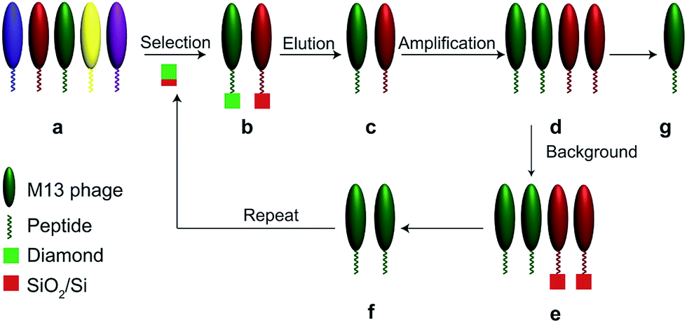 | ||
| Fig. 1 Schematic illustration of the generalized screening protocol for identifying phage displayed peptides binding to diamond deposited on SiO2/Si. (a) a phage display library, (b) phages bind to both the diamond and SiO2/Si, (c) phages binding to both the diamond and SiO2/Si are eluted from the substrate, (d) amplified phages binding to both the diamond and SiO2/Si, (e) amplified phages binding to both the diamond and SiO2/Si are incubated with plain SiO2/Si for negative screening, (f) amplified phages which only bind to diamond, and (g) phages which recognize only the diamond surface are collected. | ||
Table 1 shows the sequences of the screened phage displayed peptides obtained from phage display technology with three rounds of biopanning process. The natures of the peptides screened from phage display process (with C-terminus amidated to avoid the free charge) were analysed. As shown in the table, the screened diamond-binding peptides contain alternating hydrophobic and hydrophilic residues. Besides, all of these screened peptides carry net positive charge at neutral pH, and exhibit high values of pI. The analysis of the amino acids in the peptide sequences shows that arginine, asparagine, threonine and valine are the frequent binding amino acid residues, and three out of these amino acid residues (arginine, asparagine, and threonine) are hydrophilic while valine is hydrophobic. The result indicates that diamond binds to both hydrophobic and hydrophilic amino acid residues with irregular affinities (see Fig. S1 in ESI†).
| Sequence | Average hydrophilicity | Hydrophilic ratio | Isoelectric point | Net charge |
|---|---|---|---|---|
| GVGGLTTVNYSR | −0.4 | 25% | 11.3 | 2 |
| NVVRNVFPALDH | −0.3 | 33% | 11 | 1.1 |
| ISYQTRHTFPTI | −0.5 | 25% | 11.3 | 2.1 |
| HKPPRQKPKAQQ | 1 | 58% | 14 | 5.1 |
| NVDYNRKDRIDR | 1.3 | 75% | 10.4 | 2 |
One peptide-displaying phage that recognized diamond (GVGGLTTVNYSR) was isolated and amplified to form a single colony and selected for further investigation. First, fluorescent characterization for the binding of this phage to plain diamond surface and SiO2/Si substrates was investigated. This was accomplished by incubating the chip of diamond on SiO2/Si sequentially to (1) peptide-displaying phages (GVGGLTTVNYSR), (2) blocking buffer, (3) anti-M13 phage coat protein biotin conjugated antibody, and (4) avidin-FITC, with buffer washing steps in between. The fluorescent color intensity on the surface resulting from FITC is proportional to the binding of the phage-displayed peptides. As shown in Fig. 2a, the fluorescent characterization of diamond surface and SiO2 surface shows that diamond surface shows a much higher fluorescence intensity compared with SiO2/Si surface due to diamond-binding phage displayed peptides. In addition, fluorescence characterization using M13 phage without any phage-displayed peptide was performed on diamond and SiO2/Si substrates, as a control experiment. As shown in Fig. 2b, there is almost no fluorescent signal for both substrates. These results indicate that screened phage-displayed peptides are specific to diamond surface and are essential for binding to diamond.
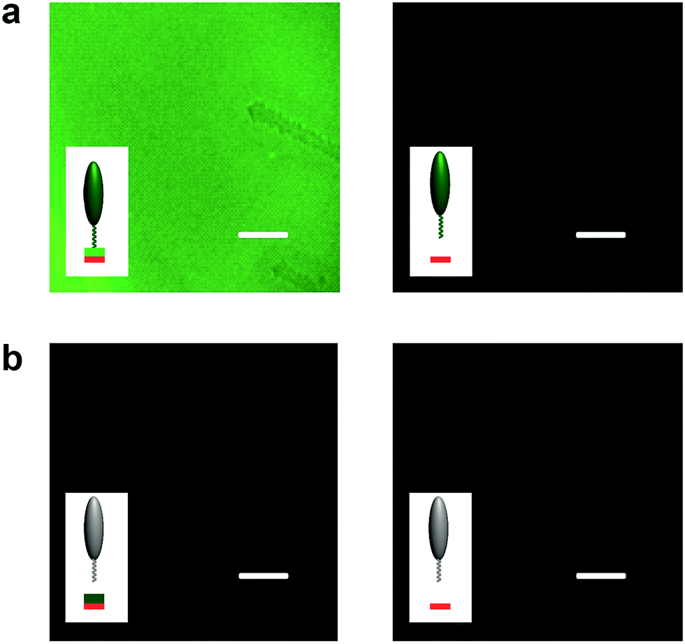 | ||
| Fig. 2 Fluorescent characterization of the binding of phage displayed peptides. (a) Peptide (GVGGLTTVNYSR) displaying phages on a diamond surface (left image) and a SiO2 surface (right image). (b) M13 phage (without displayed peptide) for a diamond surface (left image) and a SiO2 surface (right image). All scale bars: 20 μm. | ||
Further, we investigated the selective recognition of diamond micropatterns. Fig. 3a shows the schematic illustration of the generation of diamond micropatterns. A transmission electron microscope (TEM) grid with different patterns was placed on diamond (step 1). Next, a plasma oxidation was performed on the TEM grid covered diamond chip to remove the exposed diamond to generate diamond micropatterns (step 2). Finally, the TEM grid was removed to reveal the diamond patterns (see Fig. S2 in ESI†). By introducing appropriate phage displayed peptides, the phages bind to the specific locations mapped by the micropatterned diamond pattern (step 3). Fluorescent characterization was performed on these diamond micropatterns with phage-displayed peptides. The intensity of fluorescence on the diamond micropatterns was significantly higher than that on the etched surface, which is only SiO2/Si, as shown in Fig. 3b. This result clearly indicates that microstructured diamond imparts enhanced binding affinity toward the phage-displayed peptides over the host SiO2 surface.
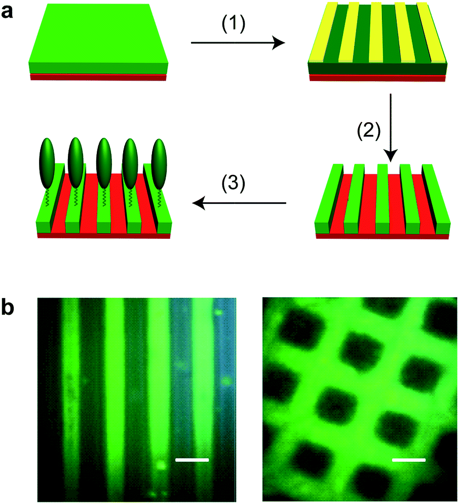 | ||
| Fig. 3 Identification of micropatterned diamond with phage displayed peptides (GVGGLTTVNYSR). (a) Schematic illustration of for the generation of diamond patterns. Step (1) covering diamond with TEM grid as shadow masks, step (2) plasma etching to remove the exposed diamond and generate diamond micropatterns, and step (3) introducing phage displayed peptides binding to diamond. Red is SiO2/Si substrate, green is diamond, and yellow is TEM grid. (b) Fluorescent characterization of the binding of phage displayed peptides on line/square diamond micropatterns. All scale bars: 20 μm. | ||
Conclusions
We have demonstrated for the first time the identification of diamond binding motifs via a combinatorial phage display peptide library. Our work assimilates the positive and negative screening of phage-displayed peptides on diamond surface and its host substrate SiO2/Si, respectively, the identification of specific diamond-binding phage displayed peptides, and the exposure the diamond and diamond micropatterns to phage displayed peptides for localized binding. These results provide the fundamental knowledge for a variety of other diamond-based studies, such as new multifunctional hybrid diamond materials, novel diamond surface and interface, diamond-based pharmaceutics, and different diamond-based biological, chemical, optical, mechanical, and electrical devices.Acknowledgements
The authors acknowledge Dan McClellan for assistant in plasma etching.References
- G. C. Chen, B. Li, Z. Q. Yan, J. Liu, F. X. Lu and H. Ye, J. Cryst. Growth, 2012, 349, 1–5 CrossRef CAS.
- O. Babchenko, Z. Remes, T. Izak, B. Rezek and A. Kromka, Phys. Status Solidi B, 2011, 248, 2736–2739 CrossRef CAS.
- D. J. Garrett, K. Ganesan, A. Stacey, K. Fox, H. Meffin and S. Prawer, J. Neural Eng., 2012, 9, 016002 CrossRef PubMed.
- G. D. Barber and W. A. Yarbrough, J. Am. Ceram. Soc., 1995, 78, 3390–3392 CrossRef CAS.
- C. H. Hsu, S. G. Cloutier, S. Palefsky and J. Xu, Nano Lett., 2010, 10, 3272–3276 CrossRef CAS PubMed.
- X. C. Xiao, J. W. Elam, S. Trasobares, O. Auciello and J. A. Carlisle, Adv. Mater., 2005, 17, 1496–1500 CrossRef CAS.
- T. Guillemet, Z. Q. Xie, Y. S. Zhou, J. B. Park, A. Veillere, W. Xiong, J. M. Heintz, J. F. Silvain, N. Chandra and Y. F. Lu, ACS Appl. Mater. Interfaces, 2011, 3, 4120–4125 CAS.
- Z. W. Wang, Y. S. Zhao, C. S. Zha, Q. Xue, R. T. Downs, R. G. Duan, R. Caracas and X. Z. Liao, Adv. Mater., 2008, 20, 3303–3307 CrossRef CAS.
- A. K. Khachatryan, S. G. Aloyan, P. W. May, R. Sargsyan, V. A. Khachatryan and V. S. Baghdasaryan, Diamond Relat. Mater., 2008, 17, 931–936 CrossRef CAS.
- J. Makowska, K. Zamojć, D. Wyrzykowski, D. Uber, M. Wierzbicka, W. Wiczk and L. Chmurzyński, Spectrochim. Acta, Part A, 2016, 153, 451–456 CrossRef CAS PubMed.
- Y. O. Kim, H. S. Jang, Y. H. Kim, J. M. You, Y. S. Park, K. Jin, O. Kang, K. T. Nam, J. W. Kim, S. M. Lee and Y. S. Lee, RSC Adv., 2015, 5, 78026–78029 RSC.
- S. Swaminathan, M. Bullough, Q. F. Li, A. H. Zhou and Y. Cui, J. R. Soc., Interface, 2014, 11, 5 Search PubMed.
- Y. Li, Z. Tang, P. N. Prasad, M. R. Knecht and M. T. Swihart, Nanoscale, 2014, 6, 3165–3172 RSC.
- Y. N. Tan, J. Y. Lee and D. I. C. Wang, J. Am. Chem. Soc., 2010, 132, 5677–5686 CrossRef CAS PubMed.
- Y. Cui, S. N. Kim, S. E. Jones, L. L. Wissler, R. R. Naik and M. C. McAlpine, Nano Lett., 2010, 10, 4559–4565 CrossRef CAS PubMed.
- Y. Cui, S. N. Kim, R. R. Naik and M. C. McAlpine, Acc. Chem. Res., 2012, 45, 696–704 CrossRef CAS PubMed.
- T. Yu, Y. Gong, T. Lu, L. Wei, Y. Li, Y. Mu, Y. Chen and K. Liao, RSC Adv., 2012, 2, 1466–1476 RSC.
- T. Yu, Y. Gong, T. Lu, L. Wei, Y. Li, Y. Mu, Y. Chen and K. Liao, RSC Adv., 2012, 2, 1466–1476 RSC.
- Y. Cui, A. Pattabiraman, B. Lisko, S. C. Collins and M. C. McAlpine, J. Am. Chem. Soc., 2010, 132, 1204–1205 CrossRef CAS PubMed.
- S. Swaminathan and Y. Cui, RSC Adv., 2012, 2, 12724–12727 RSC.
- S. Swaminathan and Y. Cui, Mater. Sci. Eng., C, 2013, 33, 3082–3084 CrossRef CAS PubMed.
- S. Swaminathan and Y. Cui, RSC Adv., 2016, 6(18), 14589–14592 RSC.
Footnote |
| † Electronic supplementary information (ESI) available. See DOI: 10.1039/c6ra06582a |
| This journal is © The Royal Society of Chemistry 2016 |