A combined experimental–theoretical study of the acid–base behavior of mangiferin: implications for its antioxidant activity†
Gabriela Mendoza-Sarmiento,
Alberto Rojas-Hernández *,
Annia Galano and
Atilano Gutiérrez
Universidad Autónoma Metropolitana-Iztapalapa, Departamento de Química, Área de Química Analítica, Apartado Postal: 55-534, 09340 México, D. F., Mexico. E-mail: suemi918@xanum.uam.mx
First published on 18th May 2016
Abstract
Acidity constants of mangiferin (H4MGF) in DMSO/H2O (80%/20%, v/v) were determined by UV-Visible and 1H and 13C NMR spectroscopies. UV-Visible absorption spectra in the 4.2 ≤ pH ≤ 11.7 range were fitted using the computational program SQUAD, for the refining of pKa values, obtaining as results: pKa1 = 7.337 ± 0.001, pKa2 = 8.936 ± 0.001, and pKa3 = 10.297 ± 0.028. The sigmoidal curves of the chemical shifts of 1H and 13C in a similar pH range were fitted using the same model refined by SQUAD for UV-Visible data. The behavior of the chemical shifts as a function of pH allowed us to assign the deprotonation order. A theoretical DFT study has been followed confirming the deprotonation order determined experimentally, as well as a proposed mechanism of antioxidant activity, that considers the fractions of the different species at physiological pH. It was found that mangiferin is an excellent peroxyl radical scavenger in aqueous solution, significantly surpassing the activity of Trolox, which is a well-known antioxidant frequently used as a reference in this context.
Introduction
Mangifera indica L. has been extensively used in Ayurvedic traditional medicine.1 One of the major components in this plant is a substance called mangiferin (Scheme 1), that was extracted for the first time in 1922 by W. Wiechowski. Mangiferin is distributed in several parts of the plant, such as the roots, stems, bark and fruits.2–4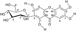 | ||
| Scheme 1 Chemical structure of mangiferin (2-β-D-glucopyranosyl-1,3,6,7-tetrahydroxyxanten-9-one, H4MGF). | ||
Several studies indicate that mangiferin has a large number of pharmacological properties including antioxidant, anti-diabetic, anti-HIV, anti-tumor, hepatoprotective, antiviral, antibacterial, and anti-cancer activities.3–27 It is clear that these pharmacological activities, particularly antioxidant properties, are related to the structure of the molecule and its protonation degree. However, only a few fundamental physicochemical studies to determine this relationship have been performed so far. Our research group has determined the four pKa values of mangiferin in water, at 25 °C, and has proposed the deprotonation order from 1H and 13C NMR spectra.28 Nevertheless, in that study only 4 NaOD additions, equivalent to the four stoichiometric amounts, were achieved, without measuring the pH of the solutions. In addition, although the antioxidant power of mangiferin has been previously documented,29 the mechanism of this activity is not completely understood yet.
Accordingly, the aims of this work are: (i) to determine the pKa values of mangiferin in a DMSO/H2O (80%/20%, v/v) mixture, where its solubility is higher than in water, using thorough UV-Visible, 1H and 13C NMR spectroscopic studies, at different pH values, as well as to confirm the deprotonation order previously proposed for mangiferin; and (ii) to investigate the possible antioxidant mechanism by a DFT theoretical study, including kinetics and the influence of pH.
Experimental section
Reagents
Equipment and experimental methods
Solutions of mangiferin 4 × 10⁻⁵ M in DMSO/H2O (80%/20%, v/v) were used, mixed with NaOH to obtain absorption spectra at different pH values. Similar experiments were also pursued using only water as a solvent.
Solutions of 0.142 M mangiferin in DMSO-d6/D2O (80%/20%, v/v) were used. The pH was adjusted with a concentrated NaOD solution that was added to mangiferin solution in such a way that the total NaOD additions only change less than 5% of the original volume.
pH measurements
The pH measurements were carried out on a pHM240 Radiometer potentiometer, using an Ag/AgCl/glass combined electrode Radiometer model pHC3359-8, for pH measurements in the 0–11 pH range in water. The pH values were corrected using the following equation:30where pHcorr is the corrected pH, pHexp is the experimental pH measured, pHcal is the calibration pH, and Ef is an empiric fitting value (related inversely to the efficiency of the potentiometric cell): Ef tends to zero when cell's efficiency tends to 100%. Ef is selected for a slope of −59.16 mV (corresponding to a temperature of 25 °C) for the curve of cell potential as a function of pHcorr.
Sigmoidal curves fitting
In order to proof the applicability of the model refined by the program SQUAD, sigmoidal curves of absorbances or chemical shifts as a function of pH were fitted assuming linear behavior of these properties with equilibrium concentration of species, as it was described in a previous work.31Computational details
All the electronic calculations were performed with Gaussian 09 package of programs.32 Geometry optimizations and frequency calculations were carried out using the M05-2X functional in conjunction with the 6-31+G(d) basis set.33 The calculations were performed in solution, using the SMD continuum model34 with water and pentyl ethanoate as solvents to mimic aqueous and lipid environments, respectively. These solvents have been modeled separately, not as a mixture, to represent both phases as they are in biological systems. The M05-2X functional was chosen because it is recommended for kinetic calculations by their developers,35 and it has been successfully used by several independent authors to that purpose.36–43 In a previous benchmark study it has also been demonstrated to be one of the best performing functionals for kinetic calculations of radical–molecule reactions in solution.44 The SMD solvent model has been chosen since its performance for describing solvation energies of both neutral and ionic species, in aqueous and also in non-aqueous solvents, is better than that achieved with other solvent models.Geometries were fully optimized without imposing any restriction. Local minima were confirmed by the absence of imaginary frequencies. Thermodynamic corrections at 298.15 K were included in the calculation of relative energies. All the reported data correspond to 1 M standard state. The rate constants (k) were calculated following the quantum mechanics based test for overall free radical scavenging activity (QM-ORSA) protocol. This computational protocol has been validated by comparison with experimental results, and its uncertainties have been proven to be no larger than those arising from experiments.45
Results and discussion
UV-Visible study of mangiferin
A 7.5 × 10⁻⁵ M mangiferin solution in DMSO/H2O (80%/20%, v/v), with an initial pH = 4.235 was added with NaOH at several concentrations and the absorption spectra were acquired (200–450 nm) at the different pH values (in the 4.2 ≤ pH ≤ 11.7 range). Fig. 1 shows some of these absorption spectra. Absorption spectra in aqueous solution were also acquired and the shape of the curves presented in Fig. 1S† (ESI) are similar to that obtained by Gómez-Zaleta et al.28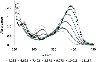 | ||
| Fig. 1 UV-Visible absorption spectra for a 7.5 × 10⁻⁵ M mangiferin solution in DMSO/H2O (80%/20%, v/v) at different pH values. | ||
In the present work the pH was studied up to 10.9 and 11.7 for the aqueous medium and the DMSO/H2O (80%/20%, v/v) mixture, respectively. From these results and the comparisons made with the absorption spectra profiles obtained by Gómez-Zaleta et al.,28 it was concluded that a model of three acid–base equilibria and four species should be enough to fit the experimental data. This is because the fully deprotonated mangiferin species should be present in very low concentrations.
Mangiferin pKa values determination by spectrophotometry
The UV-Visible absorption spectra determined in the present work were fitted using the program SQUAD,46 and the data obtained are shown in Table 1. The pKa values of mangiferin, determined in the present work for aqueous solutions, agree fair well with those reported by Gómez-Zaleta et al.,28 as well as the shape and order of magnitude of molar absorptivity coefficients, shown in Fig. 2, for the same mangiferin species, despite being in different media. The pKa values determined in the mixture of DMSO/H2O (80%/20%, v/v) have a mean difference of +0.91 compared to those obtained in water, as expected, given the lower dielectric constant of the mixture with respect to pure water.| Parameter | Reported28 in H2O | This worka in H2O | This worka in DMSO-d6/H2O |
|---|---|---|---|
| a Determined using the program SQUAD.46 | |||
| pKa1 | 6.52 ± 0.06 | 6.35 ± 0.04 | 7.337 ± 0.001 |
| pKa2 | 7.97 ± 0.06 | 7.95 ± 0.05 | 8.936 ± 0.001 |
| pKa3 | 9.44 ± 0.04 | 9.57 ± 0.05 | 10.297 ± 0.028 |
| pKa4 | 12.1 ± 0.02 | Not determined | Not determined |
| SD of fitting | σ = 4.92 × 10⁻³ | σ = 7.072 × 10⁻³ | σ = 5.935 × 10⁻³ |
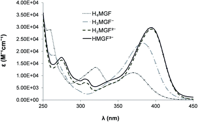 | ||
| Fig. 2 Molar absorptivity coefficients for the mangiferin species determined by the program SQUAD for the mixture DMSO/H2O (80%/20%, v/v). | ||
The fitting of sigmoid curves of absorbance as a function of pH was performed as reported by Rodríguez-Barrientos et al.,31 using the data refined by the program SQUAD, for several wavelengths related with the maxima of absorption spectra (Fig. 1). As it can be seen in Fig. 3 there is an excellent agreement among experimental data and the model.
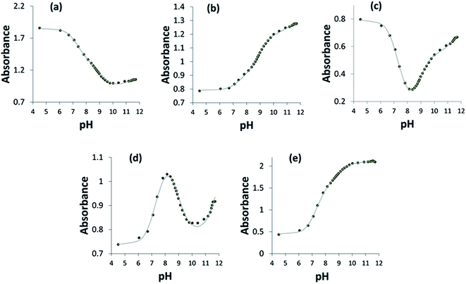 | ||
| Fig. 3 (a–e) Fitting of sigmoid curves, absorbance as a function of pH. (a) 263 nm. (b) 275 nm. (c) 308 nm. (d) 360 nm. (e) 390 nm. | ||
NMR study of mangiferin
1H and 13C NMR spectra were acquired at several pH values in order to analyze if the model that explains the UV-Visible spectroscopic behavior of mangiferin is also appropriate for predicting the NMR spectra in the DMSO/H2O (80%/20%, v/v) mixture. Fig. 4a shows a typical 1H NMR spectrum of 0.14 M mangiferin in DMSO/H2O (80%/20%, v/v) mixture at pH = 6.224, while Fig. 4b is the typical 13C NMR spectrum of mangiferin for the same solution.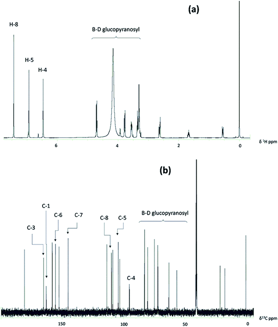 | ||
| Fig. 4 (a and b) Initial NMR spectra of 0.14 M mangiferin in DMSO/H2O (80%/20%, v/v) mixture at pH = 6.224. (a) 1H. (b) 13C. | ||
Deprotonation order of H4MGF
Comparisons of the 1H and 13C NMR chemical shifts of the xanthone ring are shown in Fig. 5. Fig. 5a shows the behavior of 1H signals of hydrogens attached to carbons in that ring. Fig. 5b presents the changes of 13C signals observed for the carbons attached to the phenolic groups of the ring; while Fig. 5c provides the behavior of its adjacent carbons. | ||
| Fig. 5 (a–c) Chemical shifts of nuclei in the xanthone ring as a function of pH. (a) 1H. (b) and (c) 13C. | ||
Fig. 5b shows that C-6 and C-7 are the first with chemical shift moved to lower frequencies at pH ≈ 7 (where first deprotonation begins) but this change is greater for C-6. After that, C-3 moves to lower frequencies when the second deprotonation begins (pH ≈ 8.2). Finally, C-7 exhibits a small change in chemical shift when the third deprotonation begins (pH ≈ 9.6). This behavior suggests that the order of acidity of the phenolic protons, based on their respective attached carbons, is as follows C-6 > C-3 > C-7 > C-1; as it has been proposed elsewhere.28 The same deprotonation order is inferred from the 1H spectra (Fig. 5a), and confirmed by the adjacent carbons (Fig. 5c). The deprotonation order is the same as the one reported by Gómez-Zaleta et al.;28 but in our case this trend is reinforced by the fact that it is based in multiple measurements of NMR spectra and pH values.
Fig. 6 shows the fit of sigmoid curves of chemical shifts as a function of pH for several relevant nuclei,31 with pKa values refined by the program SQUAD for spectrophotometric data and chemical shifts of each nucleus for the different mangiferin species, presented in Table 2, taken as refining parameters.
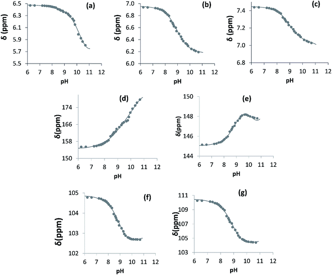 | ||
| Fig. 6 (a–g) Plots of 1H and 13C chemical shifts as a function of pH. (a) H-4. (b) H-5. (c) H-8. (d) C-6. (e) C-7. (f) C-5. (g) C-8. | ||
| Species | HMGF3− | H2MGF2− | H3MGF− | H4MGF |
|---|---|---|---|---|
| δH-4 | 7.03 | 7.078 | 7.41 | 7.45 |
| δH-5 | 6.18 | 6.25 | 6.89 | 6.96 |
| δH-8 | 5.53 | 6.34 | 6.45 | 6.48 |
| δC-5 | 102.37 | 101.89 | 104.06 | 104.36 |
| δC-6 | 181.97 | 167.84 | 157.29 | 154.69 |
| δC-7 | 146.37 | 149.18 | 145.44 | 145.05 |
| δC-8 | 104.28 | 103.32 | 109.24 | 110.00 |
Theoretical study of mangiferin
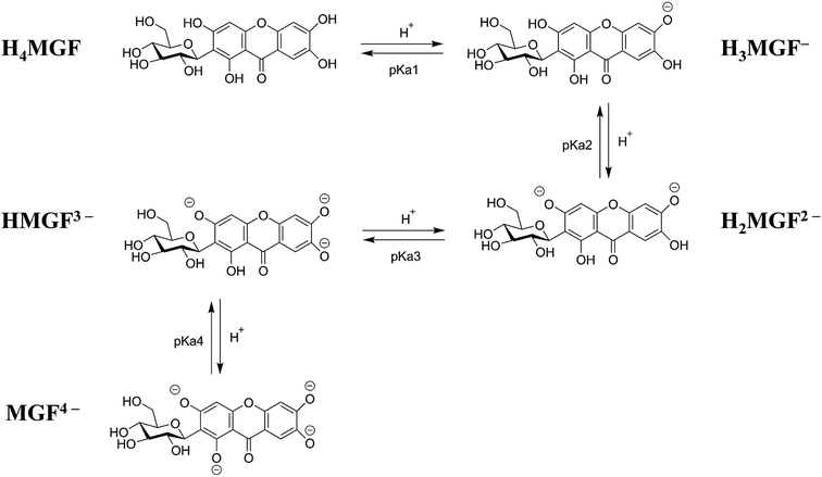
As previously discussed, depending on the pH there are 5 possible forms in which this compound can be found, the neutral one (H4MGF), and 4 anionic species (H3MGF−, H2MGF2−, HMGF3−, MGF4−). While the first and the latter are unambiguous, it is important to establish the deprotonation route for properly identifying species H3MGF−, H2MGF2− and HMGF3−. Based on comparisons among the different possible deprotonation sites (Table 2S, ESI†) it was found that site 6 is the most acidic one, followed by sites 3, 7, and 1, in that order. This deprotonation order is in line with the one found when using experimental techniques, and is proposed as the most likely deprotonation route (Scheme 2). It allowed unambiguously identifying the most probable structures for all the acid–base species of mangiferin. Then using the experimental pKa values obtained in this work, and the fourth previously reported,28 the molar fractions of the different species in aqueous solution, at physiological pH (pH = 7.4) were estimated (Table 3). It was found that, under such conditions the dominant species is the mono-anion (73.0%), while the populations of H4MGF and H2MGF2− are smaller but not negligible (6.4% and 20.4%, respectively), and those of HMGF3− and MGF4− are almost zero.
| fHjMGF | |
|---|---|
| H4MGF | 0.064 |
| H3MGF− | 0.730 |
| H2MGF2− | 0.204 |
| HMGF3− | 0.002 |
| MGF4− | ∼0.000 |
Antioxidant activity
To investigate the free radical scavenging activity of mangiferin different reaction mechanisms have been considered. As it is the case for many other free radical scavengers,47–52 the antioxidant activity of mangiferin might involve some of them. Using the neutral form of mangiferin as an example, these mechanisms can be schematically represented as:Radical Adduct Formation (RAF):
| H4MGF + ˙R → [H4MGF–R]˙ |
Hydrogen Transfer (HT):
| H4MGF + ˙R → H3MGF˙ + HR |
Single Electron Transfer (SET):
| H4MGF + ˙R → H4MGF+˙ + R− |
Sequential Proton Loss Electron Transfer (SPLET):
| H3MGF− + ˙R → H3MGF˙ + R− |
The latter was first proposed by Litwinienko and Ingold for the reactions of substituted phenols with the DPPH radical.53–56 In the present study we have chosen ˙R = ˙OOH, which is the simplest member of the peroxyl (˙OOR) family and has been suggested to be central to the toxic side effects of aerobic respiration.57 It has also been pointed out that more information on the reactivity of this species regarding biological systems is needed.57 Peroxyls are among those radicals of biological relevance that can be effectively scavenged to retard oxidative stress (OS),58 because their half-lives are not too short, which is essential for efficient interception by phenolic compounds.59 That is why ROO˙ have been proposed as major reaction partners for these compounds.29 Moreover, it has been proposed that the main antioxidant function of phenolic compounds is to trap ROO˙.60,61 In addition radicals of moderate reactivity have been recommended for studying the relative antioxidant activity of chemical compounds.62,63 This is because using highly reactive radicals might lead to miss-conclude that all the tested compounds are equally efficient as antioxidants, since such radicals usually react at diffusion-limited rates with most organic molecules.
For the reactions in aqueous solution all the above detailed mechanisms have been taken into account, while in non-polar solution, only RAF and HT have been considered. SPLET and SET processes have not been included because non-polar (lipid) media does not provide the necessary solvation for the ionic species yielded by these processes. Thus, deprotonation is not expected to occur to a significant extent. However, just to prove this point the SET reaction energy in pentyl ethanoate solution was calculated, and found to be about 80 kcal mol−1 (Table 4). In aqueous solution, the SET and SPLET processes are very close related via deprotonation, since the first step in the SPLET mechanism is controlled by the acid equilibrium with the environment. Thus, the second step of SPLET reactions is the one controlling reactivity, and for an acid–base species with n protons it is identical to the SET reaction of the species with n − 1 protons. The endergonicity of these reactions in aqueous solution is reduced, compared to lipid media, but still remains significantly large for H4MGF and H3MGF− (∼35 and 15 kcal mol−1, respectively). However, as the deprotonation degree increases so does the thermochemical viability of electron transfers from mangiferin species to the ˙OOH radical, with the reactions involving HMGF3− and MGF4− being significantly exergonic (−10.8 and −15.6 kcal mol−1, respectively). Therefore, it is expected that the relative importance of this kind of reaction increases with the pH.
| PEa | Water | ||||
|---|---|---|---|---|---|
| H4MGF | H4MGF | H3MGF− | H2MGF2− | HMGF3− | |
| a PE = pentyl ethanoate; na = non available, deprotonated site, nf = not found. | |||||
| SET | 79.94 | 35.20 | 15.19 | 10.40 | −10.81 |
| SPLET | 15.19 | 10.40 | −10.81 | −15.62 | |
![[thin space (1/6-em)]](https://www.rsc.org/images/entities/char_2009.gif) |
|||||
| HT | |||||
| Site 1 | 14.62 | 7.43 | 7.48 | 4.74 | −11.32 |
| Site 3 | 17.26 | 12.53 | 5.27 | na | na |
| Site 6 | 6.75 | 3.98 | na | na | na |
| Site 7 | −3.50 | −3.35 | −6.94 | −8.78 | na |
| Site 1p | 9.61 | 7.72 | 7.58 | 4.34 | 4.15 |
| Site 2p | 14.87 | 11.42 | 11.28 | 2.65 | 3.80 |
| Site 3p | 13.07 | 9.48 | 9.48 | 8.85 | 8.56 |
| Site 4p | 15.55 | 11.26 | 11.43 | 5.50 | 5.04 |
| Site 5p | 14.19 | 11.57 | 11.33 | 10.40 | 10.05 |
| Site 6p | 15.05 | 12.07 | 11.98 | 11.83 | 11.91 |
![[thin space (1/6-em)]](https://www.rsc.org/images/entities/char_2009.gif) |
|||||
| RAF | |||||
| Site 1 | 17.97 | 14.98 | 14.74 | 15.03 | 10.49 |
| Site 2 | 18.04 | 15.92 | 15.84 | 14.29 | 13.85 |
| Site 3 | 19.20 | 18.05 | 16.02 | 19.32 | 12.30 |
| Site 4 | 16.75 | 15.98 | 15.18 | 10.30 | 9.54 |
| Site 4a | 18.58 | 16.98 | 16.36 | 14.64 | 15.92 |
| Site 4b | 12.98 | 11.56 | 13.26 | 11.42 | −0.55 |
| Site 5 | 15.91 | 15.83 | 14.22 | 12.77 | 12.96 |
| Site 6 | 13.43 | 11.66 | 17.46 | 15.47 | 3.31 |
| Site 7 | 14.73 | 11.93 | 8.33 | 7.42 | 5.85 |
| Site 8 | 13.41 | 10.09 | 8.71 | 9.29 | −3.44 |
| Site 8a | 27.83 | 25.92 | 23.13 | 21.46 | nf |
Regarding H transfers, they are all significantly endergonic when they take place from the sugar moiety, regardless of the polarity of the environment, which rules out the possible role of this part of the molecule on the antioxidant activity of mangiferin. Only HT pathways involving site 7 were found to be exergonic for the mangiferin species that are expected to be the most abundant under physiological conditions, i.e., H4MGF in lipid media and H4MGF, H3MGF− and H2MGF2− in aqueous solution. In the last case, for HMGF3−, where site 7 is deprotonated, HT from site 1 becomes exergonic; while for MGF4− the HT mechanism is not expected to be important since all the phenolic OH groups are deprotonated. However, these species are predicted to be in negligible concentrations under physiological conditions based on their molar fractions (Table 3).
All the RAF pathways were found to be endergonic, regardless of the solvent polarity, except those corresponding to sites 4b and 8 in HMGF3−, in aqueous solution. Thus, the RAF mechanism is not expected to significantly contribute to the overall peroxyl scavenging activity of mangiferin, since (as above mentioned) the availability of this species is expected to be very low under physiological conditions. For the same reason the RAF reactions involving MGF4− were not included in the modeling. The RAF reaction at site 8a in HMGF3−, and at site 8b in all the studied species were initially considered as possible pathways. However, any attempt to locate the corresponding products invariably led to structures that are weak-bonded complexes rather than to proper radical adducts.
The reaction pathways described above as endergonic were not included in the kinetic study, albeit they might take place at a significant rate, because they would be reversible and therefore the formed products will not be observed. However, it should be noted that they might still represent significant reaction channels if their products rapidly react further. This would be particularly important if these later stages are sufficiently exergonic to provide a driving force, and if their reaction barriers are low. That is expected to be the case for the SET and SPLET mechanisms since they yield intermediates that are usually very reactive radicals. Therefore they were also included in the kinetic analyses.
The rate constants for each reaction pathway, as well as the overall rate coefficients (koverall) are reported in Table 5. The Gibbs free energies of activation (ΔG≠) used to obtain the rate constant of each individual path are provided as ESI (Table 3S†). The koverall value, in lipid solution, is equal to the rate constant of pathway 7, since it is the only one predicted as thermochemically viable. In aqueous solution, on the other hand, the electron transfers and the molar fractions of the different acid–base species, at the pH of interest (physiological pH, 7.4), were also considered. The values of koverall obtained this way are expected to be directly related to the observable ones.
According to the estimated rate constants the environment plays an important role on the antioxidant activity of mangiferin. It was found that, in aqueous solution, the rate constants of both the HT and SET reactions increases with the deprotonation degree, reaching the diffusion limit after the second deprotonation. Therefore, it is predicted that the peroxyl radical scavenging activity of mangiferin is enhanced by pH. Moreover, the deprotonated species are expected to play a crucial role in such activity. In addition it is expected to be higher in aqueous solution than in lipid media since the anionic species are promoted by polar, protic, solvents. Regarding the site reactivity, HT from site 7 in H4MGF and H3MGF− are the fastest reaction paths, while processes involving electron transfer reactions become the most rapid for the species with a higher degree of deprotonation. However, it should be noted that for these species the other reaction pathways, including the viable RAF channels, are also very fast with rate constants close to, or within, the diffusion limited regime. This analysis indicates that the pH influences not only the reactivity but also the relative importance of the reaction mechanisms involved.
To explore in more details the contributions of the different mechanisms and reaction pathways to the overall HOO˙ scavenging activity of mangiferin, the branching ratios (Γ) have been estimated, according to:
As mentioned before in non-polar, lipid, media all the reactivity of mangiferin towards ˙OOH can be attributed to the HT reaction from site 7. In aqueous solution, on the other hand, there are a variety of elementary chemical reactions that are fast enough to be of potential importance. However, after including the molar fractions of the different acid–base species, it becomes evident that the HT from site 7 in H2MGF2− is responsible for most of the peroxyl radical scavenging activity of mangiferin at physiological pH (Table 6). A smaller, but still not negligible contribution is predicted for the SET reaction of HMGF3−, i.e. the SPLET pathway of H2MGF2−. Thus, the contribution of this species alone to the overall reactivity of mangiferin towards ˙OOH in aqueous solution was found to be higher than 96%. Accordingly, it is proposed that H2MGF2− is the key species in the peroxyl radical scavenging activity of mangiferin.
Since the pH seems to be one of the key factors influencing the reactivity of mangiferin, this point has been addressed in more detail. The dependence of the rate coefficients with the pH is shown in Fig. 7. It was found that at pH below 4 the reactivity of mangiferin towards ˙OOH is rather low and constant. It increases from pH 4 to 9, in line with the increasing of the H2MGF2− population, and remains almost unchanged and within the diffusion-limit regime at pH ≥ 9. Accordingly it can be safely concluded that as pH regulates the population of the different acid–base species of mangiferin, it also influences the free radical scavenging of this compound.
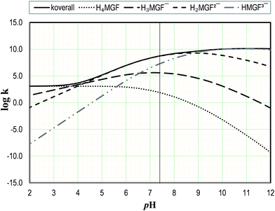 | ||
| Fig. 7 Influence of pH on the rate constants for the reaction between mangiferin acid–base species and ˙OOH. | ||
To analyze the potential protective effects of mangiferin against peroxyl induced oxidation, its overall rate coefficients have been compared to that corresponding to the ˙OOH damage to polyunsaturated fatty acids, which has been estimated to be in the range 1.18–3.05 × 10³ M−1 s−1.27 Based on the calculated koverall values of the reaction between mangiferin and this radical, it can be stated that this compound is a rather modest protector in lipid media. However, in aqueous solution it is expected to be very efficient for that purpose, since it reacts about 5 orders of magnitude faster than the threshold value.
To quantify such protection, mangiferin has been compared with Trolox,64 which is frequently used as a reference antioxidant. It is assumed that since Trolox is well known for its antioxidant activity, any compound with higher activity than Trolox should also be efficient for this purpose. Mangiferin was found to react with ˙OOH about 2.3 and 6000 times faster than Trolox, in non-polar and polar media, respectively. Therefore it can be concluded that it is a better peroxyl radical scavengers than the reference compound, and is expected to offer antioxidant protection to biological targets. Regarding the structure activity relationship (SAR), the increased activity of mangiferin with respect to Trolox can be attributed to the presence of a larger number of phenolic OH groups in mangiferin. This is in line with the previously described behavior for other polyphenols,65 and also with the results reported by Dar et al.5 for a series of mangiferin derivatives. In that study it was found that as the phenolic OH groups are acetylated or replaced by methyl groups the antioxidant activity of the modified compounds decreases.
Compared to other known antioxidants, in lipid media, the peroxyl radical scavenging activity of mangiferin was found to be higher than those of ascorbic acid15 and melatonin,66 but lower than those of caffeic acid67 and resveratrol.68 In aqueous solution, at physiological pH, it is a better protector than gallic acid,69 dopamine,70 ellagic acid,71 and α-mangostina.72 In fact, under such conditions mangiferin is predicted to be among the best peroxyl radical scavengers identified so far, with an antioxidant capacity only surpassed by that of piceatannol.73 In the particular case of α-mangostina, which has a similar ring structure and only one less OH group, the higher activity of mangiferin is attributed to the presence of the catechol moiety in ring B. This has been justified by the ability of the neighbor OH to form intra-molecular hydrogen bonding with the reacting site.65 This hypothesis is supported by the fact that the most reactive site, towards free radicals, in mangiferin is site 7. Therefore, some SAR characterization can be envisaged that supports the excellent free radical activity of mangiferin. Namely, (i) the relative large number of phenolic sites, i.e., four; and (ii) the presence of the catechol group.
Conclusions
A thorough study of the acidity constants of mangiferin was carried out in water and in a DMSO–H2O (80–20% v/v) mixture, by UV-Vis and 1H, 13C-NMR spectroscopies. The NMR studies allowed to determine the deprotonation order of its phenol groups. A theoretical study on the deprotonation order lead to the same conclusion that the experimental ones.A theoretical study of the antioxidant activity of mangiferin, performed using the QM-ORSA protocol, allowed to identify this compound as an efficient peroxyl radical scavenger. Such activity is predicted to be moderate in lipid media and excellent in aqueous solution at physiological pH. The structural key features to the antioxidant activity of mangiferin seem to be the number of phenolic sites (four) and the presence of a catechol moiety.
The acid–base equilibria were found to play an important role on the protective effects of mangiferin against oxidative stress, being H2MGF2− the key species in the peroxyl radical scavenging activity of this compound. The HT from site 7, and the SPLET processes were identified as main reaction mechanisms involved in such activity.
Acknowledgements
We gratefully acknowledge the Laboratorio de Visualización y Cómputo Paralelo at Universidad Autónoma Metropolitana-Iztapalapa for computing time. This work was partially supported by projects SEP-CONACyT 167491 and 237997. One of us (GM-S) wants to acknowledge CONACyT for the stipend to follow doctoral studies.References
- P. Scartezzini and E. Speroni, Review on some plants of Indian traditional medicine with antioxidant activity, J. Ethnopharmacol., 2000, 71, 23–43 CrossRef CAS PubMed.
- N. Rainha, K. Koci, A. Varela Coelho, E. Lima, J. Baptista and M. Fernandes-Ferreira, HPLC-UV-ESI-MS analysis of phenolic compounds and antioxidant properties of Hypericum undulatum shoot cultures and wild-growing plants, Phytochemistry, 2013, 86, 83–91 CrossRef CAS PubMed.
- T. Miura, H. Ichiki, N. Iwamoto, M. Kato, M. Kubo, H. Sasaki, M. Okada, T. Ishida, Y. Seino and K. Tanigawa, Antidiabetic activity of the rhizome of Anemarrhena asphodeloides and active components, mangiferin and its glucoside, Biol. Pharm. Bull., 2001, 24, 1009–1011 CAS.
- G. V. Srinivasan, D. Ravi, K. V. Tushar and I. Balachandran, Quantitative determination of mangiferin from the roots of Salacia fruticosa and the antioxidant studies, Journal of Tropical Medicinal Plants, 2009, 10, 213–218 Search PubMed.
- A. Dar, S. Faizi, S. Naqvi, T. Roome, S. Zikr-Ur-Rehman, M. Ali, S. Firdous and S. T. Moin, Analgesic and antioxidant activity of mangiferin and its derivatives: the structure activity relationship, Biol. Pharm. Bull., 2005, 28, 596–600 CAS.
- F. García, R. Delgado, F. Ubeira and J. Leiro, Modulation of rat macrophage function by the Mangifera indica L. extracts vimang and mangiferin, Int. Immunopharmacol., 2002, 2, 797–806 CrossRef.
- D. García, M. Escalante, R. Delgado, F. Ubeira and J. Leiro, Anthelminthic and antiallergic activities of Mangifera indica L. Stem bark components Vimang and mangiferin, Phytother. Res., 2003, 17, 1203–1208 CrossRef PubMed.
- G. Garrido, R. Delgado, Y. Lemus, J. R. García and A. J. Núñez-Sellés, Protection against septic shock and suppression of tumor necrosis factor alpha and nitric oxide production on macrophages and microglia by a standard aqueous extract of Mangifera indica L. (VIMANG®). Role of mangiferin isolated from the extract, Pharmacol. Res., 2004, 50, 165–172 CrossRef CAS PubMed.
- G. Garrido, D. González, Y. Lemus, D. García, L. Lodeiro, G. Quintero, C. Delporte, A. J. Núñez-Sellés and R. Delgado, In vivo and in vitro anti-inflammatory activity of Mangifera indica L. extract (VIMANG®), Pharmacology, 2004, 50, 143–149 Search PubMed.
- B. B. Garrido-Suárez, G. Garrido, R. Delgado, F. Bosch and M. C. Rabí, A Mangifera indica L. extract could be used to treat neuropathic pain and implication of mangiferin, Molecules, 2010, 15, 9035–9045 CrossRef PubMed.
- G. C. Jagetia and V. A. Venkatesha, Effect of mangiferin on radiation-induced micronucleus formation in cultured human peripheral blood lymphocytes, Environ. Mol. Mutagen., 2005, 46, 12–21 CrossRef CAS PubMed.
- G. C. Jagetia and M. S. Baliga, Radioprotection by mangiferin in DBAxC57BL mice: a preliminary study, Phytomedicine, 2005, 12, 209–2015 CrossRef CAS PubMed.
- J. Lei, C. Zhou, H. Hu, L. Hu, M. Zhao, Y. Yang, Y. Chuai, J. Ni and J. Cai, Mangiferin aglycone attenuates radiation-induced damage on human intestinal epithelial cells, J. Cell. Biochem., 2012, 113, 2633–2642 CrossRef CAS PubMed.
- H. Li, J. Huang, B. Yang, T. Xiang, X. Yin, W. Peng, W. Cheng, J. Wan, F. Luo, H. Li and G. Ren, Mangiferin exerts antitumor activity in breast cancer cells by regulating matrix metalloproteinases, epithelial to mesenchymal transition, and β-catenin signaling pathway, Toxicol. Appl. Pharmacol., 2013, 272, 180–190 CrossRef CAS PubMed.
- T. Miura, N. Iwamoto, M. Kato, H. Ichiki, M. Kubo, Y. Komatsu, T. Ishida, M. Okada and M. Tanigawa, The suppressive effect of mangiferin with exercise on blood lipids in type 2 diabetes, Biol. Pharm. Bull., 2001, 24, 1091–1092 CAS.
- T. Miura, H. Ichiki, I. Hashimoto, N. Iwamoto, M. Kato, M. Kubo, E. Ishihara, Y. Komatsu, M. Okada, T. Ishida and K. Tanigawa, Antidiabetic activity of a xanthone compound mangiferin, Phytomedicine, 2001, 8, 85–87 CrossRef CAS PubMed.
- S. Muruganandan, S. Gupta, M. Kataria, J. Lal and P. K. Gupta, Mangiferin protects the streptozotocin-induced oxidative damage to cardiac and renal tissues in rats, Toxicology, 2002, 176, 165–173 CrossRef CAS PubMed.
- S. Muruganandan, K. Srinivasan, S. Gupta, P. K. Gupta and J. Lal, Effect of mangiferin on hyperglycemia and atherogenicity in streptozotocin diabetic rats, J. Ethnopharmacol., 2005, 97, 497–501 CrossRef CAS PubMed.
- A. J. Núñez-Sellés, R. Delgado-Hernández, G. Garrido-Garrido, D. García-Rivera, M. Guevara-García and G. L. Pardo-Andreu, The paradox of natural products as pharmaceuticals: experimental evidences of a mango stem bark extract, Pharmacol. Res., 2007, 55, 351–358 CrossRef PubMed.
- G. L. Pardo Andreu, R. Delgado, J. A. Velho, C. Curti and A. E. Vercesi, Mangiferin, a natural occurring glucosyl xanthone, increases susceptibility of rat liver mitochondria to calcium-induced permeability transition, Arch. Biochem. Biophys., 2005, 439, 184–193 CrossRef PubMed.
- N. Prabhu-Sukumaran and C. S. Shyamala-Devi, Efficacy of mangiferin on serum and heart tissue lipids in rats subjected to isoproterenol induced cardiotoxicity, Toxicology, 2006, 228, 135–139 CrossRef PubMed.
- W. Rui-Rui, G. Yue-Dong, M. Chun-Hui, Z. Xing-Jie, H. Cheng-Gang, H. Jing-Fei and Z. Yong-Tang, Mangiferin, an anti-HIV-1 agent targeting protease and effective against resistant strains, Molecules, 2011, 16, 4264–4277 CrossRef PubMed.
- G. M. Sanchez, L. Re, A. Giuliani, A. J. Nuñez-Sellés, G. P. Davison and O. S. Leon-Fernández, Protective effects of Mangifera indica L. extract, mangiferin and selected antioxidants against TPA-induced biomolecules oxidation and peritoneal macrophage activation in mice, Pharmacol. Res., 2000, 42, 565–573 CrossRef CAS PubMed.
- S. K. Singh, Y. Kumar, S. Sadish Kumar, V. K. Sharma, K. Dua and A. Samad, Antimicrobial evaluation of mangiferin analogues, Indian J. Pharm. Sci., 2009, 71, 325–328 CrossRef PubMed.
- J. V. Van der Merwe, E. Joubert, M. Manley, D. de Beer, C. J. Malherbe and W. C. A. Gelderblom, Mangiferin glucuronidation: important hepatic modulation of antioxidant activity, Food Chem. Toxicol., 2012, 50, 808–815 CrossRef CAS PubMed.
- L. Yao-Wu, Z. Xia, Y. Qian-Qian, L. Qian, W. Jian-Yun, L. Hui-Pu, W. Ya-Qin, Y. Jia-Le and Y. Xiao-Xing, Suppression of methylglyoxal hyperactivity by mangiferin can prevent diabetes-associated cognitive decline in rats, Psychopharmacology, 2013, 228, 585–594 CrossRef PubMed.
- N. Yoshimi, K. Matsunaga, M. Katayama, Y. Yamada, T. Kuno, Z. Qiao, A. Hara, J. Yamahara and H. Mori, The inhibitory effects of mangiferin, a naturally occurring glucosylxanthone, in bowel carcinogenesis of male F344 rats, Cancer Lett., 2001, 163, 163–170 CrossRef CAS PubMed.
- B. Gómez-Zaleta, M. T. Ramírez-Silva, A. Gutiérrez, E. González-Vergara, M. Güizado-Rodríguez and A. Rojas-Hernández, UV/vis, 1H, and 13C NMR spectroscopic studies to determine mangiferin pKa values, Spectrochim. Acta, Part A, 2006, 64, 1002–1009 CrossRef PubMed.
- L. Márquez, B. García-Bueno, J. L. M. Madrigal and J. C. Leza, Mangiferin decreases inflammation and oxidative damage in rat brain after stress, Eur. J. Nutr., 2012, 51, 729–739 CrossRef PubMed.
- J. M. Islas-Martínez, D. Rodríguez-Barrientos, A. Galano, E. Ángeles, L. A. Torres, F. Olvera, M. T. Ramírez-Silva and A. Rojas-Hernández, Deprotonation mechanism of new antihypertensive piperidinylmethylphenols: a combined and theoretical study, J. Phys. Chem. B, 2009, 113, 11765–11774 CrossRef PubMed.
- D. Rodríguez-Barrientos, A. Rojas-Hernández, A. Gutiérrez, R. Moya-Hernández, R. Gómez-Balderas and M. T. Ramírez-Silva, Determination of pKa values of tenoxicam from 1H NMR chemical shifts and of oxicams from electrophoretic mobilities (CZE) with the aid of programs SQUAD and HYPNMR, Talanta, 2009, 80, 754–776 CrossRef PubMed.
- M. J. Frisch, G. W. Trucks, H. B. Schlegel, G. E. Scuseria, M. A. Robb, J. R. Cheeseman, G. Scalmani, V. Barone, B. Mennucci, G. A. Petersson, H. Nakatsuji, M. Caricato, X. Li, H. P. Hratchian, A. F. Izmaylov, J. Bloino, G. Zheng, J. L. Sonnenberg, M. Hada, M. Ehara, K. Toyota, R. Fukuda, J. Hasegawa, M. Ishida, T. Nakajima, Y. Honda, O. Kitao, H. Nakai, T. Vreven, J. A. Montgomery Jr, J. E. Peralta, F. Ogliaro, M. Bearpark, J. J. Heyd, E. Brothers, K. N. Kudin, V. N. Staroverov, R. Kobayashi, J. Normand, K. Raghavachari, A. Rendell, J. C. Burant, S. S. Iyengar, J. Tomasi, M. Cossi, N. Rega, J. M. Millam, M. Klene, J. E. Knox, J. B. Cross, V. Bakken, C. Adamo, J. Jaramillo, R. Gomperts, R. E. Stratmann, O. Yazyev, A. J. Austin, R. Cammi, C. Pomelli, J. W. Ochterski, R. L. Martin, K. Morokuma, V. G. Zakrzewski, G. A. Voth, P. Salvador, J. J. Dannenberg, S. Dapprich, A. D. Daniels, Ö. Farkas, J. B. Foresman, J. V. Ortiz, J. Cioslowski and D. J. Fox, Gaussian 09, Revision B.01, Gaussian, Inc., Wallingford CT, 2009 Search PubMed.
- Y. Zhao, N. E. Schultz and D. G. Truhlar, Exchange–correlation functional with broad accuracy for metallic and nonmetallic compounds, kinetics, and noncovalent interactions, J. Chem. Phys., 2005, 123, 161103, DOI:10.1063/1.2126975.
- A. V. Marenich, C. J. Cramer and D. G. Truhlar, Universal solvation model based on solute electron density and on a continuum model of the solvent defined by the bulk dielectric constant and atomic surface tensions, J. Phys. Chem. B, 2009, 113, 6378–6396 CrossRef CAS PubMed.
- Y. Zhao, N. E. Schultz and D. G. Truhlar, Design of density functionals by combining the method of constraint satisfaction with parametrization for thermochemistry, thermochemical kinetics, and noncovalent interactions, J. Chem. Theory Comput., 2006, 2, 364–382 CrossRef PubMed.
- H. Ando, B. P. Fingerhut, K. E. Dorfman, J. D. Biggs and S. Mukamel, Femtosecond stimulated raman spectroscopy of the cyclobutane thymine dimer repair mechanism: A computational study, J. Am. Chem. Soc., 2014, 136, 14801–14810 CrossRef CAS PubMed.
- T. V. Alves, M. M. Alves, O. Roberto-Neto and F. R. Ornellas, Direct dynamics investigation of the reaction S(3p) + CH4 → CH3 + SH(2π), Chem. Phys. Lett., 2014, 591, 103–108 CrossRef CAS.
- K. P. Prasanthkumar and J. R. Alvarez-Idaboy, An experimental and theoretical study of the kinetics and mechanism of hydroxyl radical reaction with 2-aminopyrimidine, RSC Adv., 2014, 4, 14157–14164 RSC.
- M. Altarawneh and B. Z. Dlugogorski, A mechanistic and kinetic study on the formation of PBDD/Fs from PBDEs, Environ. Sci. Technol., 2013, 47, 5118–5127 CrossRef CAS PubMed.
- W. Li, Z. Su and C. Hu, Mechanism of ketone allylation with allylboronates as catalyzed by zinc compounds: A DFT study, Chem.–Eur. J., 2013, 19, 124–134 CrossRef CAS PubMed.
- M. Dargiewicz, M. Biczysko, R. Improta and V. Barone, Solvent effects on electron-driven proton-transfer processes: adenine-thymine base pairs, Phys. Chem. Chem. Phys., 2012, 14, 8981–8989 RSC.
- D. Henao, J. Murillo, P. Ruiz, J. Quijano, B. Mejía, L. Castañeda and R. Notario, A computational study of the thermolysis of β-hydroxy ketones in gas phase and in m-xylene solution, J. Phys. Org. Chem., 2012, 25, 883–887 CrossRef CAS.
- J. Murillo, D. Henao, E. Vélez, C. Castaño, J. Quijano, J. Gaviria and E. Zapata, Thermal decomposition of 4-hydroxy-2-butanone in m-xylene solution: experimental and computational study, Int. J. Chem. Kinet., 2012, 44, 407–413 CrossRef CAS.
- A. Galano and J. R. Alvarez-Idaboy, Kinetics of radical-molecule reactions in aqueous solution: A benchmark study of the performance of density functional methods, J. Comput. Chem., 2014, 35, 2019–2026 CrossRef CAS PubMed.
- A. Galano and J. R. Alvarez-Idaboy, A computational methodology for accurate predictions of rate constants in solution: Application to the assessment of primary antioxidant activity, J. Comput. Chem., 2013, 34, 2430–2445 CrossRef CAS PubMed.
- D. J. Leggett, Computational Methods for the Determination of Formation Constants, Plenum Press, New York, 1985 Search PubMed.
- M. Belcastro, T. Marino, N. Russo and M. Toscano, Structural and electronic characterization of antioxidants from marine organisms, Theor. Chem. Acc., 2006, 115, 361–369 CrossRef CAS.
- M. Leopoldini, N. Russo, S. Chiodo and M. Toscano, Iron chelation by the powerful antioxidant flavonoid quercetin, J. Agric. Food Chem., 2006, 54, 6343–6351 CrossRef CAS PubMed.
- M. Leopoldini, F. Rondinelli, N. Russo and M. Toscano, Pyranoanthocyanins: A theoretical investigation on their antioxidant activity, J. Agric. Food Chem., 2010, 58, 8862–8871 CrossRef CAS PubMed.
- M. Leopoldini, N. Russo and M. Toscano, The molecular basis of working mechanism of natural polyphenolic antioxidants, Food Chem., 2011, 125, 288–306 CrossRef CAS.
- A. Perez-Gonzalez and A. Galano, OH radical scavenging activity of edaravone: mechanism and kinetics, J. Phys. Chem. B, 2011, 115, 1306–1314 CrossRef CAS PubMed.
- S. G. Chiodo, M. Leopoldini, N. Russo and M. Toscano, The inactivation of lipid peroxide radical by quercetin. A theoretical insight, Phys. Chem. Chem. Phys., 2010, 12, 7662–7670 RSC.
- G. Litwinienko and K. U. Ingold, Abnormal solvent effects on hydrogen atom abstractions. 1. The reactions of phenols with 2,2-diphenyl-1-picrylhydrazyl (DPPH˙) in alcohols, J. Org. Chem., 2003, 68, 3433–3438 CrossRef CAS PubMed.
- G. Litwinienko and K. U. Ingold, Abnormal solvent effects on hydrogen atom abstraction. 2. Resolution of the curcumin antioxidant controversy, the role of sequential proton loss electron transfer, J. Org. Chem., 2004, 69, 5888–5896 CrossRef CAS PubMed.
- G. Litwinienko and K. U. Ingold, Abnormal solvent effects on hydrogen atom abstraction. 3. Novel kinetics in sequential proton loss electron transfer chemistry, J. Org. Chem., 2005, 70, 8982–8990 CrossRef CAS PubMed.
- G. Litwinienko and K. U. Ingold, Solvent effects on the rates and mechanisms of reaction of phenols with free radicals, Acc. Chem. Res., 2007, 40, 222–230 CrossRef CAS PubMed.
- A. D. N. De Grey, HO2˙: the forgotten radical, DNA Cell Biol., 2002, 21, 251–257 CrossRef CAS PubMed.
- P. Terpinc and H. Abramovic, A kinetic approach for evaluation of the antioxidant activity of selected phenolic acids, Food Chem., 2010, 121, 366–371 CrossRef CAS.
- H. Sies, Oxidative stress: oxidants and antioxidants, Exp. Physiol., 1997, 82, 291–295 CrossRef CAS PubMed.
- T. Masuda, K. Yamada, T. Maekawa, Y. Takeda and H. Yamaguchi, Antioxidant mechanism studies on ferulic acid: isolation and structure identification of the main antioxidation product from methyl ferulate, Food Sci. Technol. Res., 2006, 12, 173–177 CrossRef CAS.
- T. Masuda, K. Yamada, T. Maekawa, Y. Takeda and H. Yamaguchi, Antioxidant mechanism studies on ferulic acid:
![[thin space (1/6-em)]](https://www.rsc.org/images/entities/char_2009.gif) identification of oxidative coupling products from methyl ferulate and linoleate, J. Agric. Food Chem., 2006, 54, 6069–6074 CrossRef CAS PubMed.
identification of oxidative coupling products from methyl ferulate and linoleate, J. Agric. Food Chem., 2006, 54, 6069–6074 CrossRef CAS PubMed. - R. C. Rose and A. M. Bode, Biology of free radical scavengers: an evaluation of ascorbate, FASEB J., 1993, 7, 1135–1142 CAS.
- A. Galano, D. X. Tan and R. J. Reiter, Melatonin as a natural ally against oxidative stress: a physicochemical examination, J. Pineal Res., 2011, 51, 1–16 CrossRef CAS PubMed.
- M. E. Alberto, N. Russo, A. Grand and A. Galano, A physicochemical examination of the free radical scavenging activity of trolox: mechanism, kinetics and influence of the environment, Phys. Chem. Chem. Phys., 2013, 15, 4642–4650 RSC.
- E. Bendary, R. R. Francis, H. M. G. Ali, M. I. Sarwat and S. El Hady, Antioxidant and structure–activity relationships (SARs) of some phenolic and anilines compounds, Ann. Agric. Sci., Ser. E, 2013, 58, 173–181 Search PubMed.
- A. Galano, On the direct scavenging activity of melatonin towards hydroxyl and a series of peroxyl radicals, Phys. Chem. Chem. Phys., 2011, 13, 7178–7188 RSC.
- J. R. León-Carmona, J. R. Alvarez-Idaboy and A. Galano, On the peroxyl scavenging activity of hydroxycinnamic acid derivatives: mechanisms, kinetics, and importance of the acid–base equilibrium, Phys. Chem. Chem. Phys., 2012, 14, 12534–12543 RSC.
- C. Iuga, J. R. Alvarez-Idaboy and N. Russo, Antioxidant activity of trans-resveratrol toward hydroxyl and hydroperoxyl radicals: a quantum chemical and computational kinetics study, J. Org. Chem., 2012, 77, 3868–3877 CrossRef CAS PubMed.
- T. Marino, A. Galano and N. Russo, Radical scavenging ability of gallic acid toward oh and ooh radicals. Reaction mechanism and rate constants from the density functional theory, J. Phys. Chem. B, 2014, 118, 10380–10389 CrossRef CAS PubMed.
- C. Iuga, J. R. Alvarez-Idaboy and A. Vivier-Bunge, ROS initiated oxidation of dopamine under oxidative stress conditions in aqueous and lipidic environments, J. Phys. Chem. B, 2011, 115, 12234–12246 CrossRef CAS PubMed.
- A. Galano, M. Francisco Marquez and A. Pérez-González, Ellagic acid: An unusually versatile protector against oxidative stress, Chem. Res. Toxicol., 2014, 27, 904–918 CrossRef CAS PubMed.
- A. Martínez, A. Galano and R. Vargas, Free radical scavenger properties of α-mangostin: thermodynamics and kinetics of HAT and RAF mechanisms, J. Phys. Chem. B, 2011, 115, 12591–12598 CrossRef PubMed.
- M. Cordova-Gomez, A. Galano and J. R. Alvarez-Idaboy, Piceatannol, a better peroxyl radical scavenger than resveratrol, RSC Adv., 2013, 3, 20209–20218 RSC.
Footnote |
| † Electronic supplementary information (ESI) available. See DOI: 10.1039/c6ra06328d |
| This journal is © The Royal Society of Chemistry 2016 |


