Production of GDP-L-fucose from exogenous fucose through the salvage pathway in Mortierella alpina

Hongchao Wangab, Chen Zhanga, Qin Yangab, Jinghan Fenga, Haiqin Chenab, Zhennan Guab, Hao Zhangab, Wei Chenab and Yong Q. Chen*abc
aState Key Laboratory of Food Science and Technology, School of Food Science and Technology, Jiangnan University, Wuxi, Jiangsu 214122, P. R. China. E-mail: yqchen@jiangnan.edu.cn; Fax: +86 510 85197239; Tel: +86 510 85197239
bSynergetic Innovation Center of Food Safety and Nutrition, Wuxi, Jiangsu 214122, P. R. China
cDepartment of Cancer Biology, Wake Forest School of Medicine, Winston-Salem, NC 27157, USA

Received 7th March 2016 , Accepted 24th April 2016

First published on 26th April 2016


Abstract

GDP-L-fucose is an essential donor for the biosynthesis of fucosyloligosaccharides, participating in a variety of biological and pathological processes. Mortierella alpina can accumulate lipids in quantities up to 50% of its dry weight and has been proved to possess a GDP-L-fucose de novo synthesis pathway. Analysis of the M. alpina genome suggests that there is a putative GDP-L-fucose pyrophosphorylase (GFPP) gene playing a role in the salvage pathway of GDP-L-fucose, which has never been found in fungi before. To explore the molecular mechanisms of salvage reactions for free fucose in fungi, GFPP was expressed heterologously in Escherichia coli and the recombinant enzyme was purified to homogeneity. The enzymatic activity was investigated by liquid chromatography and mass spectrometry. Characterization of the GFPP in M. alpina indicated this fungus can convert fucose to GDP-L-fucose through the salvage pathway. The addition of fucose at the initial stage of cell multiplication exerted no impact on the GDP-L-fucose content in cells, whereas the addition of fucose (10 nM) after nitrogen exhaustion led to an increase of GDP-L-fucose production by 50%. Furthermore, medium supplementation with a combination of fucose and Mg2+ (10 nM) led to a 2.7-fold increase in the yield of GDP-L-fucose in M. alpina (0.57 mg per g cell). Additionally, the transcript level of GFPP is upregulated by the addition of fucose and Mg2+, which highlights the functional significance of GFPP in GDP-L-fucose biosynthesis. To our knowledge, this study is the first to report a comprehensive characterization of GFPP in a fungus.

Introduction

Fucose is an important deoxyhexose which is frequently bound at the terminal site of oligosaccharides.1,2 It decorates N- and O-linked glycoproteins and glycolipids and can be covalently linked to some threonine or serine residues in proteins.3,4 Fucosylated carbohydrate structures participate in a variety of biological and pathological processes in eukaryotic organisms, such as inflammation, tissue development, recognition sites of selectins, fertilization, cell adhesion, and metastasis of malignant cells.5 Fucosylation requires the activated nucleotide form of fucose, GDP-L-fucose, as the donor of fucose, and fucosyltransferase, which catalyses the transfer of fucose into the glycan acting as an acceptor.6,7

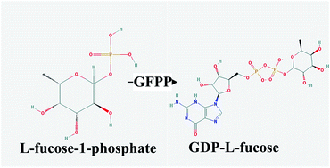
There are currently two major strategies for the synthesis of GDP-L-fucose in the cytoplasm: the de novo route, starting from GDP-D-mannose, and the salvage pathway, using fucose as the starting material (Fig. 1).8 The de novo synthesis of GDP-L-fucose is catalyzed by GDP-D-mannose 4,6-dehydratase (GMD) and GDP-keto-6-deoxymannose 3,5-epimerase/4-reductase (GMER). This pathway is present in most animal cells and tissues, as well as in plants and microorganisms.5 The salvage pathway utilizes fucose obtained from extracellular origin or from intracellular degradation of glycoproteins and glycolipids.9 In the salvage biosynthetic pathway, fucose is first phosphorylated by fucokinase (FUK) to form fucose-1-phosphate, which is then converted to GDP-L-fucose by GDP-L-fucose pyrophosphorylase (GFPP). GFPP was identified in the human,7 pig,10 mouse,9 Arabidopsis11 and Bacteroides fragilis.12 It is highly probable that such salvage reactions utilizing free fucose from culture medium or degradation of cell wall polysaccharides and glycoproteins also occur in fungi. However, little is known about the molecular mechanisms of salvage reactions in this organism.


image file: c6ra06031e-f1.tif
Fig. 1 De novo (A) and salvage (B) pathways of GDP-L-fucose synthesis.

Microbial synthesis of GDP-L-fucose was suggested to be influenced by a number of factors. One of them was the supply of precursors. The specific product formation rate of GDP-L-fucose in Corynebacterium glutamicum engineered by introducing the GMD and GMER genes was much higher when grown on precursor mannose than on glucose alone.13 Fucose is another precursor of GDP-L-fucose in the salvage pathway. When a bifunctional FUK/GFPP enzyme from Bacteroides fragilis was expressed in Saccharomyces cerevisiae, the transformed yeast cells grown in the presence of fucose generated 5.4 mg l−1 of GDP-L-fucose in vivo. Mortierella alpina is a well-known polyunsaturated fatty acid (PUFA)-producing oleaginous fungi, which can accumulate lipids to a level as high as 50% of their cell mass when under nitrogen starvation.14–16 We have characterized the GDP-L-fucose de novo synthesis pathway in M. alpina.17 Apart from these genes for the GDP-L-fucose synthesis pathway, a gene encoding GFPP accounting for the free fucose salvage pathway was also found in the genome of M. alpina,18 suggesting this fungus can convert exogenous fucose to GDP-L-fucose. According to a search in the Kyoto Encyclopedia of Genes and Genomes,19 GFPP is rarely found in microorganisms, and the only reported such case is from human symbiont Bacteroides.12

To clarify the molecular mechanism of free fucose utilization in fungi, we characterized the function of the gene encoding GFPP in the GDP-L-fucose salvage pathway in M. alpina in vitro. Kinetic parameters of GFPP and the effects of temperature, pH, and metal ions on GFPP activity were investigated. Multiple sequence alignment and phylogenetic analysis of the GFPP protein with other homologous proteins were performed. In order to increase the yield of GDP-L-fucose, the effects of exogenous fucose on GDP-L-fucose production were also investigated in M. alpina.

Results

Gene searching

To clarify the molecular mechanism of free fucose metabolism in M. alpina, the genes involved in GDP-L-fucose biosynthesis were investigated in the genome of this organism. Based on a search of the M. alpina genomic sequence (GenBank accession number ADAG01000000),18 a putative GFPP gene was found in the M. alpina genome (GenBank BankIt submission number 1825641) (Table S1). This revealed the presence of a salvage pathway of GDP-L-fucose in this fungus.

Expression and purification of GFPP

GFPP was expressed as a His-tagged fusion protein in the BL21 (DE3) gold strain of E. coli by IPTG induction, and purified to near homogeneity by nickel ion affinity chromatography. GFPP encodes a polypeptide of 608 amino acids with a theoretical isoelectric point (pI) of 5.87. The molecular mass of GFPP was estimated to be about 69 kDa by SDS-PAGE (Fig. S1), which corresponds well with the calculated mass.

Activity of GFPP

L-Fucose-1-phosphate was incubated with the purified GFPP protein and the reaction products were identified by liquid chromatography and mass spectrometry (LC-MS), as shown in Fig. 2. Extracted ion chromatogram (XIC) extraction (588.08 m/z, GDP-L-fucose) of GFPP reaction product presented a peak at 17.5 min (Fig. 2F), corresponding well to the position of the GDP-L-fucose standard (Fig. 2B). In the sample incubated in the absence of the GFPP, this peak was not observed (Fig. 2D). A peak was observed at 18 min both in sample incubated with and without GFPP (Fig. 2C and E), corresponding well to the position of the L-fucose-1-phosphate standard (Fig. 2A). These results confirm that the recombinant protein shows GFPP activity.
image file: c6ra06031e-f2.tif
Fig. 2 LC-MS chromatographs of GFPP reaction products: (A) extracted ion chromatogram (XIC) of L-fucose-1-phosphate standard (m/z 243.03); (B) XIC of GDP-L-fucose standard (m/z 588.08); (C) XIC of the blank reaction (m/z 243.03); (D) XIC of blank reaction (m/z 588.08); (E) XIC of the GFPP reaction (m/z 243.03); (F) XIC of the GFPP reaction (m/z 588.08). (G) XIC of the GDP-L-fucose extraction (m/z 588.08).

Properties of GFPP

GFPP was active at each of the temperatures tested (in the range of 4–55 °C), with the greatest activity detected at 30 °C (Fig. 3A). One hundred percent activity was defined as a GFPP reaction occurring at 30 °C under the aforementioned conditions. The optimal temperature for GFPP was in agreement with data previously obtained for Arabidopsis.11 GFPP retained 40% of its initial activity after 1.5 h of exposure at 12 °C, and lost more than 50% of its initial activity after 1 h of exposure at 20 °C, 28 °C and 37 °C (Fig. 3C). GFPP lost nearly 100% of its activity after 0.5 h of exposure above 50 °C (data not show). This indicated that GFPP was unstable at high temperature.
image file: c6ra06031e-f3.tif
Fig. 3 Effect of temperature, pH and metal ions on the activity and stability of GFPP. Each point represents the average of three independent measurements. Panel (A) and (B) show the effect of pH and temperature on the activity of GFPP. Panel (C) and (D) represented the effect of temperature and pH on the stability of GFPP. The pH stability was analyzed by pre-incubating the enzyme buffers of different pH values at 4 °C for 2 h and measuring the remaining activity at 30 °C and pH 7.0. Panel (E) represented the effect of Mg2+ on GFPP activity. Values are means of three replicates ± standard deviation.

The pH optimum for this enzyme was about pH 7.0 in 100 mM Tris–HCl buffer (Fig. 3B), which is similar to the optimum pH for recombinant Arabidopsis and pig GFPP.11,20 In addition, less than 30% activity of GFPP was retained after incubation at pH 3 and pH 12 at 4 °C for 2 h (Fig. 3D), indicating that GFPP was not stable under acidic or alkaline conditions.

Of the metal ions tested for activity with the GFPP, only Mg2+ showed significant activity (Fig. S4D), whereas K+, Mn2+, Co2+, Ca2+, Cu2+, Zn2+, Ni2+, Hg2+, and Fe2+ had little effect (Fig. S4). In addition, even in presence of Mg2+, GFPP lost all its activity when EDTA was added as metal ion chelating agent (Fig. S4X). Therefore, the recombinant GFPP is a strictly metal-dependent enzyme, which is consistent with pig GFPP.20 The effect of Mg2+ concentration on catalytic activity of metal-dependent GFPP was also investigated (Fig. 3E). No activity could be detected when the Mg2+ concentration was below 1 nM, probably because the enzyme needed a certain amount of Mg2+ concentration to effectively bind the metal cofactor. Mg2+ showed optimal activity at 4 mM and was inhibitory at higher concentrations.

The kinetics of the reaction catalyzed by GFPP present a good fit with the Lineweaver and Burk model (Fig. S5). The Km and Vmax values for L-fucose-1-phosphate were determined to be 0.075 mM and 0.071 μM min−1.

Amino acid sequence alignment of M. alpina GFPP protein and other homologous proteins

The predicted peptide sequence of GFPP does not contain a secretory signal or transmembrane domain, suggesting that GFPP is located inside the cell. The sequence of M. alpina GFPP shares low but significant similarities with GFPP from Mus musculus (26%), Homo sapiens (26%), Dictyostelium discoideum (22%), and Medicago truncatula (13%) at the amino acid level. GFPP shares little primary sequence identity with other nucleotide-sugar metabolizing enzymes. A Prosite analysis of GFPP revealed no exact matches to consensus sequences involved in nucleotide-sugar binding. Multiple sequence alignment of the M. alpina GFPP and other characterized GFPP proteins revealed the presence of certain conserved motifs and residues (Fig. S2), including a region (from His125 to Lys141) similar to the conserved pyrophosphorylase consensus motif for nucleotide sugar pyrophosphorylases;11,21 two canonical GTP binding motifs:22 Asn-Cys-Leu-Asp (568–572) and Asp-Ile-Val-Gly (590–593); two residues (Ser487 and Val488) that resemble Walker A sites;23,24 two residues (Pro96 and Cys305) involved in catalytic sites; two residues (Ala309 and Asp312) involved in GTP binding;23 and Asp93, which is important for substrate recognition and enzymatic activity.23 GFPP does not contain the T(V/L)RD motif identified as a canonical guanylate recognition motif in human,25 the GXGTRXLPXTK motif,26 nor does it contain recognition motifs identified in other nucleotide-sugar metabolizing proteins.27 A phylogenetic tree was generated with GFPP sequences and other nucleotide-sugar metabolizing enzymes (Fig. S3). M. alpina GFPP clusters as a distinct group in the phylogenetic tree from GFPP in human, mouse and D. discoideum.

Production of GDP-L-fucose from exogenous fucose

GDP-L-fucose was extracted from the mycelia and analyzed by LC-MS (Table S3). The retention times of both the purified product (Fig. 2G) and commercial GDP-L-fucose standard (Fig. 2B) were 17.5 min, indicating that GDP-L-fucose was successfully extracted from M. alpina. After 3 days cultivation, the nitrogen was exhausted. When 10 nM of fucose was added at the initial stage of cell multiplication (1 day), the amount of GDP-L-fucose in cells did not change compared to M. alpina grown on medium without fucose (Fig. 4A). Interestingly, the addition of fucose (10 nM) after nitrogen exhaustion (3 days) increased the yield of GDP-L-fucose in M. alpina by about 50% (Fig. 4A), indicating the exogenous fucose was converted to GDP-L-fucose by the salvage pathway. Introduction of exogenous Mg2+ (10 nM) in the medium alone led to an increase of GDP-L-fucose production by about 20% (Fig. 4). M. alpina grown in medium containing exogenous fucose (10 nM) and Mg2+ (10 nM) presented a 2.7-fold increase in the productivity of GDP-L-fucose (Fig. 4A). This could be due to the fact that the activity of GFPP is Mg2+-dependent. We also investigated the effect of exogenous fucose concentration on the yield of GDP-L-fucose (Fig. 4B). When the concentration of exogenous fucose was up to 20 nM in the presence of Mg2+ (10 nM), the yield of GDP-L-fucose reached a plateau of 0.65 mg per g cell.
image file: c6ra06031e-f4.tif
Fig. 4 Effect of exogenous fucose (A) and its concentrations (B) on the production of GDP-L-fucose in M. alpina. M. alpina was cultivated in Kendrick medium at 28 °C for 8 days. The empty bars mean no fucose or Mg2+ was added to the medium (control). The grey bars mean 10 nM fucose was added to the medium at the initial stage of cell multiplication (1 day). The left-hatched bars mean 10 nM fucose was added to the medium after nitrogen exhaustion (3 days). The right-hatched bars mean 10 nM Mg2+ was added to the medium after nitrogen exhaustion (3 days). The flat-hatched bars mean 10 nM fucose and Mg2+ were both added to the medium after nitrogen exhaustion (3 days).

qPCR analysis

Real-time quantitative PCR (qRT-PCR) was performed to investigate the effect of exogenous fucose and Mg2+ on the transcript levels of genes involved in GDP-L-fucose biosynthesis. Total RNA samples were extracted from the mycelia when exogenous fucose and Mg2+ were both added to the medium at the concentration of 10 nM after nitrogen exhaustion (3 days). Compared to the samples grown in medium without exogenous fucose and Mg2+, the expression of GFPP was up-regulated 1.6 fold (Fig. 5D), indicating its key role in GDP-L-fucose biosynthesis. By contrast, the addition of exogenous fucose and Mg2+ did not lead to a significantly change in the expression levels of GMD, GMER, and FUK (Fig. 5A–C).
image file: c6ra06031e-f5.tif
Fig. 5 The effect of exogenous fucose and Mg2+ on the transcript levels of the GMD (A), GMER (B), FUK (C), and GFPP (D) genes involved in GDP-L-fucose biosynthesis. Each point represents the average of three independent measurements.

Discussion

GFPP in M. alpina is a monomeric enzyme of 609 amino acids, which is unique among the family of nucleotide-sugar pyrophosphorylases.28 Unlike other such enzymes, the reaction catalyzed by M. alpina GFPP is strictly Mg2+-dependent. It is assumed that GFPP binds a Mg2+-GTP complex, but there are no studies yet that investigated enzyme-bound divalent cations.23 Because nucleotide-sugar metabolizing enzymes that catalyze reversible reactions (like GFPP) need to coordinate phosphate species from nucleoside triphosphate, sugar-phosphate, and pyrophosphate, metal ligation between these species and glutamate or aspartate residues is most likely a key feature of the GFPP active site.23 The mechanism of metal dependence of these enzymes having a metal coordinating region may be a fascinating topic worthy of deep investigation.

Quantitative analyses of the cellular GDP-L-fucose pool indicate that more than 90% of the GDP-L-fucose is derived from the de novo pathway.29,30 Based on the data of our previous transcriptome analysis (Table S1),15 the M. alpina GFPP gene was transcribed at relatively high levels in all growth phases during lipid biosynthesis. Also, several studies have shown that the enzymes accounting for the salvage pathway are expressed at relatively high levels in the brain, liver, kidney, testis, and ovary.10,20 An EST library search (http://www.ncbi.nlm.nih.gov/UniGene) highlighted the wide expression of GFPP involved in the salvage pathway of GDP-L-fucose. These analyses of GFPP expression indicate that not only the de novo pathway, but also the salvage pathway, could have an essential role in the synthesis of GDP-L-fucose in the cytosol. The molecular defect of GDP-L-fucose transporter leads to leukocyte adhesion deficiency type 2 (LADII), which is characterized by defective selectin ligand formation, recurrent infection, and mental retardation.31 Provision of oral fucose restored fucosylation in LADII patients, indicating that the salvage pathway maintains the capacity to generate GDP-L-fucose concentrations sufficient to overcome the defective GDP-L-fucose import in this disease.32 The existence of the salvage GDP-L-fucose biosynthetic pathway has also proven useful for correction of defects in the de novo biosynthesis pathway of GDP-L-fucose in mice and mutant cell lines.8 Thus, though the salvage pathway makes a minor contribution to cellular GDP-fucose pools under normal conditions, it has considerable importance for the study of fucosylation processes.

In the case of cultured cells, free fucose for the salvage pathway is obtained from culture medium and is transported across the plasma membrane into the cytoplasm. Furthermore, free fucose can be obtained from intracellular degradation of glycoproteins and glycolipids in lysosomes by fucosidases.33 Since GMD is competitively inhibited by GDP-L-fucose, significant accumulation of GDP-L-fucose cannot occur through the de novo synthesis pathway in the absence of exogenous fucose.34,35 Conservation of the GDP-L-fucose salvage pathway in M. alpina has permitted direct uptake and conversion of exogenous fucose from the medium in order to synthesize GDP-L-fucose. When exogenous fucose was added to the culture medium after nitrogen exhaustion, cell multiplication of M. alpine stopped15,36 and fucose could be directly converted into GDP-L-fucose instead of being used as an energy source. Since M. alpina GFPP is strictly Mg2+ dependent, introduction of exogenous Mg2+ in the medium would be expected to enhance the activity of GFPP. This could account for the fact that supplementing the medium with a combination of fucose (10 nM) and Mg2+ (10 nM) led to a 2.7-fold increase in the yield of GDP-L-fucose in M. alpina. Furthermore, because cell biomass was not affected by the accumulation of GDP-L-fucose (Table S3), M. alpina is thought to be quite tolerant to high concentrations of nucleotide sugar GDP-L-fucose. Therefore, high levels of nucleotide sugars can be obtained from exogenous fucose through the salvage pathway in M. alpina.

Apart from the salvage pathway of GDP-L-fucose from fucose, the existence of additional pathways of fucose metabolism is unclear. In bacteria, fucose catabolism varies according to the species, and can be robust enough to allow some bacteria to use fucose as a carbon source. The steps in this pathway include entry of free fucose through the cell wall by a permease protein, isomerization of fucose to form fuculose, phosphorylation of fuculose to form fuculose-1-phosphate, and an aldolase reaction to form lactaldehyde and dihydroxyacetone phosphate from fuculose-1-phosphate.37 In contrast, the pathway for utilization of free fucose as an energy source has not been identified in higher organisms and fungi.8 Interestingly, a putative fucose dehydrogenase gene needed for bacteria fucose catabolism is present in the M. alpina genome (Table S1). It remains to be determined if additional GDP-fucose biosynthetic or fucose catabolism pathways are active under specific circumstances in M. alpina.

Experimental

Strains and growth conditions

M. alpina (ATCC 32222) was inoculated on potato dextrose agar and incubated at 28 °C for 14 days. Spores were gently scraped off the surface with a sterile loop, and then passed through a 40-micron cell strainer. 1 ml of spore suspension was added into 50 ml of Kendrick medium38 in a 250 ml flask, and shaken at 200 rpm, 28 °C for 4 days. Mycelia were collected, weighed and blended in fresh medium (0.25 g ml−1) using a Braun hand blender for 8 pulses of 5 s each. 2 ml of mycelial suspension was inoculated into 200 ml of medium in a 1000 ml flask and shaken at 200 rpm, 28 °C for 36 h. The blending procedure was repeated once and 2 ml of mycelial suspension was inoculated into 200 ml of medium in a 1000 ml flask and shaken at 200 rpm, 28 °C for 8 days. The mycelia were collected by filtration through sterile cheesecloth, and frozen immediately in liquid nitrogen for RNA extraction. Ammonium concentration in the medium was determined as described previously.15

Gene searching

Predicted genes in the genome of M. alpina (GenBank accession number ADAG00000000) were annotated by BLAST39 searches against protein databases with E-value 1 × 10⁻⁵: NR (http://www.ncbi.nlm.nih.gov), Swiss-Prot and UniRef100,40 KOGs and COGs,41 BRENDA,42 KEGG,43 and by InterProScan44 with default parameter settings. Pathway mapping was conducted by associating EC assignment and KO assignment with KEGG metabolic pathways based on BLAST search results.

Cloning and plasmid construction

Total RNA was extracted using Trizol Reagent (Invitrogen) according to the manufacturer's instructions. Total RNA was purified and reverse transcribed with the PrimeScript RT reagent kit (Takara Bio, Inc.) following the manufacturer's instructions before PCR amplification of GFPP gene using the primer pairs shown in Table 1. The PCR condition used was as follows: 0.5 min denaturing at 94 °C, 0.5 min annealing at 50 °C, and 2.0 min amplification at 68 °C, 25 cycles, and the final volume in each well was 50 μl. The PCR amplified product was cloned into pET28a+ to give rise to a plasmid construct, pw712 (containing GFPP), which was designed to express the recombinant enzyme fused to His6 tags at the N terminus.
Table 1 Primers used in this study
Gene Primer sequencea
a Restriction cleavage sites are underlined.
GFPP CTAGCTAGCATGAGCAGCAACAACCAGG
GFPP CCCAAGCTTCTATTCGTATTCTCGAGCTTTGC
GFPP (qPCR) AACACCCAGCTACTGCCATACC
GFPP (qPCR) CCTCCACTGCCAATCTTTG
GMD (qPCR) CGAGAAGGGCTACCAGGTTC
GMD (qPCR) GAGACCACAGGTACGGATGG
GMER (qPCR) GAACCGTGCCTACAACCAGC
GMER (qPCR) CGAAGGGTCCAGATGAAGAGC
18S rDNA CTATTGGCGGAGGTCTATTCGT
18S rDNA GCACGCATTCGGATAATTGGT


Protein expression and purification

The plasmid construct was transfected into the BL21 (DE3) gold strain of E. coli, which was grown overnight at 37 °C with shaking in LB medium containing 50 μg ml−1 kanamycin. 5 ml of the overnight culture was inoculated into 500 ml of fresh LB medium and grown until the OD600 reached 0.5. The recombinant GFPP protein was induced by treatment with 0.01 mM isopropyl β-D-thiogalactopyranoside (IPTG) at 15 °C for 48 h. Cells were harvested by centrifugation after the IPTG induction, washed with binding buffer (50 mM Tris–HCl, 300 mM NaCl and 10 mM imidazole; pH 8.0), resuspended in the same buffer (supplemented with 1 mM phenylmethanesulfonyl fluoride and 1 mg ml−1 lysozyme), and then sonicated. Cell debris were removed by centrifugation at 12[thin space (1/6-em)]000 rpm for 30 min, and the supernatants (containing the soluble proteins) were collected. The recombinant enzyme was purified by nickel ion affinity chromatography using a HisTrap Sepharose Fast Flow column according to the manufacturer's instructions (GE Healthcare). The column was washed with 45 ml of wash buffer (50 mM Tris–HCl, 300 mM NaCl, and 25 mM imidazole; pH 8.0) and eluted with 10 ml of elution buffer (50 mM Tris–HCl, 300 mM NaCl and 500 mM imidazole; pH 8.0). The purified recombinant enzyme was dialyzed overnight at 4 °C against 50 mM Tris–HCl buffer containing 20% glycerol (pH 8.0). Protein was measured by the Bradford method using bovine serum albumin as the standard, and stored at −80 °C.

Enzyme activity assays

The GFPP activity of the recombinant enzyme was determined by monitoring the formation of GDP-L-fucose in the presence of L-fucose-1-phosphate and GTP. The GFPP activity was assayed using the method described earlier45 with minor modifications. The reaction mixture for GFPP contained 50 mM Tris–HCl (pH 7.0), 2 mM MgCl2, 0.2 mM L-fucose-1-phosphate, 2 mM GTP and 0.32 μg ml−1 purified GFPP protein in a total volume of 50 μl. After incubation at 30 °C for 30 min, the reaction was terminated by addition of 50 μl ethanol; this assay method was defined as the standard assay condition for GFPP activity. The reaction products were prepared for LC-MS after centrifugation.

LC-MS

The LC was performed on a Thermo Scientific™ Dionex™ UltiMate™ 3000 RSLC system, equipped with a WPS3000 autosampler, a HPG3400 binary pump, and a TCC3000 column oven. The Q Exactive™ Hybrid Quadrupole-Orbitrap Mass Spectrometer (Thermo Fisher Scientific Inc., MA, USA) was coupled to acquire the MS data. A heated electrospray ionization (HESI-II) source was operated at negative mode (2.8 kV, 250 °C). Full scan with an m/z range of 55–700 and selected ion monitoring (SIM) scan of 243.03 (L-fucose-1-phosphate) and 588.08 (GDP-L-fucose) with an isolation width of 1 m/z were performed simultaneously. The maximum injection time and the resolution for both scan modes were 200 ms and 70[thin space (1/6-em)]000 FWHM. The maximum target capacity of the C-trap was 1 × 10⁶ ions for full scan and 2 × 10⁵ ions for SIM scan. The LC-MS was controlled by Xcalibur 2.2 (Thermo Fisher Scientific Inc., USA). The chromatographic separation was operated on a ZIC®-HILIC column (100 mm × 2.1 mm, 3.5 μm) (Merck, Darmstadt, Germany) which was maintained at 37 °C. The mobile phase consisted of solvent A (5 mM CH3COONH4 in water) and solvent B (Acetonitrile), following the gradient elution program shown in Table S2. The amount of GDP-L-fucose was calculated from the peak areas by reference to external GDP-L-fucose standard.

Determination of temperature and pH optima and metal ions requirements

The optimum temperature of GFPP activity was measured by assaying the enzyme samples over the range of 4–65 °C for 30 min at pH 7.0. Three different buffer systems, 100 mM sodium acetate–acetic acid (pH 3–6), 100 mM Tris–HCl (pH 7–8), and 100 mM glycine–NaOH (pH 10–12), were used for measuring the optimum pH of enzyme activity. The thermal stability of GFPP was determined by incubating the enzyme in Tris–HCl buffer (100 mM, pH 7.0) at various temperatures. At given time intervals, GFPP samples were withdrawn and the residual activity was measured under standard assay conditions. In order to measure the pH stability, GFPP was incubated at pH 3–12 at 4 °C for up to 2 h, and the remaining GFPP activity was determined at time intervals under standard assay conditions. To determine the effects of cations, the samples were incubated in the presence of the different metal ions at a concentration of 2 mM, including KCl, MgCl2, MnCl2, FeCl2, CuCl2, and CoCl2.

Measurement of kinetic parameters

To measure the Km and Vmax values for GFPP using L-fucose-1-phosphate, reactions were carried out using various concentrations of L-fucose-1-phosphate (0.03–1 mM) in conjunction with a fixed concentration of GTP (1 mM). The GFPP reactions were performed in Tris–HCl pH 7.0 at 30 °C for 30 min in a final volume of 50 μl. The kinetic parameters were obtained at various concentrations of one substrate at a fixed concentration of the other based on the Lineweaver and Burk equation. The amount of GDP-L-fucose-1-phosphate was calculated from the peak areas by reference to GDP-L-fucose-1-phosphate standard. This experiment was replicated 3 times.

Production of GDP-L-fucose from exogenous fucose

M. alpina was cultivated in Kendrick medium at 28 °C for 8 days. The GDP-L-fucose in M. alpina was extracted using the method of Liu with minor modifications.12 Cells were homogenized in liquid nitrogen with a pestle. The homogenate was resuspended in 1 ml of ice-cold 1 M formic acid containing 10% 1-butanol (v/v, a lipid solvent) and kept on ice water for 30 min. The supernatant was then cleared of cell debris by centrifugation at 6000 × g for 10 min at 4 °C. To recover free monosaccharides completely, the precipitate was homogenized again and centrifuged, and the resulting supernatant was combined with the soluble fraction, which was then applied to LC-MS analysis.

Real-time quantitative PCR (qPCR)

Total RNA extraction was performed with Trizol Reagent (Invitrogen) according to the manufacturer's instructions. Then the samples were subjected to RNase-free DNase I digestion and then purified using an RNease Mini kit (Qiagen). Total RNA was reverse transcribed with the iScript cDNA synthesis kit (Bia-Rad) according to the manufacturer's guidelines. qPCR was performed with 4 μl of reverse-transcribed cDNA using a CFX96 Real-Time PCR System (Bio-Rad) and iTaq Universal SYBR Green Supermix (Bio-Rad) following the manufacturer's instructions. The primer pairs used for qPCR are shown in Table 1. The PCR cycling conditions were: 10 s at 95 °C followed by 30 s at 55 °C for a total of 40 cycles. The 18S rDNA gene was used as the reference gene. Fold changes in gene expression were calculated using the method described earlier.36

Conclusions

In conclusion, GFPP in the GDP-L-fucose salvage pathway has been characterized at the molecular level in a fungus, M. alpina. The fucose catabolism in M. alpina deserves further investigation. Our work offers a simple and convenient strategy to produce GDP-L-fucose by adding fucose and Mg2+ to the culture medium after nitrogen exhaustion. The transcript level of GFPP is upregulated by the addition of fucose and Mg2+, which highlights the functional significance of GFPP in GDP-L-fucose biosynthesis. Though M. alpina is noteworthy for its production of various PUFAs, it could be used as an efficient source of GDP-L-fucose.

Acknowledgements

This study was supported in part by the National Science Foundation of China (NSFC) (31400038, 31530056, and 31471128), the Program for New Century Excellent Talents (NCET-13-0831), and the Program for Changjiang Scholars and Innovative Research Team in University (IRT1249). This study is supported by the Jiangsu province “Collaborative Innovation Center for Food safety and quality control” industry development program.

References

  1. J. R. Munro, S. Narasimhan, S. Wetmore, J. R. Riordan and H. Schachter, Arch. Biochem. Biophys., 1975, 169, 269–277 CrossRef CAS PubMed.
  2. J. R. Munro, S. Narasimhan, S. Wetmore, J. R. Riordan and H. Schachter, Arch. Biochem. Biophys., 1975, 169, 269–277 CrossRef CAS PubMed.
  3. R. J. Harris and M. W. Spellman, Glycobiology, 1993, 3, 219–224 CrossRef CAS PubMed.
  4. G. Walz, A. Aruffo, W. Kolanus, M. Bevilacqua and B. Seed, Science, 1990, 250, 1132–1135 CAS.
  5. B. Ma, J. L. Simala-Grant and D. E. Taylor, Glycobiology, 2006, 16, 158R–184R CrossRef CAS PubMed.
  6. D. J. Becker and J. B. Lowe, Biochim. Biophys. Acta, 1999, 1455, 193–204 CrossRef CAS.
  7. S. Hinderlich, M. Berger, A. Blume, H. Chen, D. Ghaderi and C. Bauer, Biochem. Biophys. Res. Commun., 2002, 294, 650–654 CrossRef CAS PubMed.
  8. D. J. Becker and J. B. Lowe, Glycobiology, 2003, 13, 41R–53R CrossRef CAS PubMed.
  9. J. Niittymaki, P. Mattila, C. Roos, L. Huopaniemi, S. Sjoblom and R. Renkonen, Eur. J. Biochem., 2004, 271, 78–86 CrossRef PubMed.
  10. S. H. Park, I. Pastuszak, R. Drake and A. D. Elbein, J. Biol. Chem., 1998, 273, 5685–5691 CrossRef CAS PubMed.
  11. T. Kotake, S. Hojo, N. Tajima, K. Matsuoka, T. Koyama and Y. Tsumuraya, J. Biol. Chem., 2008, 283, 8125–8135 CrossRef CAS PubMed.
  12. T. W. Liu, H. Ito, Y. Chiba, T. Kubota, T. Sato and H. Narimatsu, Glycobiology, 2011, 21, 1228–1236 CrossRef CAS PubMed.
  13. Y. W. Chin, J. B. Park, Y. C. Park, K. H. Kim and J. H. Seo, Bioprocess Biosyst. Eng., 2013, 36, 749–756 CrossRef CAS PubMed.
  14. E. Sakuradani, A. Ando, J. Ogawa and S. Shimizu, Appl. Microbiol. Biotechnol., 2009, 84, 1–10 CrossRef CAS PubMed.
  15. H. Chen, G. Hao, L. Wang, H. Wang, Z. Gu, L. Liu, H. Zhang, W. Chen and Y. Q. Chen, Sci. Rep., 2015, 5, 11247 CrossRef CAS PubMed.
  16. H. Wang, B. Yang, G. Hao, Y. Feng, H. Chen, L. Feng, J. Zhao, H. Zhang, Y. Q. Chen, L. Wang and W. Chen, Microbiology, 2011, 157, 3059–3070 CrossRef CAS PubMed.
  17. Y. Ren, A. V. Perepelov, H. Wang, H. Zhang, Y. A. Knirel, L. Wang and W. Chen, Biochem. Biophys. Res. Commun., 2010, 391, 1663–1669 CrossRef CAS PubMed.
  18. L. Wang, W. Chen, Y. Feng, Y. Ren, Z. Gu, H. Chen, H. Wang, M. J. Thomas, B. Zhang, I. M. Berquin, Y. Li, J. Wu, H. Zhang, Y. Song, X. Liu, J. S. Norris, S. Wang, P. Du, J. Shen, N. Wang, Y. Yang, W. Wang, L. Feng, C. Ratledge and Y. Q. Chen, PLoS One, 2011, 6, e28319 CAS.
  19. M. Kanehisa, Trends Genet., 1997, 13, 375–376 CrossRef CAS PubMed.
  20. I. Pastuszak, C. Ketchum, G. Hermanson, E. J. Sjoberg, R. Drake and A. D. Elbein, J. Biol. Chem., 1998, 273, 30165–30174 CrossRef CAS PubMed.
  21. T. Kotake, D. Yamaguchi, H. Ohzono, S. Hojo, S. Kaneko, H. K. Ishida and Y. Tsumuraya, J. Biol. Chem., 2004, 279, 45728–45736 CrossRef CAS PubMed.
  22. T. E. Dever, M. J. Glynias and W. C. Merrick, Proc. Natl. Acad. Sci. U. S. A., 1987, 84, 1814–1818 CrossRef CAS.
  23. S. Quirk and K. L. Seley, Biochemistry, 2005, 44, 13172–13178 CrossRef CAS PubMed.
  24. J. E. Walker, M. Saraste, M. J. Runswick and N. J. Gay, EMBO J., 1982, 1, 945–951 CAS.
  25. G. J. Praefcke, M. Geyer, M. Schwemmle, H. Robert Kalbitzer and C. Herrmann, J. Mol. Biol., 1999, 292, 321–332 CrossRef CAS PubMed.
  26. A. R. Marques, P. B. Ferreira, I. Sa-Correia and A. M. Fialho, Mol. Genet. Genomics, 2003, 268, 816–824 CAS.
  27. P. R. Salamone, I. H. Kavakli, C. J. Slattery and T. W. Okita, Proc. Natl. Acad. Sci. U. S. A., 2002, 99, 1070–1075 CrossRef CAS PubMed.
  28. S. Quirk and K. L. Seley-Radtke, Acta Crystallogr., Sect. F: Struct. Biol. Cryst. Commun., 2006, 62, 392–394 CrossRef CAS PubMed.
  29. P. D. Yurchenco and P. H. Atkinson, Biochemistry, 1975, 14, 3107–3114 CrossRef CAS PubMed.
  30. P. D. Yurchenco and P. H. Atkinson, Biochemistry, 1977, 16, 944–953 CrossRef CAS PubMed.
  31. A. Hidalgo, S. Ma, A. J. Peired, L. A. Weiss, C. Cunningham-Rundles and P. S. Frenette, Blood, 2003, 101, 1705–1712 CrossRef CAS PubMed.
  32. T. Marquardt, K. Luhn, G. Srikrishna, H. H. Freeze, E. Harms and D. Vestweber, Blood, 1999, 94, 3976–3985 CAS.
  33. J. C. Michalski and A. Klein, Biochim. Biophys. Acta, 1999, 1455, 69–84 CrossRef CAS.
  34. P. Mattila, J. Rabina, S. Hortling, J. Helin and R. Renkonen, Glycobiology, 2000, 10, 1041–1047 CrossRef CAS PubMed.
  35. K. Nakayama, Y. Maeda and Y. Jigami, Glycobiology, 2003, 13, 673–680 CrossRef CAS PubMed.
  36. H. Wang, H. Chen, G. Hao, B. Yang, Y. Feng, Y. Wang, L. Feng, J. Zhao, Y. Song, H. Zhang, Y. Q. Chen, L. Wang and W. Chen, Appl. Environ. Microbiol., 2013, 79, 3225–3233 CrossRef CAS PubMed.
  37. Y. Zhu and E. C. Lin, J. Bacteriol., 1988, 170, 2352–2358 CAS.
  38. A. Kendrick and C. Ratledge, Eur. J. Biochem., 1992, 209, 667–673 CrossRef CAS PubMed.
  39. S. W. White, J. Zheng, Y. M. Zhang and C. O. Rock, Annu. Rev. Biochem., 2005, 74, 791–831 CrossRef CAS PubMed.
  40. C. H. Wu, R. Apweiler, A. Bairoch, D. A. Natale, W. C. Barker, B. Boeckmann, S. Ferro, E. Gasteiger, H. Huang, R. Lopez, M. Magrane, M. J. Martin, R. Mazumder, C. O'Donovan, N. Redaschi and B. Suzek, Nucleic Acids Res., 2006, 34, D187–D191 CrossRef CAS PubMed.
  41. R. L. Tatusov, N. D. Fedorova, J. D. Jackson, A. R. Jacobs, B. Kiryutin, E. V. Koonin, D. M. Krylov, R. Mazumder, S. L. Mekhedov, A. N. Nikolskaya, B. S. Rao, S. Smirnov, A. V. Sverdlov, S. Vasudevan, Y. I. Wolf, J. J. Yin and D. A. Natale, BMC Bioinf., 2003, 4, 41 CrossRef PubMed.
  42. A. Chang, M. Scheer, A. Grote, I. Schomburg and D. Schomburg, Nucleic Acids Res., 2009, 37, D588–D592 CrossRef CAS PubMed.
  43. M. Kanehisa, S. Goto, S. Kawashima, Y. Okuno and M. Hattori, Nucleic Acids Res., 2004, 32, D277–D280 CrossRef CAS PubMed.
  44. E. Quevillon, V. Silventoinen, S. Pillai, N. Harte, N. Mulder, R. Apweiler and R. Lopez, Nucleic Acids Res., 2005, 33, W116–W120 CrossRef CAS PubMed.
  45. T. Kotake, S. Hojo, N. Tajima, K. Matsuoka, T. Koyama and Y. Tsumuraya, J. Biol. Chem., 2008, 283, 8125–8135 CrossRef CAS PubMed.

Footnote

Electronic supplementary information (ESI) available: Tables S1–S3. See DOI: 10.1039/c6ra06031e

This journal is © The Royal Society of Chemistry 2016
Click here to see how this site uses Cookies. View our privacy policy here.