On the adhesion of hierarchical electrospun fibrous structures and prediction of their pull-off strength

Rahul Sahay, Hashina Parveen, Anupama Sargur Ranganath, V. Anand Ganesh and Avinash Baji*
Engineering Product Development Pillar, Singapore University of Technology and Design (SUTD), 8 Somapah Rd, Singapore – 487372, Singapore. E-mail: avinash_baji@sutd.edu.sg; Fax: +65 6779 5161; Tel: +65 6499 4502

Received 4th March 2016 , Accepted 6th May 2016

First published on 9th May 2016


Abstract

In this study, we used electrospinning combined with template wetting method to fabricate hierarchical poly(methyl methacrylate) (PMMA) fibrous structures. The adhesion performance of these samples was investigated using a nanoindenter. A flat circular diamond indenter of 10 μm diameter was brought in contact with the sample and then retracted back. The force required to detach the indenter from the sample was determined to be the pull-off force. The effect of indentation depth on the pull-off force was also investigated. Following this, an empirical relationship image file: c6ra05757h-t1.tif 
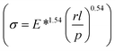
to predict the pull-off strength (σ) was established for a given fibril radius (r), fibril height (l), preload (p), and effective Young's modulus (E*). The pull-off force values recorded for hierarchical PMMA fibrous structure were also used to validate the empirical relationship. The empirical relationship demonstrated good correlation between the recorded pull-off strength and system parameters. We believe that this empirical relationship will be helpful in designing high strength synthetic dry-adhesives as the relationship can be used to predict the pull-off strength a priori.


1 Introduction

Bio-inspired materials ranging from bio-nanomaterials, to hybrid organic/inorganic materials and composites1–4 have found applications in a wide variety of fields including neural memory devices, drug delivery, adhesives, and biosensors.5–7 In the last decade, much of attention has been focused on fabricating synthetic durable adhesives that can be reused multiple times.8–11 These synthetic adhesives are fabricated by mimicking the structure and adhesion mechanisms that are found in nature. For instance, synthetic dry-adhesives are fabricated by taking cues from the hierarchical structures found on the feet of geckos. Each foot of a gecko possesses arrays of millions of hair-like setae (diameter ∼20 μm, height ∼130 μm). Each of these seta branches out into finer structures called spatulae (width ∼200 nm, thickness ∼5 nm). These structures make intimate physical contact with any given surface due to their nanometer sized spatulae and the hierarchical design of the structures. When properly oriented, preloaded and dragged, each of these hierarchical structures (a single seta with ∼1000 spatulae) can generate 200 μN of shear adhesion and 40 μN of normal adhesion respectively.12,13 Although, the animal can generate large adhesion and can cling on to any given surface, they can also be easily detached when they are peeled off at certain angle.12–16 The fact that natural materials depend on their geometry rather than their surface chemistry paved the way for fabrication of synthetic biomimetic adhesives.9,10,17–19 Fabrication of such hierarchical structures that can be repeatedly attached and detached from surfaces while maintaining their adhesive strength are appropriate for climbing robot applications.

It is well known that adhesion of these synthetic dry-adhesives depend on geometrical parameters. For example, Sitti et al.20 in their study demonstrated that pull-off force increases when fibril radius (r) is reduced. They attribute this to the increase in contact area (A). Similarly, Greiner et al.21 demonstrated that pull-off force increases when the height of the fibril (l) is increased or when the effective Young's modulus (E*) of the structures is decreased. They attribute this to enhanced compliance of fibrils to a given surface that results in an increase in contact area. In yet another study, Murphy et al.22 used nanoindenter to measure the pull-off force and demonstrated the pull-off force to increase with the preload (p). They also attribute this to the increase in contact area. As the indenter is pressed into the fibrils, the fibrils deform elastically allowing neighboring fibrils to come into contact with the indenter tip, leading to an increase in contact area.

Other studies23–25 have carefully investigated the effect of fiber size, preload etc. on pull-off force. Here, we compile the data presented in literature and demonstrate that by performing dimensional analysis, the pull-off force can be correlated to the parameters such as radius of the fibril (r), contact area (A), height of the fibril (l), preload (p), and effective Young's modulus (E*). We use electrospinning26–30 to fabricate hierarchical poly(methyl methacrylate) (PMMA) structures (see Fig. 1) and examine their adhesive performance. We also use our experimental results to verify the validity of the empirical relationship.


image file: c6ra05757h-f1.tif
Fig. 1 (A) Schematic depicting the fabrication of PMMA fibers using electrospinning; and (B) schematic depicting the AAO template wetting method that is used to produce hierarchical PMMA fibrous structures.

2 Experimental section

2.1. Materials

Poly(methyl methacrylate) (PMMA, Mw = 120[thin space (1/6-em)]000) and N,N-dimethyl formamide (DMF) were purchased from Sigma-Aldrich, Singapore. Anodized aluminum oxide (AAO) templates (pore diameter ∼100 nm, thickness ∼ 0.5 mm) were purchased from Shanghai Shangmu Technology Co., Ltd, China.

2.2. Electrospinning

The solution for electrospinning was prepared by dissolving PMMA in DMF. The concentration of PMMA in DMF was chosen as 29 wt%. The solution was stirred for 24 h at 25 °C to prepare a homogeneous colorless solution. The solution was then electrospun using a Nanospinner 24 (Inovenso, Turkey) to fabricate PMMA fibrous membrane. The humidity and the temperature within the electrospinning chamber were maintained at ∼60% and ∼25 °C respectively. Electrospinning31–34 was conducted by applying 1 kV cm−1 electric field between the metallic needle (inner diameter = 1 mm and outer diameter = 3 mm) and the collector. The flow rate of the polymer solution through the needle was maintained at 1.5 ml h−1. A rotating drum (rotational speed ∼3000 rpm) covered with an aluminum foil was used to collect aligned fibers. The obtained as-spun fibers were then dried at 80 °C for 24 h to remove any residual solvent.

2.3. Fabrication of hierarchical structures

The electrospun PMMA fibers collected on the aluminum foil were placed on AAO template and then sandwiched between 2 brass plates (cross-sectional area = 7 × 7 cm2 and thickness = 0.6 cm) as shown in Fig. 1. A dead weight was then placed on top of the brass plates to apply ∼3.46 kPa pressure on the fiber–AAO template setup. This setup ensured good physical contact between the PMMA fibers and the AAO template. Following this, the setup was heated above the glass transition temperature of PMMA to enable the flow of PMMA within the porous channels of the AAO and to grow nanometer sized pillars on the surface of the fibers.

2.4. Microstructure characterization and pull-off force measurements

The morphology of the fibrous membranes with and without nanopillars were examined using a scanning electron microscope (SEM, JEOL, JSM-6700F). The samples were examined at an accelerating voltage of 5 kV.

TI 950 triboindenter (Hysitron Inc., Minneapolis, MN, USA) with ∼1 nN load resolution, ∼0.04 nm z-axis displacement resolution and ∼0.05 μm as diamond tip positioning system resolution in the xy directions was used to measure the adhesion performance of the samples. Measurements were made using a calibrated 60° conical flat diamond indenter of 10 μm diameter. The measurements were carried out under displacement control. Load vs. tip displacement curves recorded during the indentation tests were then used to measure the pull-off force between the diamond tip and the samples.

Test samples with 0.5 cm2 cross-sectional area and ∼150 μm thickness were mounted on a steel magnetic stub. As the electrospun fibrous membrane consists of aligned fibers with void spaces, care was taken to ensure that the tip only indented the fibrous region of the sample. This was achieved by visualizing the selected indentation region using an optical microscope connected to the triboindenter. Care was also taken to ensure that the indentation depth was less than one-tenth of sample's thickness to eliminate substrate effect.35,36 Further, to investigate the effect of indentation depth, the sample was indented at a chosen spot and the depth of indentation was increased from 50 to 200 nm. Sufficient time (∼1 h) was given between consecutive test to allow fiber (with and without surface pillars) to relax back to its initial unstrained state. The indentation locations for subsequent tests were chosen such that they were at least 20 μm (two times the contact radius) away from the previous test location.

3 Results and discussion

Fig. 2 shows the SEM images of the neat PMMA fibers. It is evident from the SEM images that smooth, uniform and bead free fibers are obtained. Fibers are seen to be highly aligned and closely packed. This is attributed to the high rotational speed of the collector (3000 rpm) used during electrospinning. The average diameter (d) of the fibers is determined to be ∼3.2 μm from these SEM images. Hierarchical structures are obtained by growing nanopillars on the surface of the PMMA fibers. For this purpose, the electrospun PMMA fibers are deposited on the AAO templates and the setup is placed inside an oven for thermal annealing. The effect of annealing temperature and time is also investigated. The thermal annealing temperature is varied from 135 °C to 155 °C and the annealing time is varied from 0.5 h to 18 h. The height of nanopillars grown on the surface of the fibers is seen to increase with annealing time and temperature. Fig. 3 shows the representative SEM images of the samples that are annealed at 150 °C for two different annealing times viz. 1 h and 18 h. It is evident from Fig. 3 that tall and uniformly distributed nanopillars are obtained when the annealing time is increased from 1 h to 18 h. Samples that are annealed at 150 °C for 18 h are seen to have uniformly distributed vertical nanopillars on the surface of PMMA fibers. The average height of pillars is determined to be ∼400 nm. These samples are used to investigate the adhesion performance of hierarchical structures.
image file: c6ra05757h-f2.tif
Fig. 2 SEM images of aligned PMMA fibers. The inset shows the magnified image of the fibers.

image file: c6ra05757h-f3.tif
Fig. 3 SEM images of (A) PMMA hierarchical structures obtained by annealing the samples at 150 °C for 18 h; and (B) PMMA fibrous structures obtained by annealing the samples at 150 °C for 1 h. It is clear from these SEM images that hierarchical fibrous structures are obtained when the samples are annealed at 150 °C for 18 h.

In the next step, the adhesion performance of neat PMMA fibers and PMMA fibers with surface nanopillars (hierarchical fibrous structures) is investigated using a nanoindenter. Typically, the indenter tip is driven into the sample by applying a predefined force under load control or by controlling the displacement of the transducer under displacement control. In our study, a pre-calibrated transducer is actuated under displacement control. A withdrawal height of 100 nm is defined to ensure that the tip is free of any residual contact or surface forces. The surface is detected by approaching the sample and touching its surface with a 2 μN force. Following this, the flat circular diamond tip indents the sample's surface with a predefined transducer displacement. During this stage, the load is seen to increase with displacement of transducer into the sample surface. The tip indents the sample until the predefined transducer displacement is reached. The indenter is then withdrawn from the sample's surface and the force required to completely detach the indenter from the surface of the sample is recorded as pull-off force.

Fig. 4A shows the typical force–displacement curves recorded under ambient conditions using the nanoindenter. These load–displacement curves are used to estimate the pull-off force between the indenter and the sample. The adhesion performance of both neat PMMA fibers and hierarchical fibrous structures are investigated. Typically, the indentation measurements depend on a number of experimental parameters such as loading rate, and withdrawal rate. In our study, the loading rate is kept constant at 10 nm s−1 and the withdrawal rate at 500 nm s−1. This enabled us to plot load as a function of indentation depth of the transducer while keeping other parameters constant (see Fig. 4A). The pull-off force is estimated from the unloading part of the load–indentation depth curves. Fig. 4B shows the schematic of the flat indenter that is brought in contact with the hierarchical PMMA fibrous structures.


image file: c6ra05757h-f4.tif
Fig. 4 (A) Load–displacement curves recorded using a nanoindenter on hierarchical fibrous structures as a function of indentation depth. The pull-off force required to detach the indenter from the sample's surface is estimated from these load–displacement curves. The pull-off force is seen to increase with an increase in the indentation depth; and (B) schematic illustrating the 10 μm diameter flat circular diamond indenter that is brought in contact with the nanopillars present on the surface of PMMA fibers.

The recorded pull-off forces for both neat PMMA fibers and PMMA hierarchical fibrous structures are plotted as a function of indentation depth in Fig. 5. Fig. 5 indicates that the pull-off force increases with indentation depth for both the samples. However, higher pull-off forces are recorded for hierarchical samples compared to the neat PMMA samples. High pull-off force recorded for hierarchical structures is also reported by others22,37,38 compared to pull-off force recorded for their neat counterparts.


image file: c6ra05757h-f5.tif
Fig. 5 Pull-off force vs. indentation depth recorded for hierarchical fibrous structures and neat PMMA fibers. Error bars represent standard deviation.

For hierarchical fibrous structures, nanopillars on PMMA fibers reduce the effective modulus of the structure. This allows soft flexible nanopillars on PMMA fibers to adapt to the irregularities present on the surface of the indenter tip, which leads to increase in contact area. The contact area is given by (h × w), where h is the diameter of indenter and w is the contact width (see Fig. 4B). Contact width is calculated from the relation w ≈ 2(pd/2E*)1/3.39 It is clear from this expression that contact width increases with preload (p). As the indenter is pressed deeper into the surface of the samples, the nanopillars present on the surface of the PMMA fibers elastically deform and allow neighboring pillars to come into contact with the indenter tip, leading to an increase in contact area. This explains why the pull-off force increases with indentation depth.22,40 The absolute values of the measured pull-off force increases from 2.1 to 3.7 μN as the indentation depth increases from 50 to 200 nm.

The experimental results are comparable with pull-off force recorded for vertically oriented carbon nanotubes (VACNTs) and poly(dimethyl-siloxane) (PDMS).41 Chen et al.41 measured ∼50 μN and ∼60 μN pull-off force for VACNTs and PDMS respectively for ∼3500 nm indentation depth. Their results show that the recorded pull-force is roughly one order of magnitude larger than the pull-off force reported in this study. However, it should be pointed out that the indentation depth is also an order of magnitude larger than the indentation depth used in our study. In our study, we noticed that when the indentation depth is increased beyond 200 nm, the pull-off force values reduce. The reduction in the pull-off force is due to the buckling of nanopillars present on the surface of the PMMA fibers. The nanopillars lose contact with the indenter tip when they buckle. This reduces the contact area and results in lower pull-off force values.37

For neat PMMA fibers, the measured pull-off force is seen to marginally increase from 0.4 to 1.0 μN when the indentation depth is increased from 50 to 200 nm. The neat samples fail to intimately come in contact with the surface of the indenter as the fiber's diameter does not match the surface roughness of the indenter. This explains the lower values of pull-off force recorded for neat samples and also explains why there is no appreciable increase in pull-off force when the indentation depth is increased. This is in agreement with the Johnson–Kendall–Roberts (JKR) theory, which predicts that pull-off force is independent of preload/indentation depth for neat samples.42

Following this, we also performed dimensional analysis to correlate pull-off force with the parameters outlined in Section 1.

 
image file: c6ra05757h-t2.tif(1)

Eqn (1) shows that the pull-off force ‘F’ is a function of radius of the fibril (r), contact area (A), height of the fibril (l), preload (p), and effective Young's modulus (E*). Pull-off strength (σ) is determined by dividing pull-off force with the contact area. Thus, the pull-off strength is now a function of r, l, p and E*.

The dimensional analysis is applied to the eqn (1) considering r, l and p as the repeating variables. Thus, it gives rise to two dimensionless numbers viz. dimensionless pull-off strength image file: c6ra05757h-t3.tif and dimensionless Young's modulus image file: c6ra05757h-t4.tif. In order to investigate the empirical relationship between these dimensionless numbers, we use the results reported by Greiner et al.43 and plot dimensionless pull-off strength vs. dimensionless Young's modulus in Fig. 6. Dimensionless pull-off strength vs. dimensionless Young's modulus is plotted for different aspect ratio fibrils (λ) in Fig. 6. It is evident that the data points for all aspect ratios collapse on a straight line demonstrating good correlation between the dimensionless pull-off strength and dimensionless Young's modulus. The empirical relationship between the dimensionless pull-off strength and dimensionless Young's modulus is determined by applying a linear regression to fit a straight line to the data. The equation for the straight line is determined to be:

 
image file: c6ra05757h-t5.tif(2)
where 1.54 is the slope of the fitted line. Using eqn (2), the expression for pull-off strength is determined as:
 
image file: c6ra05757h-t6.tif(3)


image file: c6ra05757h-f6.tif
Fig. 6 Plot of dimensionless pull-off strength vs. dimensionless Young's modulus. The graph depicts a very good correlation between dimensionless numbers for majority of the data set. The data set used for this plot is obtained from Greiner et al.43

Thus, if the parameters such as r, l, E* and p are known, the pull-strength can be determined a priori using eqn (3).

To evaluate the validity of this empirical relationship, we determine the dimensionless pull-off strength and dimensionless Young's modulus for our hierarchical PMMA fibrous samples and plot them in Fig. 7. Pull-off strength is estimated by dividing the recorded pull-off force by the contact area. To determine the contact area, the indenter tip is assumed to be perfectly aligned on an aligned PMMA fiber (see Fig. 4B). The contact area is given by the expression (h × w). E* for PMMA is taken as 0.12 GPa.47 The radius of pillar r ∼ 50 nm, and height of pillar l ∼ 400 nm are measured from the SEM images. Preload (p) is the maximum load acting on the sample for a given indentation depth, which is obtained from the plot shown in Fig. 4A. We also determine the dimensionless pull-off strength and dimensionless Young's modulus for materials used in literature for dry-adhesive applications. Table 1 lists the materials reported in literature and their parameters such as recorded pull-off strength, preload, radius of fibrils, height of the fibrils, and effective Young modulus. The dimensionless pull-off strength vs. dimensionless Young's modulus for the materials that are listed in Table 1 is plotted in Fig. 7 along with our results. Similar correlation trend (eqn (3)) is evident between the dimensionless pull-off strength and dimensionless Young's modulus for the data plotted in Fig. 7. Thus, we believe that this empirical relationship can be extended to other materials and structures and will enable the researchers to estimate the pull-off force values based on the geometrical parameters and preload values.


image file: c6ra05757h-f7.tif
Fig. 7 Dimensionless pull-off strength vs. dimensionless Young's modulus plotted for the following materials: (1) PVS,37 (2) PDMS,21 (3) PDMS,43 (4) VACNTs,41 (5) PU,44 (6) PVS,38 (7) PDMS,45 (8) PA,46 (9) PDMS,21 (10) PU22 and (11) PMMA.
Table 1 Summary of materials used in literature for fabrication of synthetic adhesives and their recorded pull-off strength values. The parameters such as preload, radius of fibrils, height of the fibrils, and effective Young modulus are also shown in the table
Material Effective Young modulus (E*) (GPa) Pull-off strength (σ) (kN m−2) Preload (p) (mN) Radius (r) (μm) Height (l) (μm) Reference
Polyvinylsiloxane (PVS) 0.003 32.4 600 125 400 37
Poly(dimethylsiloxane) (PDMS) 0.00143 2.15 2 5 5 21
Poly(dimethylsiloxane) (PDMS) 0.0015 10 1 2.5 20 43
Vertically oriented carbon nanotubes (VACNTs) 0.01 26 0.8 0.008 100 41
Polyurethane (PU) 0.003 50 5 35 100 44
Polyvinylsiloxane (PVS) 0.003 60.60 60 60 100 38
Poly(dimethylsiloxane) (PDMS) 4.4 × 10⁻⁴ 5 0.4 10 80 45
Polyimide (PI) 0.003 30 0.1 2 2 46
Poly(dimethylsiloxane) (PDMS) 0.00193 2.15 1 0.1 4 21
Polyurethane (PU) 0.003 15.625 64 25 100 22


4 Conclusion

In this study, hierarchical PMMA structures for dry-adhesive application are fabricated using electrospinning and template wetting method. The adhesive performance of these structures is investigated using nanoindentation. Hierarchical PMMA structures exhibited higher pull-off force compared to neat PMMA samples. Enhanced adhesion is attributed to increase in contact area due to reduction in effective modulus of the hierarchical sample. These results indicate that the hierarchical PMMA structures can potentially be used as reusable adhesive for wide variety of applications. Following this, an empirical relationship between dimensionless pull-off force and dimensionless Young's modulus is determined to predict the pull-off strength of synthetic dry-adhesives. The empirical relationship showed good correlation between the pull-off strength and system parameters. We believe that our empirical relationship will aid in the fabrication of high strength and durable synthetic dry-adhesives.

Acknowledgements

The authors would like to acknowledge the support of SUTD-MIT International Design Centre (Project No. IDG31400101). The authors also acknowledge the financial support of SUTD start-up grant (Grant No. SRG-EPD-2013–055).

References

  1. N. Ashammakhi, A. Ndreu, L. Nikkola, I. Wimpenny and Y. Yang, Regener. Med., 2008, 3, 547–574 CrossRef CAS PubMed.
  2. E. Munch, M. E. Launey, D. H. Alsem, E. Saiz, A. P. Tomsia and R. O. Ritchie, Science, 2008, 322, 1516–1520 CrossRef CAS PubMed.
  3. X. Yao, Y. Song and L. Jiang, Adv. Mater., 2011, 23, 719–734 CrossRef CAS PubMed.
  4. Z. Su, J. Li, Z. Ouyang, M. M. Arras, G. Wei and K. D. Jandt, RSC Adv., 2014, 4, 14833–14839 RSC.
  5. S. Igarashi, J. Tanaka and H. Kobayashi, J. Nanosci. Nanotechnol., 2007, 7, 814–817 CrossRef CAS PubMed.
  6. U. K. Parashar, P. S. Saxena and A. Srivastava, Dig. J. Nanomater. Biostruct., 2008, 3, 81–87 Search PubMed.
  7. A. K. Geim, S. V. Dubonos, I. V. Grigorieva, K. S. Novoselov, A. A. Zhukov and S. Y. Shapoval, Nat. Mater., 2003, 2, 461–463 CrossRef CAS PubMed.
  8. N. Cañas, M. Kamperman, B. Völker, E. Kroner, R. M. McMeeking and E. Arzt, Acta Biomater., 2012, 8, 282–288 CrossRef PubMed.
  9. H. E. Jeong and K. Y. Suh, Nano Today, 2009, 4, 335–346 CrossRef CAS.
  10. H. Ko, Z. X. Zhang, J. C. Ho, K. Takei, R. Kapadia, Y. L. Chueh, W. Z. Cao, B. A. Cruden and A. Javey, Small, 2010, 6, 22–26 CrossRef CAS PubMed.
  11. R. Sahay, H. Y. Low, A. Baji, S. Foong and K. L. Wood, RSC Adv., 2015, 5, 50821–50832 RSC.
  12. K. Autumn, S. Hsieh, D. Dudek, J. Chen, C. Chitaphan and R. Full, J. Exp. Biol., 2006, 209, 260–272 CrossRef CAS PubMed.
  13. K. Autumn, C. Majidi, R. Groff, A. Dittmore and R. Fearing, J. Exp. Biol., 2006, 209, 3558–3568 CrossRef CAS PubMed.
  14. K. Autumn, Y. A. Liang, S. T. Hsieh, W. Zesch, W. P. Chan, T. W. Kenny, R. Fearing and R. J. Full, Nature, 2000, 405, 681–685 CrossRef CAS PubMed.
  15. K. Autumn and A. M. Peattie, Integr. Comp. Biol., 2002, 42, 1081–1090 CrossRef PubMed.
  16. K. Autumn, M. Sitti, Y. A. Liang, A. M. Peattie, W. R. Hansen, S. Sponberg, T. W. Kenny, R. Fearing, J. N. Israelachvili and R. J. Full, Proc. Natl. Acad. Sci. U. S. A., 2002, 99, 12252–12256 CrossRef CAS PubMed.
  17. A. E. Filippov and V. Popov, Phys. Lett. A, 2006, 358, 309–312 CrossRef CAS.
  18. N. J. Glassmaker, A. Jagota and C.-Y. Hui, Acta Biomater., 2005, 1, 367–375 CrossRef PubMed.
  19. A. Peyvandi, S. U. Abideen, Y. Huang, I. Lee, P. Soroushian and J. Lu, Appl. Surf. Sci., 2014, 289, 586–591 CrossRef CAS.
  20. M. Sitti and R. S. Fearing, J. Adhes. Sci. Technol., 2003, 17, 1055–1073 CrossRef CAS.
  21. C. Greiner, E. Arzt and A. del Campo, Adv. Mater., 2009, 21, 479–482 CrossRef CAS.
  22. M. P. Murphy, S. Kim and M. Sitti, ACS Appl. Mater. Interfaces, 2009, 1, 849–855 CAS.
  23. J. Y. Chung and M. K. Chaudhury, J. Adhes., 2005, 81, 1119–1145 CrossRef CAS.
  24. A. J. Crosby, M. Hageman and A. Duncan, Langmuir, 2005, 21, 11738–11743 CrossRef CAS PubMed.
  25. C. Greiner, A. del Campo and E. Arzt, Langmuir, 2007, 23, 3495–3502 CrossRef CAS PubMed.
  26. R. Sahay, C. Teo and Y. Chew, J. Fluid Mech., 2013, 735, 150–175 CrossRef.
  27. D. H. Reneker, A. L. Yarin, H. Fong and S. Koombhongse, J. Appl. Phys., 2000, 87, 4531–4547 CrossRef CAS.
  28. A. L. Yarin, W. Kataphinan and D. H. Reneker, J. Appl. Phys., 2005, 98, 1–12 CrossRef.
  29. A. L. Yarin, S. Koombhongse and D. H. Reneker, J. Appl. Phys., 2001, 90, 4836–4846 CrossRef CAS.
  30. Z. Su, J. Ding and G. Wei, RSC Adv., 2014, 4, 52598–52610 RSC.
  31. Z. Su, J. Li, Q. Li, T. Ni and G. Wei, Carbon, 2012, 50, 5605–5617 CrossRef CAS.
  32. Y. Li, P. Zhang, Z. Ouyang, M. Zhang, Z. Lin, J. Li, Z. Su and G. Wei, Adv. Funct. Mater., 2016, 26, 2122–2134 CrossRef CAS.
  33. P. Zhang, X. Zhao, X. Zhang, Y. Lai, X. Wang, J. Li, G. Wei and Z. Su, ACS Appl. Mater. Interfaces, 2014, 6, 7563–7571 CAS.
  34. P. Zhang, X. Zhao, Y. Ji, Z. Ouyang, X. Wen, J. Li, Z. Su and G. Wei, J. Mater. Chem. B, 2015, 3, 2487–2496 RSC.
  35. M. Wang, K. M. Liechti, J. M. White and R. M. Winter, J. Mech. Phys. Solids, 2004, 52, 2329–2354 CrossRef CAS.
  36. B. Oommen and K. Van Vliet, Thin Solid Films, 2006, 513, 235–242 CrossRef CAS.
  37. A. Peressadko and S. N. Gorb, J. Adhes., 2004, 80, 247–261 CrossRef CAS.
  38. S. Gorb, M. Varenberg, A. Peressadko and J. Tuma, J. R. Soc., Interface, 2007, 4, 271–275 CrossRef CAS PubMed.
  39. M. F. Ashby, Materials Selection in Mechanical Design, Elsevier Science, 2004 Search PubMed.
  40. T. W. Kim and B. Bhushan, J. R. Soc., Interface, 2008, 5, 319–327 CrossRef PubMed.
  41. B. Chen, P. Goldberg Oppenheimer, T. A. V. Shean, C. T. Wirth, S. Hofmann and J. Robertson, J. Phys. Chem. C, 2012, 116, 20047–20053 CAS.
  42. K. Johnson, K. Kendall and A. Roberts, Proc. R. Soc. London, Ser. A, 1971, 324, 301–313 CrossRef CAS.
  43. C. Greiner, A. del Campo and E. Arzt, Langmuir, 2007, 23, 3495–3502 CrossRef CAS PubMed.
  44. M. P. Murphy, B. Aksak and M. Sitti, Small, 2009, 5, 170–175 CrossRef CAS PubMed.
  45. A. Parness, D. Soto, N. Esparza, N. Gravish, M. Wilkinson, K. Autumn and M. Cutkosky, J. R. Soc., Interface, 2009, 2009, 0048 Search PubMed.
  46. J. Davies, S. Haq, T. Hawke and J. P. Sargent, Int. J. Adhes. Adhes., 2009, 29, 380–390 CrossRef CAS.
  47. W. Jiang, L. Wang, H. Liu, H. Ma, H. Tian, B. Chen, Y. Shi, L. Yin and Y. Ding, RSC Adv., 2014, 4, 42002–42008 RSC.

This journal is © The Royal Society of Chemistry 2016
Click here to see how this site uses Cookies. View our privacy policy here.